|
Intercondylar Eminence
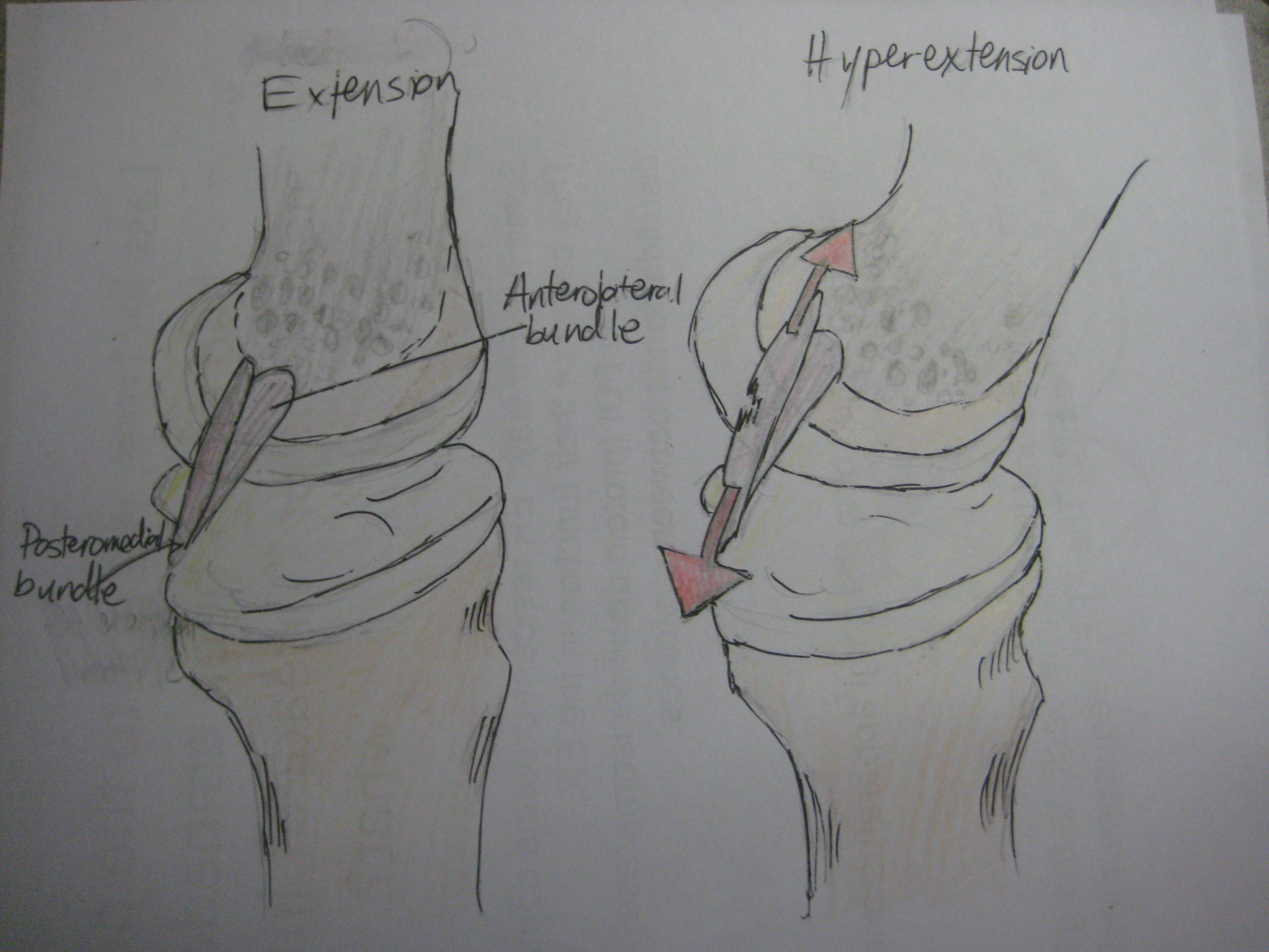
The intercondylar area is the separation between the medial and lateral condyle on the upper extremity of the tibia. The anterior and posterior cruciate ligaments and the menisci attach to the intercondylar area. The intercondyloid eminence is composed of the medial and lateral intercondylar tubercles, and divides the intercondylar area into an anterior and a posterior area. Structure Anterior area The anterior intercondylar area (or anterior intercondyloid fossa) is an area on the tibia, a bone in the lower leg. Together with the posterior intercondylar area it makes up the intercondylar area. The intercondylar area is the separation between the medial and lateral condyle located toward the proximal portion of the tibia. The intercondylar eminence composed of the medial and lateral intercondylar tubercle divides the intercondylar area into anterior and posterior part. The anterior intercondylar area is the location where the anterior cruciate ligament attaches to the ti ... [...More Info...] [...Related Items...] OR: [Wikipedia] [Google] [Baidu] |
Posterior Intercondylar Area
The intercondylar area is the separation between the medial and lateral condyle on the upper extremity of the tibia. The anterior and posterior cruciate ligaments and the menisci attach to the intercondylar area. The intercondyloid eminence is composed of the medial and lateral intercondylar tubercles, and divides the intercondylar area into an anterior and a posterior area. Structure Anterior area The anterior intercondylar area (or anterior intercondyloid fossa) is an area on the tibia, a bone in the lower leg. Together with the posterior intercondylar area it makes up the intercondylar area. The intercondylar area is the separation between the medial and lateral condyle located toward the proximal portion of the tibia. The intercondylar eminence composed of the medial and lateral intercondylar tubercle divides the intercondylar area into anterior and posterior part. The anterior intercondylar area is the location where the anterior cruciate ligament attaches to t ... [...More Info...] [...Related Items...] OR: [Wikipedia] [Google] [Baidu] |
Intercondylar Eminence
The intercondylar area is the separation between the medial and lateral condyle on the upper extremity of the tibia. The anterior and posterior cruciate ligaments and the menisci attach to the intercondylar area. The intercondyloid eminence is composed of the medial and lateral intercondylar tubercles, and divides the intercondylar area into an anterior and a posterior area. Structure Anterior area The anterior intercondylar area (or anterior intercondyloid fossa) is an area on the tibia, a bone in the lower leg. Together with the posterior intercondylar area it makes up the intercondylar area. The intercondylar area is the separation between the medial and lateral condyle located toward the proximal portion of the tibia. The intercondylar eminence composed of the medial and lateral intercondylar tubercle divides the intercondylar area into anterior and posterior part. The anterior intercondylar area is the location where the anterior cruciate ligament attaches to the ti ... [...More Info...] [...Related Items...] OR: [Wikipedia] [Google] [Baidu] |
Tibia
The tibia (; : tibiae or tibias), also known as the shinbone or shankbone, is the larger, stronger, and anterior (frontal) of the two Leg bones, bones in the leg below the knee in vertebrates (the other being the fibula, behind and to the outside of the tibia); it connects the knee with the ankle bones, ankle. The tibia is found on the anatomical terms of location#Medial, medial side of the leg next to the fibula and closer to the median plane. The tibia is connected to the fibula by the interosseous membrane of leg, forming a type of fibrous joint called a syndesmosis with very little movement. The tibia is named for the flute ''aulos, tibia''. It is the second largest bone in the human body, after the femur. The leg bones are the strongest long bones as they support the rest of the body. Structure In human anatomy, the tibia is the second largest bone next to the femur. As in other vertebrates the tibia is one of two bones in the lower leg, the other being the fibula, and is a ... [...More Info...] [...Related Items...] OR: [Wikipedia] [Google] [Baidu] |
Medial Condyle Of Tibia
The medial condyle is the medial (or inner) portion of the upper extremity of tibia. It is the site of insertion for the semimembranosus muscle. See also * Lateral condyle of tibia * Medial collateral ligament The medial collateral ligament (MCL), also called the superficial medial collateral ligament (sMCL) or tibial collateral ligament (TCL), is one of the major ligaments of the knee. It is on the medial (inner) side of the knee joint and occurs in ... Additional images File:Gray258.png, Bones of the right leg. Anterior surface. File:Gray259.png, Bones of the right leg. Posterior surface. File:Slide2bib.JPG, Right knee in extension. Deep dissection. Posterior view. File:Slide2cocc.JPG, Right knee in extension. Deep dissection. Posterior view. References External links * * * () Bones of the lower limb Tibia {{musculoskeletal-stub ... [...More Info...] [...Related Items...] OR: [Wikipedia] [Google] [Baidu] |
Lateral Condyle Of Tibia
The lateral condyle is the lateral portion of the upper extremity of tibia. It serves as the insertion for the biceps femoris muscle (small slip). Most of the tendon of the biceps femoris inserts on the fibula. See also * Gerdy's tubercle * Medial condyle of tibia Additional images File:Gray258.png, Bones of the right leg. Anterior surface. File:Slide2bib.JPG, Right knee in extension. Deep dissection. Posterior view. File:Slide2cocc.JPG, Right knee in extension. Deep dissection. Posterior view. References External links * * * () Bones of the lower limb Tibia {{musculoskeletal-stub ... [...More Info...] [...Related Items...] OR: [Wikipedia] [Google] [Baidu] |
Upper Extremity Of Tibia
The tibia (; : tibiae or tibias), also known as the shinbone or shankbone, is the larger, stronger, and anterior (frontal) of the two Leg bones, bones in the leg below the knee in vertebrates (the other being the fibula, behind and to the outside of the tibia); it connects the knee with the ankle bones, ankle. The tibia is found on the anatomical terms of location#Medial, medial side of the leg next to the fibula and closer to the median plane. The tibia is connected to the fibula by the interosseous membrane of leg, forming a type of fibrous joint called a syndesmosis with very little movement. The tibia is named for the flute ''aulos, tibia''. It is the second largest bone in the human body, after the femur. The leg bones are the strongest long bones as they support the rest of the body. Structure In human anatomy, the tibia is the second largest bone next to the femur. As in other vertebrates the tibia is one of two bones in the lower leg, the other being the fibula, and is a ... [...More Info...] [...Related Items...] OR: [Wikipedia] [Google] [Baidu] |
Anterior Cruciate Ligament
The anterior cruciate ligament (ACL) is one of a pair of cruciate ligaments (the other being the posterior cruciate ligament) in the human knee. The two ligaments are called "cruciform" ligaments, as they are arranged in a crossed formation. In the quadruped stifle joint (analogous to the knee), based on its anatomical position, it is also referred to as the cranial cruciate ligament. The term cruciate is Latin for cross. This name is fitting because the ACL crosses the posterior cruciate ligament to form an "X". It is composed of strong, fibrous material and assists in controlling excessive motion by limiting mobility of the joint. The anterior cruciate ligament is one of the four main ligaments of the knee, providing 85% of the restraining force to anterior tibial displacement at 30 and 90° of knee flexion. The ACL is the most frequently injured ligament in the knee. Structure The ACL originates from deep within the notch of the distal femur. Its proximal fibers fan out alo ... [...More Info...] [...Related Items...] OR: [Wikipedia] [Google] [Baidu] |
Posterior Cruciate Ligament
The posterior cruciate ligament (PCL) is a ligament in each knee of humans and various other animals. It works as a counterpart to the anterior cruciate ligament (ACL). It connects the posterior intercondylar area of the tibia to the Medial condyle of femur, medial condyle of the femur. This configuration allows the PCL to resist forces pushing the tibia posteriorly relative to the femur. The PCL and ACL are intracapsular ligaments because they lie deep within the knee joint. They are both isolated from the fluid-filled synovial cavity, with the synovial membrane wrapped around them. The PCL gets its name by attaching to the posterior portion of the tibia. The PCL, Anterior cruciate ligament, ACL, medial collateral ligament, MCL, and fibular collateral ligament, LCL are the four main ligaments of the knee in primates. Structure The PCL is located within the knee joint where it stabilizes the articulating bones, particularly the femur and the tibia, during movement. It originates ... [...More Info...] [...Related Items...] OR: [Wikipedia] [Google] [Baidu] |
Meniscus (anatomy)
A meniscus (: menisci or meniscuses) is a crescent-shaped fibrocartilage, fibrocartilaginous anatomy, anatomical structure that, in contrast to an articular disc, only partly divides a synovial joint#Structure, joint cavity.Platzer (2004), p 208 In humans, they are present in the knee, wrist, acromioclavicular joint, acromioclavicular, sternoclavicular joint, sternoclavicular, and temporomandibular joints; in other animals they may be present in other joints. Generally, the term "meniscus" is used to refer to the cartilage of the knee, either to the lateral meniscus, lateral or medial meniscus. Both are Cartilaginous#Fibrocartilage, cartilaginous tissues that provide structural integrity to the knee when it undergoes tension (mechanics), tension and torsion (mechanics), torsion. The menisci are also known as "semi-lunar" cartilages, referring to their half-moon, crescent shape. The term "meniscus" is from the Ancient Greek word (), meaning "crescent". Structure The menisci of t ... [...More Info...] [...Related Items...] OR: [Wikipedia] [Google] [Baidu] |
Lower Leg
The leg is the entire lower limb (anatomy), limb of the human body, including the foot, thigh or sometimes even the hip or Gluteal muscles, buttock region. The major bones of the leg are the femur (thigh bone), tibia (shin bone), and adjacent fibula. There are 30 bones in each leg. The thigh is located in between the hip and knee. The calf (leg), calf (rear) and Tibia#Structure, shin (front), or shank, are located between the knee and ankle. Legs are used for standing, many forms of human movement, recreation such as dancing, and constitute a significant portion of a person's mass. Evolution has led to the human leg's development into a mechanism specifically adapted for efficient bipedalism, bipedal gait. While the capacity to walk upright is not unique to humans, other primates can only achieve this for short periods and at a great expenditure of energy. In humans, female legs generally have greater hip anteversion and tibiofemoral angles, while male legs have longer femur a ... [...More Info...] [...Related Items...] OR: [Wikipedia] [Google] [Baidu] |


