|
Ictidosaurus Angusticeps
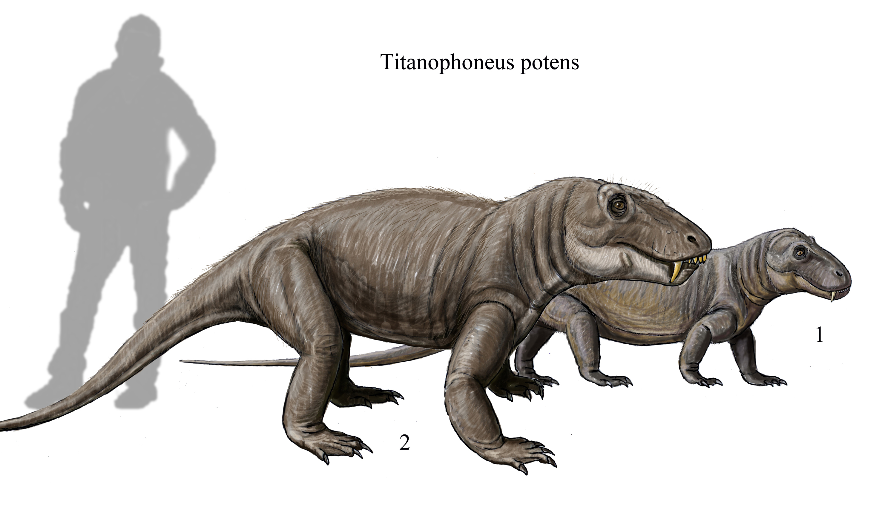
''Ictidosaurus'' was a therapsid genus found in the Abrahamskraal Formation of South Africa, which lived during the middle Permian period. Fossils of the type species were found in the ''Tapinocephalus'' (Capitanian age, 265.8-260.4 Ma), and the base of the ''Eodicynodon'' (Wordian age, 268–265.8 Ma) assembly zones, of the Karoo Basin. Older classifications of the species, along with many other specimens found in the Iziko South African Museum archives, were originally classified within therocephalian family names, in this case the Ictidosauridae, which has been reclassified as belonging to the Scylacosauridae. The type species is ''I. angusticeps''. Description Type specimen The holotype was of the ''Ictidosaurus angusticeps'' labeled SAM-PK-630 (NMQR 2910), found in the Tapinocephalus assembly zone. The skull measured 168 mm from snout to posterior of the left mandible, 41 mm across the skull in-between the canines, and 41 mm in height, and has been heavily disturbed by fr ... [...More Info...] [...Related Items...] OR: [Wikipedia] [Google] [Baidu] |
Middle Permian
The Guadalupian is the second and middle series/ epoch of the Permian. The Guadalupian was preceded by the Cisuralian and followed by the Lopingian. It is named after the Guadalupe Mountains of New Mexico and Texas, and dates between 272.95 ± 0.5 – 259.1 ± 0.4 Mya. The series saw the rise of the therapsids, a minor extinction event called Olson's Extinction and a significant mass extinction called the end-Capitanian extinction event. The Guadalupian was previously known as the Middle Permian. Name and background The Guadalupian is the second and middle series or epoch of the Permian. Previously called Middle Permian, the name of this epoch is part of a revision of Permian stratigraphy for standard global correlation. The name "Guadalupian" was first proposed in the early 1900s, and approved by the International Subcommission on Permian Stratigraphy in 1996. References to the Middle Permian still exist. The Guadalupian was preceded by the Cisuralian and followed by ... [...More Info...] [...Related Items...] OR: [Wikipedia] [Google] [Baidu] |
Holotype
A holotype is a single physical example (or illustration) of an organism, known to have been used when the species (or lower-ranked taxon) was formally described. It is either the single such physical example (or illustration) or one of several examples, but explicitly designated as the holotype. Under the International Code of Zoological Nomenclature (ICZN), a holotype is one of several kinds of name-bearing types. In the International Code of Nomenclature for algae, fungi, and plants (ICN) and ICZN, the definitions of types are similar in intent but not identical in terminology or underlying concept. For example, the holotype for the butterfly '' Plebejus idas longinus'' is a preserved specimen of that subspecies, held by the Museum of Comparative Zoology at Harvard University. In botany, an isotype is a duplicate of the holotype, where holotype and isotypes are often pieces from the same individual plant or samples from the same gathering. A holotype is not necessaril ... [...More Info...] [...Related Items...] OR: [Wikipedia] [Google] [Baidu] |
CAT-scan
A computed tomography scan (CT scan; formerly called computed axial tomography scan or CAT scan) is a medical imaging technique used to obtain detailed internal images of the body. The personnel that perform CT scans are called radiographers or radiology technologists. CT scanners use a rotating X-ray tube and a row of detectors placed in a gantry to measure X-ray attenuations by different tissues inside the body. The multiple X-ray measurements taken from different angles are then processed on a computer using tomographic reconstruction algorithms to produce tomographic (cross-sectional) images (virtual "slices") of a body. CT scans can be used in patients with metallic implants or pacemakers, for whom magnetic resonance imaging (MRI) is contraindicated. Since its development in the 1970s, CT scanning has proven to be a versatile imaging technique. While CT is most prominently used in medical diagnosis, it can also be used to form images of non-living objects. The 1979 Nob ... [...More Info...] [...Related Items...] OR: [Wikipedia] [Google] [Baidu] |
Occlusion (dentistry)
Occlusion, in a dental context, means simply the contact between teeth. More technically, it is the relationship between the maxillary (upper) and mandibular (lower) teeth when they approach each other, as occurs during chewing or at rest. Static occlusion refers to contact between teeth when the jaw is closed and stationary, while dynamic occlusion refers to occlusal contacts made when the jaw is moving. The masticatory system also involves the periodontium, the TMJ (and other skeletal components) and the neuromusculature, therefore the tooth contacts should not be looked at in isolation, but in relation to the overall masticatory system. Anatomy of Masticatory System One cannot fully understand occlusion without an in depth understanding of the anatomy including that of the teeth, TMJ, musculature surrounding this and the skeletal components. The Dentition and Surrounding Structures The human dentition consists of 32 permanent teeth and these are distributed betwee ... [...More Info...] [...Related Items...] OR: [Wikipedia] [Google] [Baidu] |
Symphysis
A symphysis (, pl. symphyses) is a fibrocartilaginous fusion between two bones. It is a type of cartilaginous joint, specifically a secondary cartilaginous joint. # A symphysis is an amphiarthrosis, a slightly movable joint. # A growing together of parts or structures. Unlike synchondroses, symphyses are permanent. Examples The more prominent symphyses are: * the pubic symphysis * sacrococcygeal symphysis * intervertebral disc between two vertebrae * in the sternum, between the manubrium and body * mandibular symphysis In human anatomy, the facial skeleton of the skull The skull is a bone protective cavity for the brain. The skull is composed of four types of bone i.e., cranial bones, facial bones, ear ossicles and hyoid bone. However two parts are more p ..., in the jaw Symphysis disorders Pubic symphysis diastasis Pubic symphysis diastasis, is an extremely rare complication that occurs in women who are giving birth. Separation of the two pubic bones during deliv ... [...More Info...] [...Related Items...] OR: [Wikipedia] [Google] [Baidu] |
Mentum
The mentum is an anatomical structure, a projecting feature that is near the mouth of a variety of animals: *In insects, the mentum is the distal part of the labium. The mentum bears the palps, glossae, paraglossae, and/or ligula. *On the human face, the mentum refers to the protruding part of the chin. *In certain sea snails, marine gastropod The gastropods (), commonly known as snails and slugs, belong to a large taxonomic class of invertebrates within the phylum Mollusca called Gastropoda (). This class comprises snails and slugs from saltwater, from freshwater, and from land. T ... mollusks, the mentum is a thin projection of the soft parts of the animal, below the mouth. It is found in the family Pyramidellidae. References Insect anatomy Human anatomy Gastropod anatomy {{animal-anatomy-stub ... [...More Info...] [...Related Items...] OR: [Wikipedia] [Google] [Baidu] |
Premaxilla
The premaxilla (or praemaxilla) is one of a pair of small cranial bones at the very tip of the upper jaw of many animals, usually, but not always, bearing teeth. In humans, they are fused with the maxilla. The "premaxilla" of therian mammal has been usually termed as the incisive bone. Other terms used for this structure include premaxillary bone or ''os premaxillare'', intermaxillary bone or ''os intermaxillare'', and Goethe's bone. Human anatomy In human anatomy, the premaxilla is referred to as the incisive bone (') and is the part of the maxilla which bears the incisor teeth, and encompasses the anterior nasal spine and alar region. In the nasal cavity, the premaxillary element projects higher than the maxillary element behind. The palatal portion of the premaxilla is a bony plate with a generally transverse orientation. The incisive foramen is bound anteriorly and laterally by the premaxilla and posteriorly by the palatine process of the maxilla. It is formed from ... [...More Info...] [...Related Items...] OR: [Wikipedia] [Google] [Baidu] |
Tooth Eruption
Tooth eruption is a process in tooth development in which the teeth enter the mouth and become visible. It is currently believed that the periodontal ligament plays an important role in tooth eruption. The first human teeth to appear, the deciduous (primary) teeth (also known as baby or milk teeth), erupt into the mouth from around 6 months until 2 years of age, in a process known as " teething". These teeth are the only ones in the mouth until a person is about 6 years old creating the primary dentition stage. At that time, the first permanent tooth erupts and begins a time in which there is a combination of primary and permanent teeth, known as the mixed dentition stage, which lasts until the last primary tooth is lost. Then, the remaining permanent teeth erupt into the mouth during the permanent dentition stage. Theories Although researchers agree that tooth eruption is a complex process, there is little agreement on the identity of the mechanism that controls eruption. There ... [...More Info...] [...Related Items...] OR: [Wikipedia] [Google] [Baidu] |
Serrations
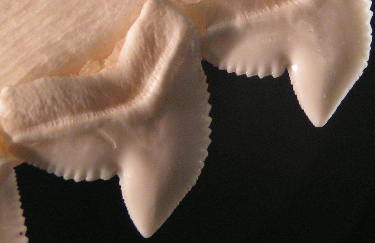
Serration is a saw-like appearance or a row of sharp or tooth-like projections. A serrated cutting edge has many small points of contact with the material being cut. By having less contact area than a smooth blade or other edge, the applied pressure at each point of contact is greater and the points of contact are at a sharper angle to the material being cut. This causes a cutting action that involves many small splits in the surface of the material being cut, which cumulatively serve to cut the material along the line of the blade. In nature, serration is commonly seen in the cutting edge on the teeth of some species, usually sharks. However, it also appears on non-cutting surfaces, for example in botany where a toothed leaf margin or other plant part, such as the edge of a carnation petal, is described as being serrated. A serrated leaf edge may reduce the force of wind and other natural elements. Probably the largest serrations on Earth occur on the skylines of mountains (t ... [...More Info...] [...Related Items...] OR: [Wikipedia] [Google] [Baidu] |
Diastema
A diastema (plural diastemata, from Greek διάστημα, space) is a space or gap between two teeth. Many species of mammals have diastemata as a normal feature, most commonly between the incisors and molars. More colloquially, the condition may be referred to as gap teeth or tooth gap. In humans, the term is most commonly applied to an open space between the upper incisors (front teeth). It happens when there is an unequal relationship between the size of the teeth and the jaw. Diastemata are common for children and can exist in adult teeth as well. In humans Causes 1. Oversized Labial Frenulum: Diastema is sometimes caused or exacerbated by the action of a labial frenulum (the tissue connecting the lip to the gum), causing high mucosal attachment and less attached keratinized tissue. This is more prone to recession or by tongue thrusting, which can push the teeth apart. 2. Periodontal Disease: Periodontal disease, also known as gum disease, can result in bone loss th ... [...More Info...] [...Related Items...] OR: [Wikipedia] [Google] [Baidu] |
Postcanine
Cheek teeth or post-canines comprise the molar and premolar teeth in mammals. Cheek teeth are multicuspidate (having many folds or tubercles). Mammals have multicuspidate molars (three in placentals, four in marsupials, in each jaw quadrant) and premolars situated between canines and molars whose shape and number varies considerably among particular groups. For example, many modern Carnivora possess carnassials, or secodont teeth. This scissor-like pairing of the last upper premolar and first lower molar is adapted for shearing meat. In contrast, the cheek teeth of deer and cattle are selenodont. Viewed from the side, these teeth have a series of triangular cusps or ridges, enabling the ruminants' sideways jaw motions to break down tough vegetable matter. Cheek teeth are sometimes separated from the incisors by a gap called a diastema. Cheek teeth in reptiles are much simpler as compared to mammals. Roles and significance Apart from helping grind the food to properly reduce the ... [...More Info...] [...Related Items...] OR: [Wikipedia] [Google] [Baidu] |


