|
Hypoglossal
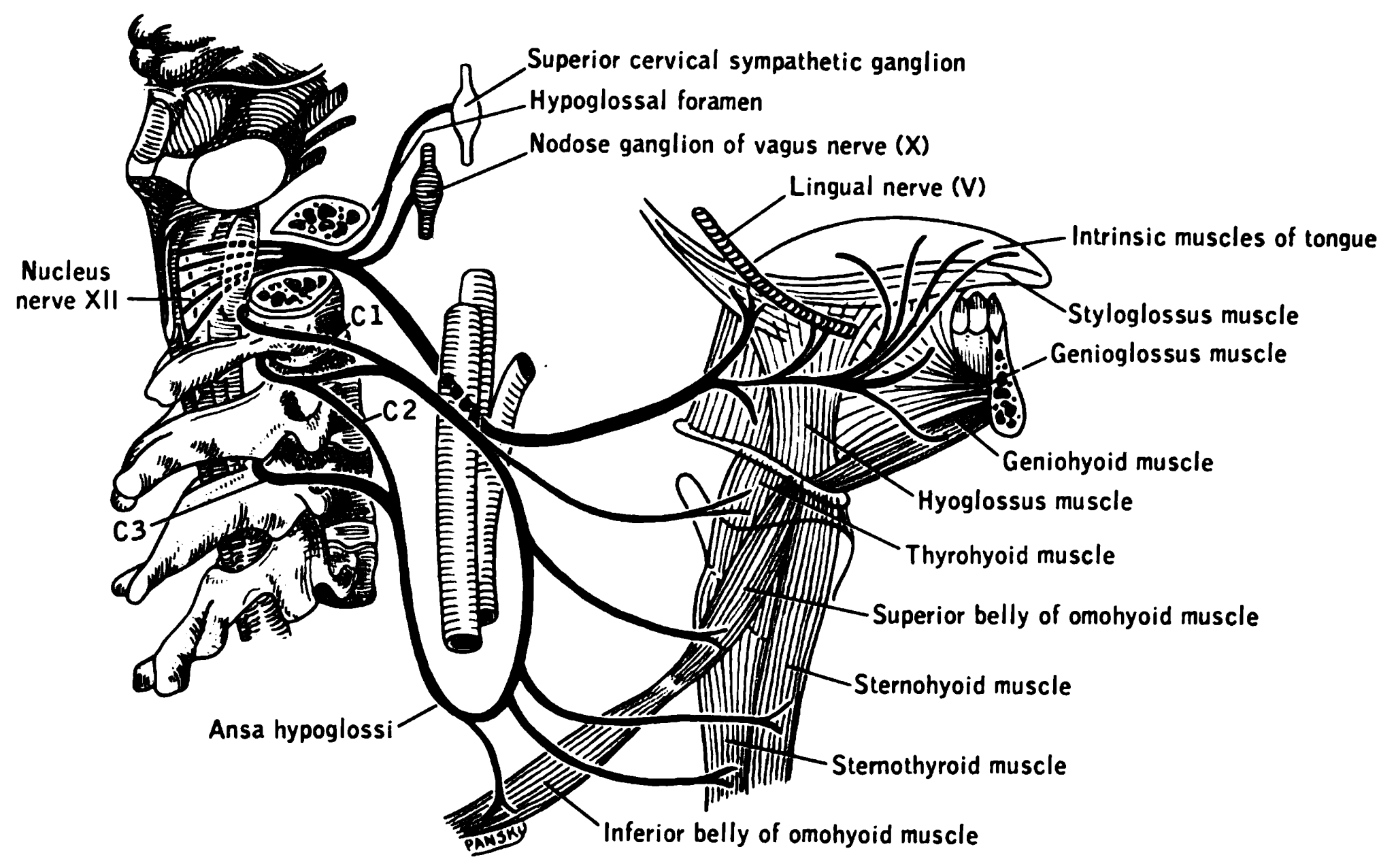
The hypoglossal nerve, also known as the twelfth cranial nerve, cranial nerve XII, or simply CN XII, is a cranial nerve that innervates all the extrinsic and intrinsic muscles of the tongue except for the palatoglossus, which is innervated by the vagus nerve. CN XII is a nerve with a solely motor function. The nerve arises from the hypoglossal nucleus in the medulla as a number of small rootlets, passes through the hypoglossal canal and down through the neck, and eventually passes up again over the tongue muscles it supplies into the tongue. The nerve is involved in controlling tongue movements required for speech and swallowing, including sticking out the tongue and moving it from side to side. Damage to the nerve or the neural pathways which control it can affect the ability of the tongue to move and its appearance, with the most common sources of damage being injury from trauma or surgery, and motor neuron disease. The first recorded description of the nerve is by Herophi ... [...More Info...] [...Related Items...] OR: [Wikipedia] [Google] [Baidu] |
Hypoglossal Nucleus
The hypoglossal nucleus is a cranial nerve nucleus, found within the medulla. Being a motor nucleus, it is close to the midline. In the open medulla, it is visible as what is known as the '' hypoglossal trigone'', a raised area (medial to the vagal trigone) protruding slightly into the fourth ventricle. The hypoglossal nucleus is located between the dorsal motor nucleus of the vagus and the midline of the medulla. Axons from the hypoglossal nucleus pass anteriorly through the medulla forming the hypoglossal nerve which exits between the pyramid and olive in a groove called the anterolateral sulcus. See also * Hypoglossal nerve Additional images File:Gray695.png, Transverse section of medulla oblongata below the middle of the olive. File:Gray697.png, Nuclei of origin of cranial motor nerves schematically represented; lateral view. File:Medulla_oblongata_-_posterior_-_very_low_mag.jpg, Micrograph showing the hypoglossal nuclei in relation to their surrounding structures. ... [...More Info...] [...Related Items...] OR: [Wikipedia] [Google] [Baidu] |
Hyoglossus
The hyoglossus, thin and quadrilateral, arises from the side of the body and from the whole length of the greater cornu of the hyoid bone, and passes almost vertically upward to enter the side of the tongue, between the styloglossus and the inferior longitudinal muscle of the tongue. It forms a part of the floor of submandibular triangle. Structure The fibers arising from the body of the hyoid bone overlap those from the greater cornu. Structures that are medial/deep to the hyoglossus are the glossopharyngeal nerve (cranial nerve 9), the stylohyoid ligament and the lingual artery and lingual vein. The lingual vein passes medial to the hyoglossus, and the lingual artery passes deep to the hyoglossus. Laterally, in between the hyoglossus muscle and the mylohyoid muscle lay several important structures (from upper to lower): sublingual gland, submandibular duct, lingual nerve, vena comitans of hypoglossal nerve, and the hypoglossal nerve. Note, posteriorly, the lingual ne ... [...More Info...] [...Related Items...] OR: [Wikipedia] [Google] [Baidu] |
Styloglossus
The styloglossus, the shortest and smallest of the three styloid muscles, arises from the anterior and lateral surfaces of the styloid process near its apex, and from the stylomandibular ligament. Passing inferiorly and anteriorly between the internal and external carotid arteries, it divides upon the side of the tongue near its dorsal surface, blending with the fibers of the longitudinalis inferior in front of the hyoglossus; the other, oblique, overlaps the Hyoglossus and decussates with its fibers. Innervation The styloglossus is innervated by the hypoglossal nerve (CN XII) like all muscles of the tongue except palatoglossus which is innervated by the pharyngeal plexus of vagus nerve The pharyngeal plexus is a network of nerve fibers innervating most of the palate and pharynx. (The larynx, which is innervated by the superior and recurrent laryngeal nerves from vagus nerve (CN X), is not included.) It is located on the sur ... (CN X). Action The styloglossus draws up ... [...More Info...] [...Related Items...] OR: [Wikipedia] [Google] [Baidu] |
Muscles Of The Tongue
The tongue is a muscular organ in the mouth of a typical tetrapod. It manipulates food for mastication and swallowing as part of the digestive process, and is the primary organ of taste. The tongue's upper surface (dorsum) is covered by taste buds housed in numerous lingual papillae. It is sensitive and kept moist by saliva and is richly supplied with nerves and blood vessels. The tongue also serves as a natural means of cleaning the teeth. A major function of the tongue is the enabling of speech in humans and vocalization in other animals. The human tongue is divided into two parts, an oral part at the front and a pharyngeal part at the back. The left and right sides are also separated along most of its length by a vertical section of fibrous tissue (the lingual septum) that results in a groove, the median sulcus, on the tongue's surface. There are two groups of muscles of the tongue. The four intrinsic muscles alter the shape of the tongue and are not attached to bon ... [...More Info...] [...Related Items...] OR: [Wikipedia] [Google] [Baidu] |
Tongue
The tongue is a muscular organ in the mouth of a typical tetrapod. It manipulates food for mastication and swallowing as part of the digestive process, and is the primary organ of taste. The tongue's upper surface (dorsum) is covered by taste buds housed in numerous lingual papillae. It is sensitive and kept moist by saliva and is richly supplied with nerves and blood vessels. The tongue also serves as a natural means of cleaning the teeth. A major function of the tongue is the enabling of speech in humans and vocalization in other animals. The human tongue is divided into two parts, an oral part at the front and a pharyngeal part at the back. The left and right sides are also separated along most of its length by a vertical section of fibrous tissue (the lingual septum) that results in a groove, the median sulcus, on the tongue's surface. There are two groups of muscles of the tongue. The four intrinsic muscles alter the shape of the tongue and are not attached to bone. ... [...More Info...] [...Related Items...] OR: [Wikipedia] [Google] [Baidu] |
Hypoglossal Canal
The hypoglossal canal is a foramen in the occipital bone of the skull. It is hidden medially and superiorly to each occipital condyle. It transmits the hypoglossal nerve. Structure The hypoglossal canal lies in the epiphyseal junction between the basiocciput and the jugular process of the occipital bone. Variation Embryonic variants sometimes lead to the presence of more than two canals as the occipital bone is formed. Development The hypoglossal canal is formed during the embryological stage of development in mammals. Function The hypoglossal canal transmits the hypoglossal nerve from its point of entry near the medulla oblongata to its exit from the base of the skull near the jugular foramen A jugular foramen is one of the two (left and right) large foramina (openings) in the base of the skull, located behind the carotid canal. It is formed by the temporal bone and the occipital bone. It allows many structures to pass, including the .... Clinical significance ... [...More Info...] [...Related Items...] OR: [Wikipedia] [Google] [Baidu] |
Carotid Sheath
The carotid sheath is an anatomical term for the fibrous connective tissue that surrounds the vascular compartment of the neck. It is part of the deep cervical fascia of the neck, below the superficial cervical fascia meaning the subcutaneous adipose tissue immediately beneath the skin. The deep cervical fascia of the neck includes four parts: * The investing layer (encloses the SCM and Trapezius) * The carotid sheath (encloses the vascular region of the neck) * The pretracheal fascia (encloses the visceral region of the neck) * The prevertebral fascia (encloses the vertebral region of the neck) Structure The carotid sheath is located at the lateral boundary of the retropharyngeal space at the level of the oropharynx on each side of the neck and deep to the sternocleidomastoid muscle. It extends from the base of the skull to the first rib and sternum, varying between C7 and T4. It merges with the axillary sheath when it reaches the subclavian vein. Contents The four major st ... [...More Info...] [...Related Items...] OR: [Wikipedia] [Google] [Baidu] |
Thyrohyoid
The thyrohyoid muscle is a small skeletal muscle on the neck. It originates from the lamina of the thyroid cartilage, and inserts into the greater cornu of the hyoid bone. It is supplied by the hypoglossal nerve, and a branch of the ventral rami of the cervical plexus, spinal nerve C1, which travels with the hypoglossal nerve. The thyrohyoid muscle depresses the hyoid bone and elevates the larynx. By controlling the position and shape of the larynx, it aids in making sound. Structure The thyrohyoid muscle is a quadrilateral muscle in shape. It appears like an upward continuation of the sternothyroid muscle. It belongs to the infrahyoid muscles group. It lies in the carotid triangle. It arises from the oblique line on the lamina of the thyroid cartilage. It is inserted into the lower border of the greater cornu of the hyoid bone. Nerve supply The thyrohyoid muscle is supplied by the hypoglossal nerve (XII). It is the only infrahyoid muscle that is not supplied by the ansa ce ... [...More Info...] [...Related Items...] OR: [Wikipedia] [Google] [Baidu] |
Cranial Nerve
Cranial nerves are the nerves that emerge directly from the brain (including the brainstem), of which there are conventionally considered twelve pairs. Cranial nerves relay information between the brain and parts of the body, primarily to and from regions of the head and neck, including the special senses of vision, taste, smell, and hearing. The cranial nerves emerge from the central nervous system above the level of the first vertebra of the vertebral column. Each cranial nerve is paired and is present on both sides. There are conventionally twelve pairs of cranial nerves, which are described with Roman numerals I–XII. Some considered there to be thirteen pairs of cranial nerves, including cranial nerve zero. The numbering of the cranial nerves is based on the order in which they emerge from the brain and brainstem, from front to back. The terminal nerves (0), olfactory nerves (I) and optic nerves (II) emerge from the cerebrum, and the remaining ten pairs arise ... [...More Info...] [...Related Items...] OR: [Wikipedia] [Google] [Baidu] |
Genioglossus
The genioglossus is one of the paired extrinsic muscles of the tongue. The genioglossus is the major muscle responsible for protruding (or sticking out) the tongue. Structure Genioglossus is the fan-shaped extrinsic tongue muscle that forms the majority of the body of the tongue. It arises from the mental spine of the mandible and its insertions are the hyoid bone and the bottom of the tongue. The genioglossus is innervated by the hypoglossal nerve, as are all muscles of the tongue except for the palatoglossus. Blood is supplied to the sublingual branch of the lingual artery, a branch of the external carotid artery. The canine genioglossus muscle has been divided into horizontal and oblique compartments. Function The left and right genioglossus muscles protrude the tongue and deviate it towards the opposite side. When acting together, the muscles depress the center of the tongue at its back. Clinical significance Contraction of the genioglossus stabilizes and enlarges the port ... [...More Info...] [...Related Items...] OR: [Wikipedia] [Google] [Baidu] |
Dura Mater
In neuroanatomy, dura mater is a thick membrane made of dense irregular connective tissue that surrounds the brain and spinal cord. It is the outermost of the three layers of membrane called the meninges that protect the central nervous system. The other two meningeal layers are the arachnoid mater and the pia mater. It envelops the arachnoid mater, which is responsible for keeping in the cerebrospinal fluid. It is derived primarily from the neural crest cell population, with postnatal contributions of the paraxial mesoderm. Structure The dura mater has several functions and layers. The dura mater is a membrane that envelops the arachnoid mater. It surrounds and supports the dural sinuses (also called dural venous sinuses, cerebral sinuses, or cranial sinuses) and carries blood from the brain toward the heart. Cranial dura mater has two layers called '' lamellae'', a superficial layer (also called the periosteal layer), which serves as the skull's inner periosteum, called t ... [...More Info...] [...Related Items...] OR: [Wikipedia] [Google] [Baidu] |
Occipital Bone
The occipital bone () is a cranial dermal bone and the main bone of the occiput (back and lower part of the skull). It is trapezoidal in shape and curved on itself like a shallow dish. The occipital bone overlies the occipital lobes of the cerebrum. At the base of skull in the occipital bone, there is a large oval opening called the foramen magnum, which allows the passage of the spinal cord. Like the other cranial bones, it is classed as a flat bone. Due to its many attachments and features, the occipital bone is described in terms of separate parts. From its front to the back is the basilar part, also called the basioccipital, at the sides of the foramen magnum are the lateral parts, also called the exoccipitals, and the back is named as the squamous part. The basilar part is a thick, somewhat quadrilateral piece in front of the foramen magnum and directed towards the pharynx. The squamous part is the curved, expanded plate behind the foramen magnum and is the largest par ... [...More Info...] [...Related Items...] OR: [Wikipedia] [Google] [Baidu] |


