|
Hyoid
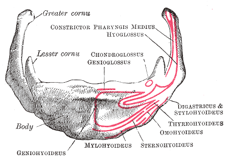
The hyoid-bone (lingual-bone or tongue-bone) () is a horseshoe-shaped bone situated in the anterior midline of the neck between the chin and the thyroid-cartilage. At rest, it lies between the base of the mandible and the third cervical vertebra. Unlike other bones, the hyoid is only distantly articulated to other bones by muscles or ligaments. It is the only bone in the human body that is not connected to any other bones. The hyoid is anchored by muscles from the anterior, posterior and inferior directions, and aids in tongue movement and swallowing. The hyoid bone provides attachment to the muscles of the floor of the mouth and the tongue above, the larynx below, and the epiglottis and pharynx behind. Its name is derived . Structure The hyoid bone is classed as an irregular bone and consists of a central part called the body, and two pairs of horns, the greater and lesser horns. Body The body of the hyoid bone is the central part of the hyoid bone. *At the front ... [...More Info...] [...Related Items...] OR: [Wikipedia] [Google] [Baidu] |
Hyoid Bone Hariadhi
The hyoid-bone (lingual-bone or tongue-bone) () is a horseshoe-shaped bone situated in the anterior midline of the neck between the chin and the thyroid-cartilage. At rest, it lies between the base of the mandible and the third cervical vertebra. Unlike other bones, the hyoid is only distantly articulated to other bones by muscles or ligaments. It is the only bone in the human body that is not connected to any other bones. The hyoid is anchored by muscles from the anterior, posterior and inferior directions, and aids in tongue movement and swallowing. The hyoid bone provides attachment to the muscles of the floor of the mouth and the tongue above, the larynx below, and the epiglottis and pharynx behind. Its name is derived . Structure The hyoid bone is classed as an irregular bone and consists of a central part called the body, and two pairs of horns, the greater and lesser horns. Body The body of the hyoid bone is the central part of the hyoid bone. *At the front, the ... [...More Info...] [...Related Items...] OR: [Wikipedia] [Google] [Baidu] |
Sternohyoid Muscle
The sternohyoid muscle is a bilaterally paired, long, thin, narrow strap muscle of the anterior neck. It is one of the infrahyoid muscles. It is innervated by the ansa cervicalis. It acts to depress the hyoid bone. The sternohyoid muscle is a flat muscle located on both sides of the neck, part of the infrahyoid muscle group. It originates from the medial edge of the clavicle, sternoclavicular ligament, and posterior side of the manubrium, and ascends to attach to the body of the hyoid bone. The sternohyoid muscle, along with other infrahyoid muscles, functions to depress the hyoid bone, which is important for activities such as speaking, chewing, and swallowing. Additionally, this muscle group contributes to the protection of the trachea, esophagus, blood vessels, and thyroid gland. The sternohyoid muscle also plays a minor role in head movements. Structure The sternohyoid muscle is one of the paired strap muscles of the infrahyoid muscles. The muscle is directed superom ... [...More Info...] [...Related Items...] OR: [Wikipedia] [Google] [Baidu] |
Hyoglossus
The hyoglossus is a thin and quadrilateral extrinsic muscle of the tongue. It originates from the hyoid bone; it inserts onto the side of the tongue. It is innervated by the hypoglossal nerve (cranial nerve XII). It acts to depress and retract the tongue. Structure It forms a part of the floor of submandibular triangle. Origin from the side of the body and from the whole length of the greater cornu of the hyoid bone. The fibers arising from the body of the hyoid bone overlap those from the greater cornu. Insertion Its fibres pass almost vertically upward to enter the side of the tongue, inserting between the styloglossus and the inferior longitudinal muscle of the tongue. Relations Structures that are medial/deep to the hyoglossus are the glossopharyngeal nerve (CN IX), the stylohyoid ligament and the lingual artery and lingual vein. The lingual vein passes medial to the hyoglossus. The lingual artery passes deep to the hyoglossus. Laterally, in between the hyo ... [...More Info...] [...Related Items...] OR: [Wikipedia] [Google] [Baidu] |
Horseshoe-shaped
Many shapes have metaphorical names, i.e., their names are metaphors: these shapes are named after a most common object that has it. For example, "U-shape" is a shape that resembles the letter U, a bell-shaped curve has the shape of the vertical cross section of a bell, etc. These terms may variously refer to objects, their cross sections or projections. Types of shapes Some of these names are "classical terms", i.e., words of Latin or Ancient Greek etymology. Others are English language constructs (although the base words may have non-English etymology). In some disciplines, where shapes of subjects in question are a very important consideration, the shape naming may be quite elaborate, see, e.g., the taxonomy of shapes of plant leaves in botany. * Astroid * Aquiline, shaped like an eagle's beak (as in a Roman nose) * Bell-shaped curve * Biconic shape, a shape in a way opposite to the hourglass: it is based on two oppositely oriented cones or truncated cones with their ba ... [...More Info...] [...Related Items...] OR: [Wikipedia] [Google] [Baidu] |
Thyrohyoid Muscle
The thyrohyoid muscle is a small skeletal muscle of the neck. Above, it attaches onto the greater cornu of the hyoid bone; below, it attaches onto the oblique line of the thyroid cartilage. It is innervated by fibres derived from the cervical spinal nerve 1 that run with the Hypoglossal nerve, hypoglossal nerve (CN XII) to reach this muscle. The thyrohyoid muscle depresses the hyoid bone and elevates the larynx during swallowing. By controlling the position and shape of the larynx, it aids in making sound. Structure The thyrohyoid muscle is a small, broad and short muscle. It is quadrilateral in shape. It may be considered a superior-ward continuation of sternothyroid muscle. It belongs to the infrahyoid muscles group and the outer laryngeal muscle group. Attachments Its superior attachment is the inferior border of the greater cornu of the hyoid bone and adjacent portions of the body of hyoid bone. Its inferior attachment is the oblique line of the thyroid cartilage (alongs ... [...More Info...] [...Related Items...] OR: [Wikipedia] [Google] [Baidu] |
Hyoglossus Muscle
The hyoglossus is a thin and quadrilateral extrinsic muscle of the tongue. It originates from the hyoid bone; it inserts onto the side of the tongue. It is innervated by the hypoglossal nerve (cranial nerve XII). It acts to depress and retract the tongue. Structure It forms a part of the floor of submandibular triangle. Origin from the side of the body and from the whole length of the greater cornu of the hyoid bone. The fibers arising from the body of the hyoid bone overlap those from the greater cornu. Insertion Its fibres pass almost vertically upward to enter the side of the tongue, inserting between the styloglossus and the inferior longitudinal muscle of the tongue. Relations Structures that are medial/deep to the hyoglossus are the glossopharyngeal nerve (CN IX), the stylohyoid ligament and the lingual artery and lingual vein. The lingual vein passes medial to the hyoglossus. The lingual artery passes deep to the hyoglossus. Laterally, in between the hyoglossus ... [...More Info...] [...Related Items...] OR: [Wikipedia] [Google] [Baidu] |
Mylohyoid Muscle
The mylohyoid muscle or diaphragma oris is a paired muscle of the neck. It runs from the Human mandible, mandible to the hyoid bone, forming the floor of the oral cavity of the human mouth, mouth. It is named after its two attachments near the molar (tooth), molar teeth. It forms the floor of the submental triangle. It elevates the hyoid bone and the tongue, important during swallowing and Speech, speaking. Structure The mylohyoid muscle is flat and triangular, and is situated immediately Anatomical terms of location#Superior and inferior, superior to the digastric muscle, anterior belly of the digastric muscle. It is a pharyngeal musculature, pharyngeal muscle (derived from the first pharyngeal arch) and classified as one of the suprahyoid muscles. Together, the paired mylohyoid muscles form a muscular floor for the oral cavity of the human mouth, mouth. The two mylohyoid muscles arise from the mandible at the mylohyoid line, which extends from the mandibular symphysis in front ... [...More Info...] [...Related Items...] OR: [Wikipedia] [Google] [Baidu] |
Geniohyoid Muscle
The geniohyoid muscle is a narrow paired muscle situated superior to the medial border of the mylohyoid muscle. It is named for its passage from the chin ("genio-" is a standard prefix for "chin") to the hyoid bone. Structure The geniohyoid is a paired short muscle that arises from the inferior mental spine, on the back of the mandibular symphysis, and runs backward and slightly downward, to be inserted into the anterior surface of the body of the hyoid bone. It lies in contact with its fellow of the opposite side. It thus belongs to the suprahyoid muscles. The muscle receives its blood supply from branches of the lingual artery. Innervation The geniohyoid muscle is innervated by fibres from the first cervical spinal nerve travelling alongside the hypoglossal nerve. Although the first three cervical nerves give rise to the ansa cervicalis, the geniohyoid muscle is said to be innervated by the first cervical nerve, as some of its efferent fibers do not contribute to ansa cerv ... [...More Info...] [...Related Items...] OR: [Wikipedia] [Google] [Baidu] |
Irregular Bone
The irregular bones are bones which, from their peculiar form, cannot be grouped as long, short, flat or sesamoid bones. Irregular bones serve various purposes in the body, such as protection of nervous tissue (such as the vertebrae protect the spinal cord), affording multiple anchor points for skeletal muscle attachment (as with the sacrum), and maintaining pharynx and trachea support, and tongue attachment (such as the hyoid bone). They consist of cancellous tissue enclosed within a thin layer of compact bone. Irregular bones can also be used for joining all parts of the spinal column together. The spine is the place in the human body where the most irregular bones can be found. There are, in all, 33 irregular bones found here. The irregular bones are: the vertebrae, sacrum, coccyx, temporal, sphenoid, ethmoid, zygomatic, maxilla, mandible, palatine, inferior nasal concha The inferior nasal concha (inferior turbinated bone or inferior turbinal/turbinate) is one of ... [...More Info...] [...Related Items...] OR: [Wikipedia] [Google] [Baidu] |
Joint
A joint or articulation (or articular surface) is the connection made between bones, ossicles, or other hard structures in the body which link an animal's skeletal system into a functional whole.Saladin, Ken. Anatomy & Physiology. 7th ed. McGraw-Hill Connect. Webp.274/ref> They are constructed to allow for different degrees and types of movement. Some joints, such as the knee, elbow, and shoulder, are self-lubricating, almost frictionless, and are able to withstand compression and maintain heavy loads while still executing smooth and precise movements. Other joints such as suture (joint), sutures between the bones of the skull permit very little movement (only during birth) in order to protect the brain and the sense organs. The connection between a tooth and the jawbone is also called a joint, and is described as a fibrous joint known as a gomphosis. Joints are classified both structurally and functionally. Joints play a vital role in the human body, contributing to movement, sta ... [...More Info...] [...Related Items...] OR: [Wikipedia] [Google] [Baidu] |
Omohyoid Muscle
The omohyoid muscle is a muscle in the neck. It is one of the infrahyoid muscles. It consists of two bellies separated by an intermediate tendon. Its inferior belly is attached to the scapula; its superior belly is attached to the hyoid bone. Its intermediate tendon is anchored to the clavicle and first rib by a fascial sling. The omohyoid is innervated by the ansa cervicalis of the cervical plexus. It acts to depress the hyoid bone. Anatomy Structure The omohyoid muscle consists of muscle bellies that meet at an angle at the muscle's intermediate tendon. Inferior belly The inferior belly is narrow and flat band. It arises from the superior border of scapula (near the scapular notch). It sometimes also arises from the superior transverse scapular ligament. It is directed anteriorly and somewhat superiorly from its origin, extending across the inferior portion of the neck. It passes posterior to the sternocleidomastoid muscle to insert at the intermediate tendon. Super ... [...More Info...] [...Related Items...] OR: [Wikipedia] [Google] [Baidu] |
Lateral Thyrohyoid Ligament
The lateral thyrohyoid ligament (lateral hyothyroid ligament) is a round elastic cord, which forms the posterior border of the thyrohyoid membrane and passes between the tip of the superior cornu of the thyroid cartilage and the extremity of the greater cornu of the hyoid bone. The internal branch of the superior laryngeal nerve The superior laryngeal nerve is a branch of the vagus nerve. It arises from the middle of the inferior ganglion of the vagus nerve and additionally receives a sympathetic branch from the superior cervical ganglion. The superior laryngeal nerve ... typical lies lateral to this ligament. Triticeal cartilage A small cartilaginous nodule (cartilago triticea), sometimes bony, is frequently found in the lateral thyrohyoid ligament. References External links * - "Larynx, anterior view" * - "Larynx, lateral view" Human head and neck Ligaments {{Portal bar, Anatomy ... [...More Info...] [...Related Items...] OR: [Wikipedia] [Google] [Baidu] |


