|
Gamma Catenin
Plakoglobin, also known as junction plakoglobin or gamma-catenin, is a protein that in humans is encoded by the ''JUP'' gene. Plakoglobin is a member of the catenin protein family and homologous to β-catenin. Plakoglobin is a cytoplasmic component of desmosomes and adherens junctions structures located within intercalated discs of cardiac muscle that function to anchor sarcomeres and join adjacent cells in cardiac muscle. Mutations in plakoglobin are associated with arrhythmogenic right ventricular dysplasia. Structure Human plakoglobin is 81.7 kDa in molecular weight and 745 amino acids long. The ''JUP'' gene contains 13 exons spanning 17 kb on chromosome 17q21. Plakoglobin is a member of the catenin family, since it contains a distinct repeating amino acid motif called the armadillo repeat. Plakoglobin is highly similar to β-catenin; both have 12 armadillo repeats as well as N-terminal and C-terminal globular domains of unknown structure. Plakoglobin was originally identifi ... [...More Info...] [...Related Items...] OR: [Wikipedia] [Google] [Baidu] |
Protein
Proteins are large biomolecules and macromolecules that comprise one or more long chains of amino acid residues. Proteins perform a vast array of functions within organisms, including catalysing metabolic reactions, DNA replication, responding to stimuli, providing structure to cells and organisms, and transporting molecules from one location to another. Proteins differ from one another primarily in their sequence of amino acids, which is dictated by the nucleotide sequence of their genes, and which usually results in protein folding into a specific 3D structure that determines its activity. A linear chain of amino acid residues is called a polypeptide. A protein contains at least one long polypeptide. Short polypeptides, containing less than 20–30 residues, are rarely considered to be proteins and are commonly called peptides. The individual amino acid residues are bonded together by peptide bonds and adjacent amino acid residues. The sequence of amino acid ... [...More Info...] [...Related Items...] OR: [Wikipedia] [Google] [Baidu] |
E-cadherin
Cadherin-1 or Epithelial cadherin (E-cadherin), (not to be confused with the APC/C activator protein CDH1) is a protein that in humans is encoded by the ''CDH1'' gene. Mutations are correlated with gastric, breast, colorectal, thyroid, and ovarian cancers. CDH1 has also been designated as CD324 (cluster of differentiation 324). It is a tumor suppressor gene. History The discovery of cadherin cell-cell adhesion proteins is attributed to Masatoshi Takeichi, whose experience with adhering epithelial cells began in 1966. His work originally began by studying lens differentiation in chicken embryos at Nagoya University, where he explored how retinal cells regulate lens fiber differentiation. To do this, Takeichi initially collected media that had previously cultured neural retina cells (CM) and suspended lens epithelial cells in it. He observed that cells suspended in the CM media had delayed attachment compared to cells in his regular medium. His interest in cell adherence was spark ... [...More Info...] [...Related Items...] OR: [Wikipedia] [Google] [Baidu] |
Sudden Cardiac Death
Cardiac arrest is when the heart suddenly and unexpectedly stops beating. It is a medical emergency that, without immediate medical intervention, will result in sudden cardiac death within minutes. Cardiopulmonary resuscitation (CPR) and possibly defibrillation are needed until further treatment can be provided. Cardiac arrest results in a rapid loss of consciousness, and breathing may be abnormal or absent. While cardiac arrest may be caused by heart attack or heart failure, these are not the same, and in 15 to 25% of cases, there is a non-cardiac cause. Some individuals may experience chest pain, shortness of breath, nausea, an elevated heart rate, and a light-headed feeling immediately before entering cardiac arrest. The most common cause of cardiac arrest is an underlying heart problem like coronary artery disease that decreases the amount of oxygenated blood supplying the heart muscle. This, in turn, damages the structure of the muscle, which can alter its function. Th ... [...More Info...] [...Related Items...] OR: [Wikipedia] [Google] [Baidu] |
Cardiomyopathy
Cardiomyopathy is a group of diseases that affect the heart muscle. Early on there may be few or no symptoms. As the disease worsens, shortness of breath, feeling tired, and swelling of the legs may occur, due to the onset of heart failure. An irregular heart beat and fainting may occur. Those affected are at an increased risk of sudden cardiac death. Types of cardiomyopathy include hypertrophic cardiomyopathy, dilated cardiomyopathy, restrictive cardiomyopathy, arrhythmogenic right ventricular dysplasia, and Takotsubo cardiomyopathy (broken heart syndrome). In hypertrophic cardiomyopathy the heart muscle enlarges and thickens. In dilated cardiomyopathy the ventricles enlarge and weaken. In restrictive cardiomyopathy the ventricle stiffens. In many cases, the cause cannot be determined. Hypertrophic cardiomyopathy is usually inherited, whereas dilated cardiomyopathy is inherited in about one third of cases. Dilated cardiomyopathy may also result from alcohol, h ... [...More Info...] [...Related Items...] OR: [Wikipedia] [Google] [Baidu] |
Gap Junction
Gap junctions are specialized intercellular connections between a multitude of animal cell-types. They directly connect the cytoplasm of two cells, which allows various molecules, ions and electrical impulses to directly pass through a regulated gate between cells. One gap junction channel is composed of two protein hexamers (or hemichannels) called connexons in vertebrates and innexons in invertebrates. The hemichannel pair connect across the intercellular space bridging the gap between two cells. Gap junctions are analogous to the plasmodesmata that join plant cells. Gap junctions occur in virtually all tissues of the body, with the exception of adult fully developed skeletal muscle and mobile cell types such as sperm or erythrocytes. Gap junctions are not found in simpler organisms such as sponges and slime molds. A gap junction may also be called a ''nexus'' or ''macula communicans''. While an ephapse has some similarities to a gap junction, by modern definition t ... [...More Info...] [...Related Items...] OR: [Wikipedia] [Google] [Baidu] |
Fibrosis
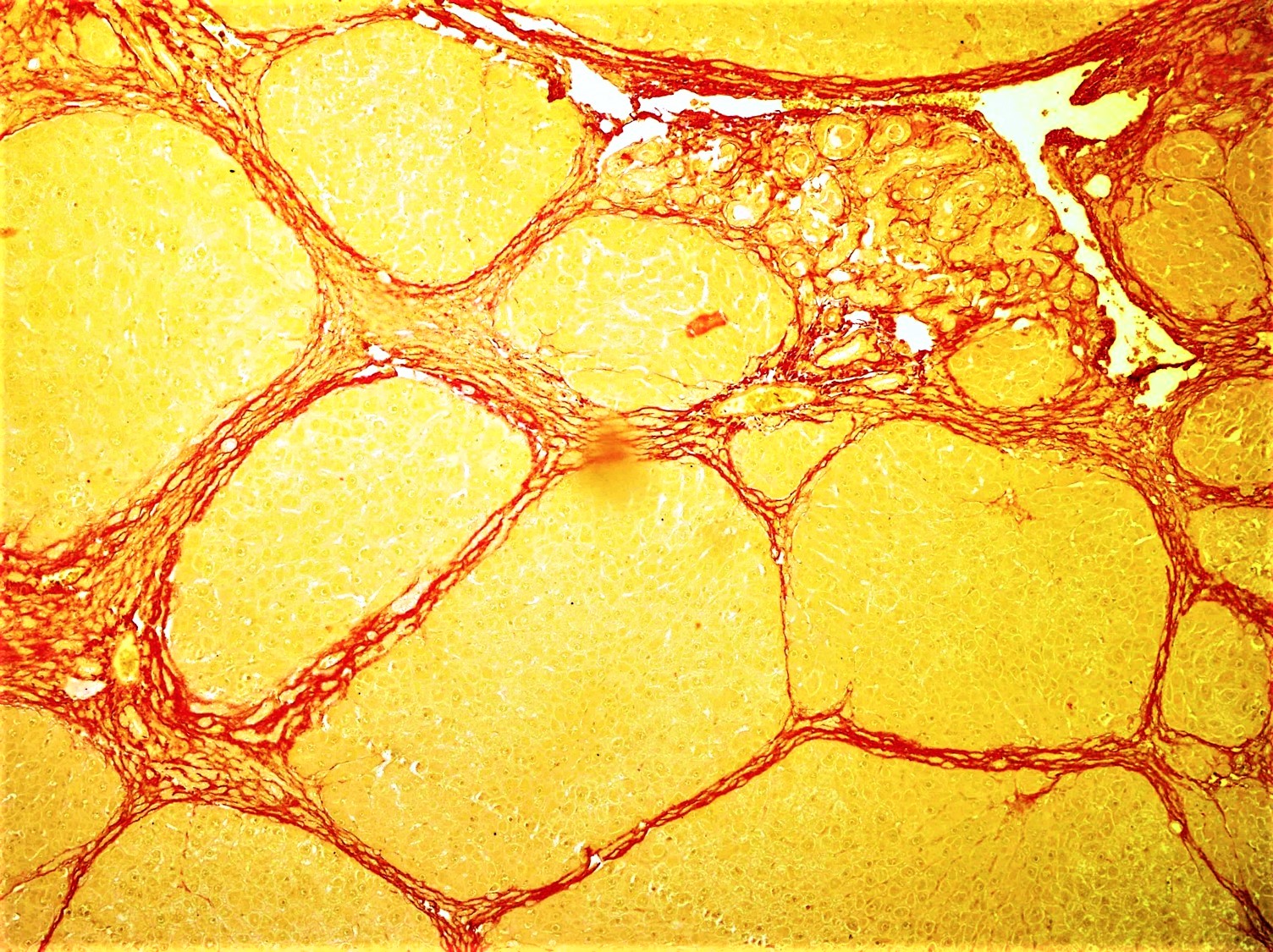
Fibrosis, also known as fibrotic scarring, is a pathological wound healing in which connective tissue replaces normal parenchymal tissue to the extent that it goes unchecked, leading to considerable tissue remodelling and the formation of permanent scar tissue. Repeated injuries, chronic inflammation and repair are susceptible to fibrosis where an accidental excessive accumulation of extracellular matrix components, such as the collagen is produced by fibroblasts, leading to the formation of a permanent fibrotic scar. In response to injury, this is called scarring, and if fibrosis arises from a single cell line, this is called a fibroma. Physiologically, fibrosis acts to deposit connective tissue, which can interfere with or totally inhibit the normal architecture and function of the underlying organ or tissue. Fibrosis can be used to describe the pathological state of excess deposition of fibrous tissue, as well as the process of connective tissue deposition in healing. Defin ... [...More Info...] [...Related Items...] OR: [Wikipedia] [Google] [Baidu] |
Cardiomyocytes
Cardiac muscle (also called heart muscle, myocardium, cardiomyocytes and cardiac myocytes) is one of three types of vertebrate muscle tissues, with the other two being skeletal muscle and smooth muscle. It is an involuntary, striated muscle that constitutes the main tissue of the wall of the heart. The cardiac muscle (myocardium) forms a thick middle layer between the outer layer of the heart wall (the pericardium) and the inner layer (the endocardium), with blood supplied via the coronary circulation. It is composed of individual cardiac muscle cells joined by intercalated discs, and encased by collagen fibers and other substances that form the extracellular matrix. Cardiac muscle contracts in a similar manner to skeletal muscle, although with some important differences. Electrical stimulation in the form of a cardiac action potential triggers the release of calcium from the cell's internal calcium store, the sarcoplasmic reticulum. The rise in calcium causes the cell's ... [...More Info...] [...Related Items...] OR: [Wikipedia] [Google] [Baidu] |
Arrhythmogenic Right Ventricular Cardiomyopathy
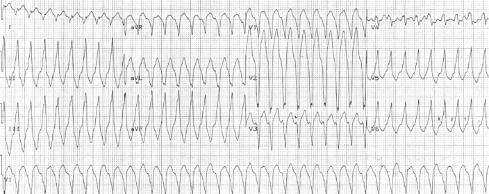
Arrhythmogenic cardiomyopathy (ACM), arrhythmogenic right ventricular dysplasia (ARVD), or arrhythmogenic right ventricular cardiomyopathy (ARVC), most commonly is an inherited heart disease. ACM is caused by genetic defects of the parts of heart muscle (also called ''myocardium'' or ''cardiac muscle'') known as desmosomes, areas on the surface of heart muscle cells which link the cells together. The desmosomes are composed of several proteins, and many of those proteins can have harmful mutations. ARVC can also develop in intense endurance athletes in the absence of desmosomal abnormalities. Exercise-induced ARVC cause possibly is a result of excessive right ventricular wall stress during high intensity exercise. The disease is a type of non-ischemic cardiomyopathy that primarily involves the right ventricle, though cases of exclusive left ventricular disease have been reported. It is characterized by hypokinetic areas involving the free wall of the ventricle, with fibrofa ... [...More Info...] [...Related Items...] OR: [Wikipedia] [Google] [Baidu] |
Cardiac
The heart is a muscular organ in most animals. This organ pumps blood through the blood vessels of the circulatory system. The pumped blood carries oxygen and nutrients to the body, while carrying metabolic waste such as carbon dioxide to the lungs. In humans, the heart is approximately the size of a closed fist and is located between the lungs, in the middle compartment of the chest. In humans, other mammals, and birds, the heart is divided into four chambers: upper left and right atria and lower left and right ventricles. Commonly the right atrium and ventricle are referred together as the right heart and their left counterparts as the left heart. Fish, in contrast, have two chambers, an atrium and a ventricle, while most reptiles have three chambers. In a healthy heart blood flows one way through the heart due to heart valves, which prevent backflow. The heart is enclosed in a protective sac, the pericardium, which also contains a small amount of fluid. The wall o ... [...More Info...] [...Related Items...] OR: [Wikipedia] [Google] [Baidu] |
Intracellular
This glossary of biology terms is a list of definitions of fundamental terms and concepts used in biology, the study of life and of living organisms. It is intended as introductory material for novices; for more specific and technical definitions from sub-disciplines and related fields, see Glossary of genetics, Glossary of evolutionary biology, Glossary of ecology, and Glossary of scientific naming, or any of the organism-specific glossaries in :Glossaries of biology. A B C D E ... [...More Info...] [...Related Items...] OR: [Wikipedia] [Google] [Baidu] |
DSC2
Desmocollin-2 is a protein that in humans is encoded by the ''DSC2'' gene. Desmocollin-2 is a cadherin-type protein that functions to link adjacent cells together in specialized regions known as desmosomes. Desmocollin-2 is widely expressed, and is the only desmocollin isoform expressed in cardiac muscle, where it localizes to intercalated discs. Mutations in ''DSC2'' have been causally linked to arrhythmogenic right ventricular cardiomyopathy. Structure Desmocollin-2 is a calcium-dependent glycoprotein that is a member of the desmocollin subfamily of the cadherin superfamily. Three different posttranslational modifications (''N''-Glycosylations, ''O''-Mannosylations and disulfide bridges) were present in the extracellular domain of desmocollin-2. The desmocollin family members are arranged as closely linked genes on human chromosome 18q12.1. Human ''DSC2'' consists of greater than 32 kb of DNA and has 17 exons, with exon 16 being alternatively spliced and encoding distinct isof ... [...More Info...] [...Related Items...] OR: [Wikipedia] [Google] [Baidu] |
DSG2
Desmoglein-2 is a protein that in humans is encoded by the ''DSG2'' gene. Desmoglein-2 is highly expressed in epithelial cells and cardiomyocytes. Desmoglein-2 is localized to desmosome structures at regions of cell-cell contact and functions to structurally adhere adjacent cells together. In cardiac muscle, these regions are specialized regions known as intercalated discs. Mutations in desmoglein-2 have been associated with arrhythmogenic right ventricular cardiomyopathy and familial dilated cardiomyopathy. Structure Desmoglein-2 is a 122.2 kDa protein composed of 1118 amino acids. Desmoglein-2 is a calcium-binding transmembrane glycoprotein component of desmosomes in vertebrate cells. Currently, four desmoglein subfamily members have been identified and all are members of the cadherin cell adhesion molecule superfamily. These desmoglein gene family members are located in a cluster on chromosome 18. This second family member, desmoglein-2 is expressed in desmosome-containing t ... [...More Info...] [...Related Items...] OR: [Wikipedia] [Google] [Baidu] |


