|
GLUT1
Glucose transporter 1 (or GLUT1), also known as solute carrier family 2, facilitated glucose transporter member 1 (SLC2A1), is a uniporter protein that in humans is encoded by the ''SLC2A1'' gene. GLUT1 facilitates the transport of glucose across the plasma membranes of mammalian cells. This gene encodes a facilitative glucose transporter that is highly expressed in erythrocytes and endothelial cells, including cells of the blood–brain barrier. The encoded protein is found primarily in the cell membrane and on the cell surface, where it can also function as a receptor for human T-cell leukemia virus (HTLV) I and II. GLUT1 accounts for 2 percent of the protein in the plasma membrane of erythrocytes. During early development, GLUT1 expression is compartmentalized across different tissues, ensuring that metabolic requirements are met in a tissue-specific manner. This tissue-specific glucose metabolism is essential for regulating the differentiation of specific lineages, su ... [...More Info...] [...Related Items...] OR: [Wikipedia] [Google] [Baidu] |
Glut1 Deficiency
GLUT1 deficiency syndrome, also known as GLUT1-DS, De Vivo disease or Glucose transporter type 1 deficiency syndrome, is an autosomal dominant genetic metabolic disorder associated with a deficiency of GLUT1, the protein that transports glucose across the blood brain barrier. Glucose Transporter Type 1 Deficiency Syndrome has an estimated birth incidence of 1 in 90,000 to 1 in 24,300. This birth incidence translates to an estimated prevalence of 3,000 to 7,000 in the U.S. Presentation GLUT1 deficiency is characterized by an array of signs and symptoms including mental and motor developmental delays, infantile seizures refractory to anticonvulsants, ataxia, dystonia, dysarthria, opsoclonus, spasticity, other paroxysmal neurologic phenomena and sometimes deceleration of head growth also known as microcephaly. The presence and severity of symptoms vary considerably between affected individuals. Individuals with the disorder generally have frequent seizures (epilepsy), often beginni ... [...More Info...] [...Related Items...] OR: [Wikipedia] [Google] [Baidu] |
Glucose Transporter
Glucose transporters are a wide group of membrane proteins that facilitate the transport of glucose across the plasma membrane, a process known as facilitated diffusion. Because glucose is a vital source of energy for all life, these transporters are present in all phyla. The GLUT or SLC2A family are a protein family that is found in most mammalian cells. 14 GLUTS are encoded by the human genome. GLUT is a type of uniporter transporter protein. Synthesis of free glucose Most non-autotrophic cells are unable to produce free glucose because they lack expression of glucose-6-phosphatase and, thus, are involved only in glucose uptake and catabolism. Usually produced only in hepatocytes, in fasting conditions, other tissues such as the intestines, muscles, brain, and kidneys are able to produce glucose following activation of gluconeogenesis. Glucose transport in yeast In ''Saccharomyces cerevisiae'' glucose transport takes place through facilitated diffusion. The transport p ... [...More Info...] [...Related Items...] OR: [Wikipedia] [Google] [Baidu] |
Uniporter
Uniporters, also known as solute carriers or facilitated transporters, are a type of membrane transport protein that passively transports solutes (small molecules, ions, or other substances) across a cell membrane. It uses facilitated diffusion for the movement of solutes down their concentration gradient from an area of high concentration to an area of low concentration. Unlike active transport, it does not require energy in the form of ATP to function. Uniporters are specialized to carry one specific ion or molecule and can be categorized as either channels or carriers. Facilitated diffusion may occur through three mechanisms: uniport, symport, or antiport. The difference between each mechanism depends on the direction of transport, in which uniport is the only transport not coupled to the transport of another solute. Uniporter carrier proteins work by binding to one molecule or substrate at a time. Uniporter channels open in response to a stimulus and allow the free flow of ... [...More Info...] [...Related Items...] OR: [Wikipedia] [Google] [Baidu] |
Glucose
Glucose is a sugar with the Chemical formula#Molecular formula, molecular formula , which is often abbreviated as Glc. It is overall the most abundant monosaccharide, a subcategory of carbohydrates. It is mainly made by plants and most algae during photosynthesis from water and carbon dioxide, using energy from sunlight. It is used by plants to make cellulose, the most abundant carbohydrate in the world, for use in cell walls, and by all living Organism, organisms to make adenosine triphosphate (ATP), which is used by the cell as energy. In energy metabolism, glucose is the most important source of energy in all organisms. Glucose for metabolism is stored as a polymer, in plants mainly as amylose and amylopectin, and in animals as glycogen. Glucose circulates in the blood of animals as blood sugar. The naturally occurring form is -glucose, while its Stereoisomerism, stereoisomer L-glucose, -glucose is produced synthetically in comparatively small amounts and is less biologicall ... [...More Info...] [...Related Items...] OR: [Wikipedia] [Google] [Baidu] |
Human T-lymphotropic Virus 2
A virus closely related to HTLV-I, human T-lymphotropic virus 2 (HTLV-II) shares approximately 70% genomic homology (structural similarity) with HTLV-I. It was discovered by Robert Gallo and colleagues. HTLV-2 is prevalent in Africa and among Indigenous peoples in Central and South America, as well as among drug users in Europe and North America. It can be passed down from mother to child through breast milk, and even genetically from either parent. HTLV-II entry in target cells is mediated by the glucose transporter GLUT1. Virology HTLV-1 and HTLV-2 share broad similarities in their overall genetic organization and expression pattern, but they differ substantially in their pathogenic properties. The virus utilizes the GLUT-1 and NRP1 cellular receptors for their entry, although HTLV-1, but not HTLV-2, is dependent on heparan sulfate proteoglycans. Cell-to-cell transmission is essential for the virus replication and occurs through the formation of a virological s ... [...More Info...] [...Related Items...] OR: [Wikipedia] [Google] [Baidu] |
Human T-lymphotropic Virus 1
Human T-cell lymphotropic virus type 1 or human T-lymphotropic virus (HTLV-I), also called the adult T-cell lymphoma virus type 1, is a retrovirus of the human T-lymphotropic virus (HTLV) family. Most people with HTLV-1 infection do not appear to develop health conditions that can be directly linked to the infection. However, there is a subgroup of people who experience severe complications. The most well characterized are adult T-cell lymphoma (ATL) and HTLV-I-associated myelopathy/ Tropical spastic paraparesis (HAM/TSP), both of which are only diagnosed in individuals testing positive to HTLV-1 infection. The estimated lifetime risk of ATL among people with HTLV-1 infection is approximately 5%, while that of HAM/TSP is approximately 2%. In 1977, Adult T-cell lymphoma (ATL) was first described in a case series of individuals from Japan. The symptoms of ATL were different from other lymphomas known at the time. The common birthplace shared amongst most of the ATL patients was s ... [...More Info...] [...Related Items...] OR: [Wikipedia] [Google] [Baidu] |
Chromosome 1
Chromosome 1 is the designation for the largest human chromosome. Humans have two copies of chromosome 1, as they do with all of the autosomes, which are the non-sex chromosomes. Chromosome 1 spans about 249 million nucleotide base pairs, which are the basic units of information for DNA.http://vega.sanger.ac.uk/Homo_sapiens/mapview?chr=1 Chromosome size and number of genes derived from this database, retrieved 2012-03-11. It represents about 8% of the total DNA in human cells. It was the last completed chromosome, sequenced two decades after the beginning of the Human Genome Project. Genes Number of genes The following are some of the gene count estimates of human chromosome 1. Because researchers use different approaches to genome annotation their predictions of the number of genes on each chromosome varies (for technical details, see gene prediction). Among various projects, the collaborative consensus coding sequence project ( CCDS) takes an extremely conservative strategy. S ... [...More Info...] [...Related Items...] OR: [Wikipedia] [Google] [Baidu] |
Human T-lymphotropic Virus
The primate T-lymphotropic viruses (PTLVs) are a group of retroviruses that infect primates, using their lymphocytes to reproduce. The ones that infect humans are known as human T-lymphotropic virus (HTLV), and the ones that infect Old World monkeys are called simian T-lymphotropic viruses (STLVs). PTLVs are named for their ability to cause adult T-cell leukemia/lymphoma, but in the case of HTLV-1 it can also cause a demyelinating disease called tropical spastic paraparesis. On the other hand, newer PTLVs are simply placed into the group by similarity and their connection to human disease remains unclear. HTLVs have evolved from STLVs by interspecies transmission. Within each species of PTLV, the HTLV is more similar to its cognate STLV than to the other HTLVs. There are currently three species of PTLVs recognized by the ICTV (P/H/STLV-1, -2, -3), plus two that are reported but unrecognized (HTLV-4, STLV-5). The first known, and still most medically important PTLV is HTLV-1, ... [...More Info...] [...Related Items...] OR: [Wikipedia] [Google] [Baidu] |
Cytoplasm
The cytoplasm describes all the material within a eukaryotic or prokaryotic cell, enclosed by the cell membrane, including the organelles and excluding the nucleus in eukaryotic cells. The material inside the nucleus of a eukaryotic cell and contained within the nuclear membrane is termed the nucleoplasm. The main components of the cytoplasm are the cytosol (a gel-like substance), the cell's internal sub-structures, and various cytoplasmic inclusions. In eukaryotes the cytoplasm also includes the nucleus, and other membrane-bound organelles.The cytoplasm is about 80% water and is usually colorless. The submicroscopic ground cell substance, or cytoplasmic matrix, that remains after the exclusion of the cell organelles and particles is groundplasm. It is the hyaloplasm of light microscopy, a highly complex, polyphasic system in which all resolvable cytoplasmic elements are suspended, including the larger organelles such as the ribosomes, mitochondria, plant plasti ... [...More Info...] [...Related Items...] OR: [Wikipedia] [Google] [Baidu] |
Integral Membrane Protein
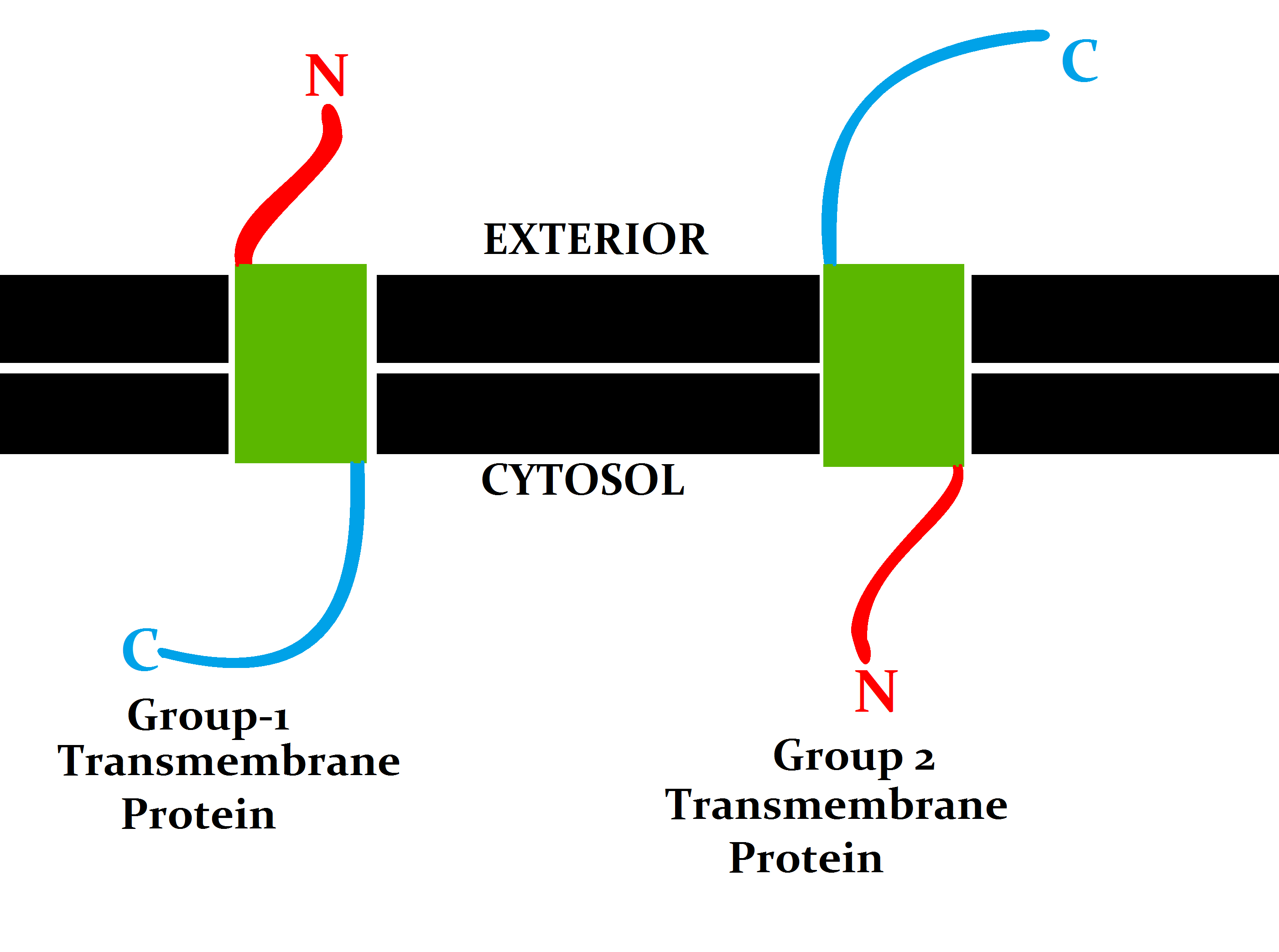
An integral, or intrinsic, membrane protein (IMP) is a type of membrane protein that is permanently attached to the biological membrane. All transmembrane proteins can be classified as IMPs, but not all IMPs are transmembrane proteins. IMPs comprise a significant fraction of the proteins encoded in an organism's genome. Proteins that cross the membrane are surrounded by annular lipids, which are defined as lipids that are in direct contact with a membrane protein. Such proteins can only be separated from the membranes by using detergents, nonpolar solvents, or sometimes denaturing agents. Proteins that adhere only temporarily to cellular membranes are known as peripheral membrane proteins. These proteins can either associate with integral membrane proteins, or independently insert in the lipid bilayer in several ways. Structure Three-dimensional structures of ~160 different integral membrane proteins have been determined at atomic resolution by X-ray crystallography or nucle ... [...More Info...] [...Related Items...] OR: [Wikipedia] [Google] [Baidu] |
Alpha Helices
An alpha helix (or α-helix) is a sequence of amino acids in a protein that are twisted into a coil (a helix). The alpha helix is the most common structural arrangement in the secondary structure of proteins. It is also the most extreme type of local structure, and it is the local structure that is most easily predicted from a sequence of amino acids. The alpha helix has a right-handed helix conformation in which every backbone N−H group hydrogen bonds to the backbone C=O group of the amino acid that is four residues earlier in the protein sequence. Other names The alpha helix is also commonly called a: * Pauling–Corey–Branson α-helix (from the names of three scientists who described its structure) * 3.613-helix because there are 3.6 amino acids in one ring, with 13 atoms being involved in the ring formed by the hydrogen bond (starting with amidic hydrogen and ending with carbonyl oxygen) Discovery In the early 1930s, William Astbury showed that there were dras ... [...More Info...] [...Related Items...] OR: [Wikipedia] [Google] [Baidu] |


