|
G9a
Euchromatic histone-lysine N-methyltransferase 2 (EHMT2), also known as G9a, is a histone methyltransferase enzyme that in humans is encoded by the ''EHMT2'' gene. G9a deposits the mono- and di-methylated states of histone H3 at lysine residue 9 (i.e., H3K9me1 and H3K9me2) and lysine residue 27 (H3K27me1 and H3K27me2). The presence of H3K9me1/2 is usually associated with gene silencing. Function A cluster of genes, BAT1-BAT5, has been localized in the vicinity of the genes for TNF alpha and TNF beta. This gene is found near this cluster; it was mapped near the gene for C2 within a 120-kb region that included a HSP70 gene pair. These genes are all within the human major histocompatibility complex class III region. This gene was thought to be two different genes, NG36 and G9a, adjacent to each other but a recent publication shows that there is only a single gene. The protein encoded by this gene is thought to be involved in intracellular protein-protein interaction. There are t ... [...More Info...] [...Related Items...] OR: [Wikipedia] [Google] [Baidu] |
H3K9me2
H3K9me2 is an epigenetic modification to the DNA packaging protein Histone H3. It is a mark that indicates the di-methylation at the 9th lysine residue of the histone H3 protein. H3K9me2 is strongly associated with transcriptional repression. H3K9me2 levels are higher at silent compared to active genes in a 10kb region surrounding the transcriptional start site. H3K9me2 represses gene expression both passively, by prohibiting acetylation as therefore binding of RNA polymerase or its regulatory factors, and actively, by recruiting transcriptional repressors. H3K9me2 has also been found in megabase blocks, termed Large Organised Chromatin K9 domains (LOCKS), which are primarily located within gene-sparse regions but also encompass genic and intergenic intervals. Its synthesis is catalyzed by G9a, G9a-like protein, and PRDM2. H3K9me2 can be removed by a wide range of histone lysine demethylases (KDMs) including KDM1, KDM3, KDM4 and KDM7 family members. H3K9me2 is important for v ... [...More Info...] [...Related Items...] OR: [Wikipedia] [Google] [Baidu] |
Addiction
Addiction is a neuropsychological disorder characterized by a persistent and intense urge to use a drug or engage in a behavior that produces natural reward, despite substantial harm and other negative consequences. Repetitive drug use can alter brain function in synapses similar to natural rewards like food or falling in love in ways that perpetuate craving and weakens self-control for people with pre-existing vulnerabilities. This phenomenon – drugs reshaping brain function – has led to an understanding of addiction as a brain disorder with a complex variety of psychosocial as well as neurobiological factors that are implicated in the development of addiction. While mice given cocaine showed the compulsive and involuntary nature of addiction, for humans this is more complex, related to behavior or personality traits. Classic signs of addiction include compulsive engagement in rewarding stimuli, ''preoccupation'' with substances or behavior, and continued use des ... [...More Info...] [...Related Items...] OR: [Wikipedia] [Google] [Baidu] |
ΔFosB
Protein fosB, also known as FosB and G0/G1 switch regulatory protein 3 (G0S3), is a protein that in humans is encoded by the FBJ murine osteosarcoma viral oncogene homolog B (''FOSB'') gene. The FOS gene family consists of four members: FOS, FOSB, FOSL1, and FOSL2. These genes encode leucine zipper proteins that can dimerize with proteins of the JUN family (e.g., c-Jun, JunD), thereby forming the transcription factor complex AP-1. As such, the FOS proteins have been implicated as regulators of cell proliferation, differentiation, and transformation. FosB and its truncated splice variants, ΔFosB and further truncated Δ2ΔFosB, are all involved in osteosclerosis, although Δ2ΔFosB lacks a known transactivation domain, in turn preventing it from affecting transcription through the AP-1 complex. The ΔFosB splice variant has been identified as playing a central, crucial role in the development and maintenance of addiction. ΔFosB overexpression (i.e., an abnormally a ... [...More Info...] [...Related Items...] OR: [Wikipedia] [Google] [Baidu] |
Epigenetic Regulation
In biology, epigenetics is the study of changes in gene expression that happen without changes to the DNA sequence. The Greek prefix ''epi-'' (ἐπι- "over, outside of, around") in ''epigenetics'' implies features that are "on top of" or "in addition to" the traditional (DNA sequence based) genetic mechanism of inheritance. Epigenetics usually involves a change that is not erased by cell division, and affects the regulation of gene expression. Such effects on cellular and physiological traits may result from environmental factors, or be part of normal development. The term also refers to the mechanism of changes: functionally relevant alterations to the genome that do not involve mutation of the nucleotide sequence. Examples of mechanisms that produce such changes are DNA methylation and histone modification, each of which alters how genes are expressed without altering the underlying DNA sequence. Further, non-coding RNA sequences have been shown to play a key role in the r ... [...More Info...] [...Related Items...] OR: [Wikipedia] [Google] [Baidu] |
Nucleus Accumbens
The nucleus accumbens (NAc or NAcc; also known as the accumbens nucleus, or formerly as the ''nucleus accumbens septi'', Latin for ' nucleus adjacent to the septum') is a region in the basal forebrain rostral to the preoptic area of the hypothalamus. The nucleus accumbens and the olfactory tubercle collectively form the ventral striatum. The ventral striatum and dorsal striatum collectively form the striatum, which is the main component of the basal ganglia. The dopaminergic neurons of the mesolimbic pathway project onto the GABAergic medium spiny neurons of the nucleus accumbens and olfactory tubercle. Each cerebral hemisphere has its own nucleus accumbens, which can be divided into two structures: the nucleus accumbens core and the nucleus accumbens shell. These substructures have different morphology and functions. Different NAcc subregions (core vs shell) and neuron subpopulations within each region ( D1-type vs D2-type medium spiny neurons) are responsible fo ... [...More Info...] [...Related Items...] OR: [Wikipedia] [Google] [Baidu] |
Neoplasm
A neoplasm () is a type of abnormal and excessive growth of tissue. The process that occurs to form or produce a neoplasm is called neoplasia. The growth of a neoplasm is uncoordinated with that of the normal surrounding tissue, and persists in growing abnormally, even if the original trigger is removed. This abnormal growth usually forms a mass, which may be called a tumour or tumor.'' ICD-10 classifies neoplasms into four main groups: benign neoplasms, in situ neoplasms, malignant neoplasms, and neoplasms of uncertain or unknown behavior. Malignant neoplasms are also simply known as cancers and are the focus of oncology. Prior to the abnormal growth of tissue, such as neoplasia, cells often undergo an abnormal pattern of growth, such as metaplasia or dysplasia. However, metaplasia or dysplasia does not always progress to neoplasia and can occur in other conditions as well. The word neoplasm is from Ancient Greek 'new' and 'formation, creation'. Types A neoplasm ... [...More Info...] [...Related Items...] OR: [Wikipedia] [Google] [Baidu] |
Leukemia
Leukemia ( also spelled leukaemia; pronounced ) is a group of blood cancers that usually begin in the bone marrow and produce high numbers of abnormal blood cells. These blood cells are not fully developed and are called ''blasts'' or '' leukemia cells''. Symptoms may include bleeding and bruising, bone pain, fatigue, fever, and an increased risk of infections. These symptoms occur due to a lack of normal blood cells. Diagnosis is typically made by blood tests or bone marrow biopsy. The exact cause of leukemia is unknown. A combination of genetic factors and environmental (non-inherited) factors are believed to play a role. Risk factors include smoking, ionizing radiation, petrochemicals (such as benzene), prior chemotherapy, and Down syndrome. People with a family history of leukemia are also at higher risk. There are four main types of leukemia—acute lymphoblastic leukemia (ALL), acute myeloid leukemia (AML), chronic lymphocytic leukemia (CLL) and chronic myelo ... [...More Info...] [...Related Items...] OR: [Wikipedia] [Google] [Baidu] |
Carcinogenesis
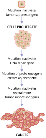
Carcinogenesis, also called oncogenesis or tumorigenesis, is the formation of a cancer, whereby normal cell (biology), cells are malignant transformation, transformed into cancer cells. The process is characterized by changes at the cellular, Genetics, genetic, and Epigenetics, epigenetic levels and abnormal cell division. Cell division is a physiological process that occurs in almost all Tissue (biology), tissues and under a variety of circumstances. Normally, the balance between proliferation and programmed cell death, in the form of apoptosis, is maintained to ensure the integrity of tissues and Organ (anatomy), organs. According to the prevailing accepted theory of carcinogenesis, the somatic mutation theory, mutations in DNA and Epigenetics, epimutations that lead to cancer disrupt these orderly processes by interfering with the programming regulating the processes, upsetting the normal balance between proliferation and cell death. This results in uncontrolled cell division ... [...More Info...] [...Related Items...] OR: [Wikipedia] [Google] [Baidu] |
KIAA0515
Protein BAT2-like is a protein that in humans is encoded by the ''BAT2L'' gene. Interactions KIAA0515 has been shown to interact with EHMT2 Euchromatic histone-lysine N-methyltransferase 2 (EHMT2), also known as G9a, is a histone methyltransferase enzyme that in humans is encoded by the ''EHMT2'' gene. G9a deposits the mono- and di-methylated states of histone H3 at lysine residue 9 .... References Further reading * * * * {{protein-stub ... [...More Info...] [...Related Items...] OR: [Wikipedia] [Google] [Baidu] |
Dorsal Root Ganglia
A dorsal root ganglion (or spinal ganglion; also known as a posterior root ganglion) is a cluster of neurons (a ganglion) in a dorsal root of a spinal nerve. The cell bodies of sensory neurons known as first-order neurons are located in the dorsal root ganglia. The axons of dorsal root ganglion neurons are known as afferents. In the peripheral nervous system, afferents refer to the axons that relay sensory information into the central nervous system (i.e. the brain and the spinal cord). Structure The neurons comprising the dorsal root ganglion are of the pseudo-unipolar type, meaning they have a cell body (soma) with two branches that act as a single axon, often referred to as a ''distal process'' and a ''proximal process''. Unlike the majority of neurons found in the central nervous system, an action potential in posterior root ganglion neuron may initiate in the ''distal process'' in the periphery, bypass the cell body, and continue to propagate along the ''proximal proces ... [...More Info...] [...Related Items...] OR: [Wikipedia] [Google] [Baidu] |
Dendritic Spine
A dendritic spine (or spine) is a small membrane protrusion from a neuron's dendrite that typically receives input from a single axon at the synapse. Dendritic spines serve as a storage site for synaptic strength and help transmit electrical signals to the neuron's cell body. Most spines have a bulbous head (the spine head), and a thin neck that connects the head of the spine to the shaft of the dendrite. The dendrites of a single neuron can contain hundreds to thousands of spines. In addition to spines providing an anatomical substrate for memory storage and synaptic transmission, they may also serve to increase the number of possible contacts between neurons. It has also been suggested that changes in the activity of neurons have a positive effect on spine morphology. Structure Dendritic spines are small with spine head volumes ranging 0.01 μm3 to 0.8 μm3. Spines with strong synaptic contacts typically have a large spine head, which connects to the dendrite via a m ... [...More Info...] [...Related Items...] OR: [Wikipedia] [Google] [Baidu] |


