|
Fundic
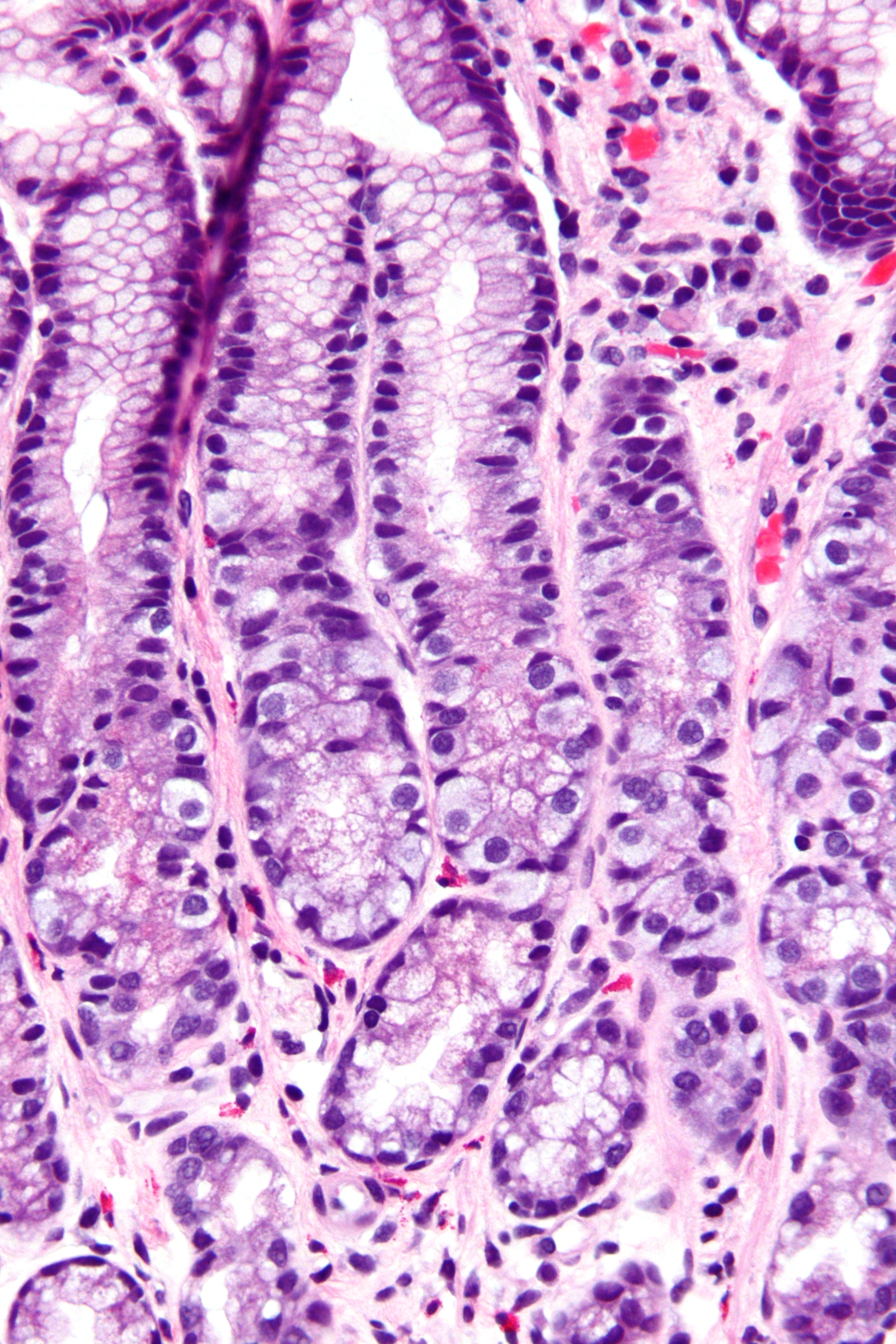
The gastric glands are glands in the lining of the stomach that play an essential role in the process of digestion. All of the glands have mucus-secreting foveolar cells. Mucus lines the entire stomach, and protects the stomach lining from the effects of hydrochloric acid released from other cells in the glands. There are two types of gland in the stomach, the oxyntic gland, and the pyloric gland. The major type of gastric gland is the oxyntic gland that is present in 80 per cent of the stomach, and is often referred to simply as the ''gastric gland''. The oxyntic gland is an exocrine gland and contains the parietal cells that produce hydrochloric acid, and intrinsic factor. Intrinsic factor is necessary for the absorption of vitamin B12. The other type of gland in the stomach is the pyloric gland found in the pyloric region taking up the remaining 20 per cent of the stomach area. The pyloric gland secretes gastrin from its G cells. Pyloric glands are similar in structure to t ... [...More Info...] [...Related Items...] OR: [Wikipedia] [Google] [Baidu] |
Stomach
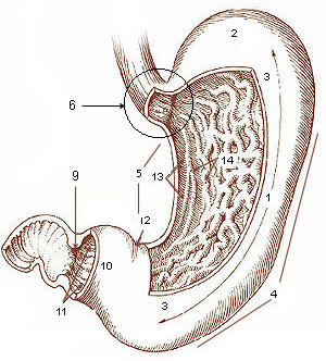
The stomach is a muscular, hollow organ in the gastrointestinal tract of humans and many other animals, including several invertebrates. The stomach has a dilated structure and functions as a vital organ in the digestive system. The stomach is involved in the gastric phase of digestion, following chewing. It performs a chemical breakdown by means of enzymes and hydrochloric acid. In humans and many other animals, the stomach is located between the oesophagus and the small intestine. The stomach secretes digestive enzymes and gastric acid to aid in food digestion. The pyloric sphincter controls the passage of partially digested food ( chyme) from the stomach into the duodenum, where peristalsis takes over to move this through the rest of intestines. Structure In the human digestive system, the stomach lies between the oesophagus and the duodenum (the first part of the small intestine). It is in the left upper quadrant of the abdominal cavity. The top of the stomach lies ... [...More Info...] [...Related Items...] OR: [Wikipedia] [Google] [Baidu] |
Fundus (stomach)
The stomach is a muscular, hollow organ in the gastrointestinal tract of humans and many other animals, including several invertebrates. The stomach has a dilated structure and functions as a vital organ in the digestive system. The stomach is involved in the gastric phase of digestion, following chewing. It performs a chemical breakdown by means of enzymes and hydrochloric acid. In humans and many other animals, the stomach is located between the oesophagus and the small intestine. The stomach secretes digestive enzymes and gastric acid to aid in food digestion. The pyloric sphincter controls the passage of partially digested food ( chyme) from the stomach into the duodenum, where peristalsis takes over to move this through the rest of intestines. Structure In the human digestive system, the stomach lies between the oesophagus and the duodenum (the first part of the small intestine). It is in the left upper quadrant of the abdominal cavity. The top of the stomach ... [...More Info...] [...Related Items...] OR: [Wikipedia] [Google] [Baidu] |
Gastric Mucosa
The gastric mucosa is the mucous membrane layer of the stomach, which contains the glands and the gastric pits. In humans, it is about 1 mm thick, and its surface is smooth, soft, and velvety. It consists of simple columnar epithelium, lamina propria, and the muscularis mucosae. Description In its fresh state, it is of a pinkish tinge at the pyloric end and of a red or reddish-brown color over the rest of its surface. In infancy it is of a brighter hue, the vascular redness being more marked. It is thin at the cardiac extremity, but thicker toward the pylorus. During the contracted state of the organ it is thrown into numerous plaits or rugae, which, for the most part, have a longitudinal direction, and are most marked toward the pyloric end of the stomach, and along the greater curvature. These folds are entirely obliterated when the organ becomes distended. When examined with a lens, the inner surface of the mucous membrane presents a peculiar honeycomb appearance fro ... [...More Info...] [...Related Items...] OR: [Wikipedia] [Google] [Baidu] |
G Cells
In anatomy, the G cell or gastrin cell, is a type of cell in the stomach and duodenum that secretes gastrin. It works in conjunction with gastric chief cells and parietal cells. G cells are found deep within the pyloric glands of the stomach antrum, and occasionally in the pancreas and duodenum. The vagus nerve innervates the G cells. Gastrin-releasing peptide is released by the post-ganglionic fibers of the vagus nerve onto G cells during parasympathetic stimulation. The peptide hormone bombesin also stimulates gastrin from G cells. Gastrin-releasing peptide, as well as the presence of amino acids in the stomach, stimulates the release of gastrin from the G cells. Gastrin stimulates enterochromaffin-like cells to secrete histamine. Gastrin also targets parietal cells by increasing the amount of histamine and the direct stimulation by gastrin, causing the parietal cells to increase HCl secretion in the stomach. G-cells frequently express PD-L1 during homeostasis which protects ... [...More Info...] [...Related Items...] OR: [Wikipedia] [Google] [Baidu] |
Parietal Cell
Parietal cells (also known as oxyntic cells) are epithelial cells in the stomach that secrete hydrochloric acid (HCl) and intrinsic factor. These cells are located in the gastric glands found in the lining of the fundus and body regions of the stomach. They contain an extensive secretory network of canaliculi from which the HCl is secreted by active transport into the stomach. The enzyme hydrogen potassium ATPase (H+/K+ ATPase) is unique to the parietal cells and transports the H+ against a concentration gradient of about 3 million to 1, which is the steepest ion gradient formed in the human body. Parietal cells are primarily regulated via histamine, acetylcholine and gastrin signalling from both central and local modulators. Structure Canaliculus A canaliculus is an adaptation found on gastric parietal cells. It is a deep infolding, or little channel, which serves to increase the surface area, e.g. for secretion. The parietal cell membrane is dynamic; the numbers o ... [...More Info...] [...Related Items...] OR: [Wikipedia] [Google] [Baidu] |
Gastric Chief Cell
A gastric chief cell (or peptic cell, or gastric zymogenic cell) is a type of gastric gland cell that releases pepsinogen and gastric lipase. It is the cell responsible for secretion of chymosin in ruminant animals. The cell stains basophilic upon H&E staining due to the large proportion of rough endoplasmic reticulum in its cytoplasm. Gastric chief cells are generally located deep in the mucosal layer of the stomach lining, in the fundus and body of the stomach.pathologyoutlines.com/topic/stomachnormalhistology.html Chief cells release the zymogen (enzyme precursor) pepsinogen when stimulated by a variety of factors including cholinergic activity from the vagus nerve and acidic condition in the stomach. Gastrin and secretin may also act as secretagogues. It works in conjunction with the parietal cell, which releases gastric acid, converting the pepsinogen into pepsin Pepsin is an endopeptidase that breaks down proteins into smaller peptides. It is produced in the ... [...More Info...] [...Related Items...] OR: [Wikipedia] [Google] [Baidu] |
G Cell
In anatomy, the G cell or gastrin cell, is a type of cell in the stomach and duodenum that secretes gastrin. It works in conjunction with gastric chief cells and parietal cells. G cells are found deep within the pyloric glands of the stomach antrum, and occasionally in the pancreas and duodenum. The vagus nerve innervates the G cells. Gastrin-releasing peptide is released by the post-ganglionic fibers of the vagus nerve onto G cells during parasympathetic stimulation. The peptide hormone bombesin also stimulates gastrin from G cells. Gastrin-releasing peptide, as well as the presence of amino acids in the stomach, stimulates the release of gastrin from the G cells. Gastrin stimulates enterochromaffin-like cells to secrete histamine. Gastrin also targets parietal cells by increasing the amount of histamine and the direct stimulation by gastrin, causing the parietal cells to increase HCl secretion in the stomach. G-cells frequently express PD-L1 during homeostasis which prot ... [...More Info...] [...Related Items...] OR: [Wikipedia] [Google] [Baidu] |
Gland
In animals, a gland is a group of cells in an animal's body that synthesizes substances (such as hormones) for release into the bloodstream ( endocrine gland) or into cavities inside the body or its outer surface ( exocrine gland). Structure Development Every gland is formed by an ingrowth from an epithelial surface. This ingrowth may in the beginning possess a tubular structure, but in other instances glands may start as a solid column of cells which subsequently becomes tubulated. As growth proceeds, the column of cells may split or give off offshoots, in which case a compound gland is formed. In many glands, the number of branches is limited, in others (salivary, pancreas) a very large structure is finally formed by repeated growth and sub-division. As a rule, the branches do not unite with one another, but in one instance, the liver, this does occur when a reticulated compound gland is produced. In compound glands the more typical or secretory epithelium is found formi ... [...More Info...] [...Related Items...] OR: [Wikipedia] [Google] [Baidu] |
Simple Tubular Glands
Tubular glands are glands with a tube-like shape throughout their length, in contrast with alveolar glands, which have a saclike secretory portion. Tubular glands are further classified as one of the following types: Additional images See also * skin - glands in skin structure * hair follicles - for hair growth References Additional images File:Gray940.png, A diagrammatic sectional view of the skin (magnified). File:Gray1169.png, Vertical section of mucous membrane of human uterus The uterus (from Latin ''uterus'', plural ''uteri'') or womb () is the organ in the reproductive system of most female mammals, including humans that accommodates the embryonic and fetal development of one or more embryos until birth. The .... File:Skin.png, Skin Glands {{anatomy-stub ... [...More Info...] [...Related Items...] OR: [Wikipedia] [Google] [Baidu] |
Brunner's Glands
Brunner's glands (or duodenal glands) are compound tubular submucosal glands found in that portion of the duodenum which is above the hepatopancreatic sphincter (i.e sphincter of Oddi). It also contains submucosa which creates special glands. The main function of these glands is to produce a mucus-rich alkaline secretion i.e. mucous (containing bicarbonate) in order to: * protect the duodenum from the acidic content of chyme (which is introduced into the duodenum from the stomach); * provide an alkaline condition for the intestinal enzymes to be active, thus enabling absorption to take place; * lubricate the intestinal walls. * They also secrete epidermal growth factor, which inhibits parietal and chief cells of the stomach from secreting acid and their digestive enzymes. This is another form of protection for the duodenum. They are the distinguishing feature of the duodenum, and are named for the Swiss physician who first described them, Johann Conrad Brunner. Structure H ... [...More Info...] [...Related Items...] OR: [Wikipedia] [Google] [Baidu] |


