|
Exostoses
An exostosis, also known as bone spur, is the formation of new bone on the surface of a bone. Exostoses can cause chronic pain ranging from mild to debilitatingly severe, depending on the shape, size, and location of the lesion. It is most commonly found in places like the ribs, where small bone growths form, but sometimes larger growths can grow on places like the ankles, knees, shoulders, elbows and hips. Very rarely are they on the skull. Exostoses are sometimes shaped like spurs, such as calcaneal spurs. Osteomyelitis, a bone infection, may leave the adjacent bone with exostosis formation. Charcot foot, the neuropathic breakdown of the feet seen primarily in diabetics, can also leave bone spurs that may then become symptomatic. They normally form on the bones of joints, and can grow upwards. For example, if an extra bone formed on the ankle, it might grow up to the shin. When used in the phrases "cartilaginous exostosis" or "osteocartilaginous exostosis", the term is consid ... [...More Info...] [...Related Items...] OR: [Wikipedia] [Google] [Baidu] |
Surfer's Ear
Surfer's ear is the common name for an exostosis or abnormal bone growth within the ear canal. Surfer's ear is not the same as swimmer's ear, although infection can result as a side effect. Irritation from cold wind and water exposure causes the bone surrounding the ear canal to develop lumps of new bony growth which constrict the ear canal. Where the ear canal is actually blocked by this condition, water and wax can become trapped and give rise to infection. The condition is so named due to its prevalence among cold water surfers. Warm water surfers are also at risk for exostosis due to the evaporative cooling caused by wind and the presence of water in the ear canal. Most avid surfers have at least some mild bone growths (exostoses), causing little to no problems. The condition is progressive, making it important to take preventive measures early, preferably whenever surfing. The condition is not limited to surfing and can occur in any activity with cold, wet, windy condition ... [...More Info...] [...Related Items...] OR: [Wikipedia] [Google] [Baidu] |
Hereditary Multiple Exostoses
Hereditary multiple osteochondromas (HMO), also known as hereditary multiple exostoses, is a disorder characterized by the development of multiple benign osteocartilaginous masses ( exostoses) in relation to the ends of long bones of the lower limbs such as the femurs and tibias and of the upper limbs such as the humeri and forearm bones. They are also known as osteochondromas. Additional sites of occurrence include on flat bones such as the pelvic bone and scapula. The distribution and number of these exostoses show a wide diversity among affected individuals. Exostoses usually present during childhood. The vast majority of affected individuals become clinically manifest by the time they reach adolescence. A small percentage of affected individuals are at risk for development of malignant transformation namely sarcomas. The incidence of hereditary multiple exostoses is around 1 in 50,000 individuals. Hereditary multiple osteochondromas is the preferred term used by the World He ... [...More Info...] [...Related Items...] OR: [Wikipedia] [Google] [Baidu] |
Buccal Exostosis
A buccal exostosis is an exostosis (bone prominence) on the buccal surface (cheek side) of the alveolar ridge of the maxilla or mandible. More commonly seen in the maxilla than the mandible, buccal exostoses are considered to be site specific. Existing as asymptomatic bony nodules, buccal exostoses don’t usually present until adult life, and some consider buccal exostoses to be a variation of normal anatomy rather than disease. Bone is thought to become hyperplastic, consisting of mature cortical and trabecular bone with a smooth outer surface. They are less common when compared with mandibular tori. Signs and symptoms Buccal exostoses are bony hamartomas, which are non- malignant, exophytic nodular outgrowths of dense cortical bone that are relatively avascular.’ Symptoms: Buccal exostoses generally tend to be asymptomatic and are usually painless. However, they may increase patient concern about poor aesthetics, inability to perform oral hygiene procedures due to difficul ... [...More Info...] [...Related Items...] OR: [Wikipedia] [Google] [Baidu] |
Bone
A bone is a Stiffness, rigid Organ (biology), organ that constitutes part of the skeleton in most vertebrate animals. Bones protect the various other organs of the body, produce red blood cell, red and white blood cells, store minerals, provide structure and support for the body, and enable animal locomotion, mobility. Bones come in a variety of shapes and sizes and have complex internal and external structures. They are lightweight yet strong and hard and serve multiple Function (biology), functions. Bone tissue (osseous tissue), which is also called bone in the mass noun, uncountable sense of that word, is hard tissue, a type of specialized connective tissue. It has a honeycomb-like matrix (biology), matrix internally, which helps to give the bone rigidity. Bone tissue is made up of different types of bone cells. Osteoblasts and osteocytes are involved in the formation and mineralization (biology), mineralization of bone; osteoclasts are involved in the bone resorption, resor ... [...More Info...] [...Related Items...] OR: [Wikipedia] [Google] [Baidu] |
Skeletal System
A skeleton is the structural frame that supports the body of an animal. There are several types of skeletons, including the exoskeleton, which is the stable outer shell of an organism, the endoskeleton, which forms the support structure inside the body, and the hydroskeleton, a flexible internal skeleton supported by fluid pressure. Vertebrates are animals with a vertebral column, and their skeletons are typically composed of bone and cartilage. Invertebrates are animals that lack a vertebral column. The skeletons of invertebrates vary, including hard exoskeleton shells, plated endoskeletons, or Sponge spicule, spicules. Cartilage is a rigid connective tissue that is found in the skeletal systems of vertebrates and invertebrates. Etymology The term ''skeleton'' comes . ''Sceleton'' is an archaic form of the word. Classification Skeletons can be defined by several attributes. Solid skeletons consist of hard substances, such as bone, cartilage, or cuticle. These can be further ... [...More Info...] [...Related Items...] OR: [Wikipedia] [Google] [Baidu] |
Pachyostosis
Pachyostosis is a non-pathological condition in vertebrate animals in which the bones experience a thickening, generally caused by extra layers of lamellar bone. It often occurs together with bone densification (osteosclerosis), reducing inner cavities. This joint occurrence is called pachyosteosclerosis. However, especially in the older literature, "pachyostosis" is often used loosely, referring to all osseous specializations characterized by an increase in bone compactness and/or volume. It occurs in both terrestrial and, especially, aquatic or semi-aquatic vertebrates. In aquatic animals, such as seacows (manatees and dugongs), ''Thalassocnus'', and plesiosaurs, pachyostosis in the thoracic region provides (or provided) ballast against the air-filled lungs. This maintains neutral buoyancy in aquatic habitats. Most giant deer showed pronounced pachyostosis of the mandible and skull. It has been suggested that this served to store minerals for antler growth. Many Pachycephalosau ... [...More Info...] [...Related Items...] OR: [Wikipedia] [Google] [Baidu] |
Pachyosteosclerosis
Pachyosteosclerosis is a combination of thickening (pachyostosis) and densification (osteosclerosis) of bones. It makes bones more heavy, but also more fragile. The condition often occurs in aquatic vertebrates, especially those living in shallow waters,Houssaye, A. (2009). "Pachyostosis" in aquatic amniotes: a review. Integrative Zoology 4(4): 325-340. creating ballast as an adaptation for maintaining neutral buoyancy and horizontal trim. It is in no way pathological. To resist bend, it frequently is found especially in ventral bones, whereas concentration near the lungs helps in maintaining trim. Examples of animals showing pachyosteosclerosis are seacows (dugongs and manatees), the extinct Plesiosauria and Mesosauria and extinct aquatic sloths.Amson, E., C. de Muizon, M. Laurin, C. Argot and V. de Buffrenil (2014). Gradual adaptation of bone structure to aquatic lifestyle in extinct sloths from Peru. Proceedings of the Royal Society B-Biological Sciences 281. See also * Pachyo ... [...More Info...] [...Related Items...] OR: [Wikipedia] [Google] [Baidu] |
Osteosclerosis
Osteosclerosis is a disorder that is characterized by abnormal hardening of bone and an elevation in bone density. It may predominantly affect the medullary portion and/or cortex of bone. Plain radiographs are a valuable tool for detecting and classifying osteosclerotic disorders. It can manifest in localized or generalized osteosclerosis. Localized osteosclerosis can be caused by Legg–Calvé–Perthes disease, sickle-cell disease and osteoarthritis among others. Osteosclerosis can be classified in accordance with the causative factor into acquired and hereditary. Types Acquired osteosclerosis * Osteogenic bone metastasis caused by carcinoma of prostate and breast * Paget's disease of bone * Myelofibrosis (primary disorder or secondary to intoxication or malignancy) * Osteosclerosing types of chronic osteomyelitis * Hypervitaminosis D * hyperparathyroidism * Schnitzler syndrome * Mastocytosis Skeletal fluorosis * Monoclonal IgM Kappa cryoglobulinemia * Hepatitis C. Heredita ... [...More Info...] [...Related Items...] OR: [Wikipedia] [Google] [Baidu] |
Osteoma
An osteoma (plural: "osteomata") is a new piece of bone usually growing on another piece of bone, typically the skull. It is a benign tumor. When the bone tumor grows on other bone it is known as "homoplastic osteoma"; when it grows on other tissue it is called "heteroplastic osteoma". Osteoma represents the most common benign neoplasm of the nose and paranasal sinuses. The cause of osteomata is uncertain, but commonly accepted theories propose embryologic, traumatic, or infectious causes. Osteomata are also found in Gardner's syndrome. Larger craniofacial osteomata may cause facial pain, headache, and infection due to obstructed nasofrontal ducts. Often, craniofacial osteoma presents itself through ocular signs and symptoms (such as proptosis). Variants * "Osteoma cutis, but there is currently no way of detecting if and when this is likely to occur. * " Fibro-osteoma" * " Chondro-osteoma" File:Osteom der Stirnhoehle Roentgen.jpg, Osteoma of the frontal sinus seen on x-ray Fi ... [...More Info...] [...Related Items...] OR: [Wikipedia] [Google] [Baidu] |
List Of Radiographic Findings Associated With Cutaneous Conditions
Many conditions of or affecting the human integumentary system have associated features that may be found by performing an x-ray or CT scan of the affected person. See also * List of cutaneous conditions * List of contact allergens * List of cutaneous conditions associated with internal malignancy * List of cutaneous conditions caused by mutations in keratins * List of cutaneous conditions caused by problems with junctional proteins * List of genes mutated in cutaneous conditions * List of histologic stains that aid in diagnosis of cutaneous conditions * List of immunofluorescence findings for autoimmune bullous conditions * List of inclusion bodies that aid in diagnosis of cutaneous conditions * List of keratins expressed in the human integumentary system * List of specialized glands within the human integumentary system * List of target antigens in pemphigoid * List of target antigens in pemphigus References * * {{DEFAULTSORT:Radiographic findings associated with cuta ... [...More Info...] [...Related Items...] OR: [Wikipedia] [Google] [Baidu] |
Ganglion Cyst
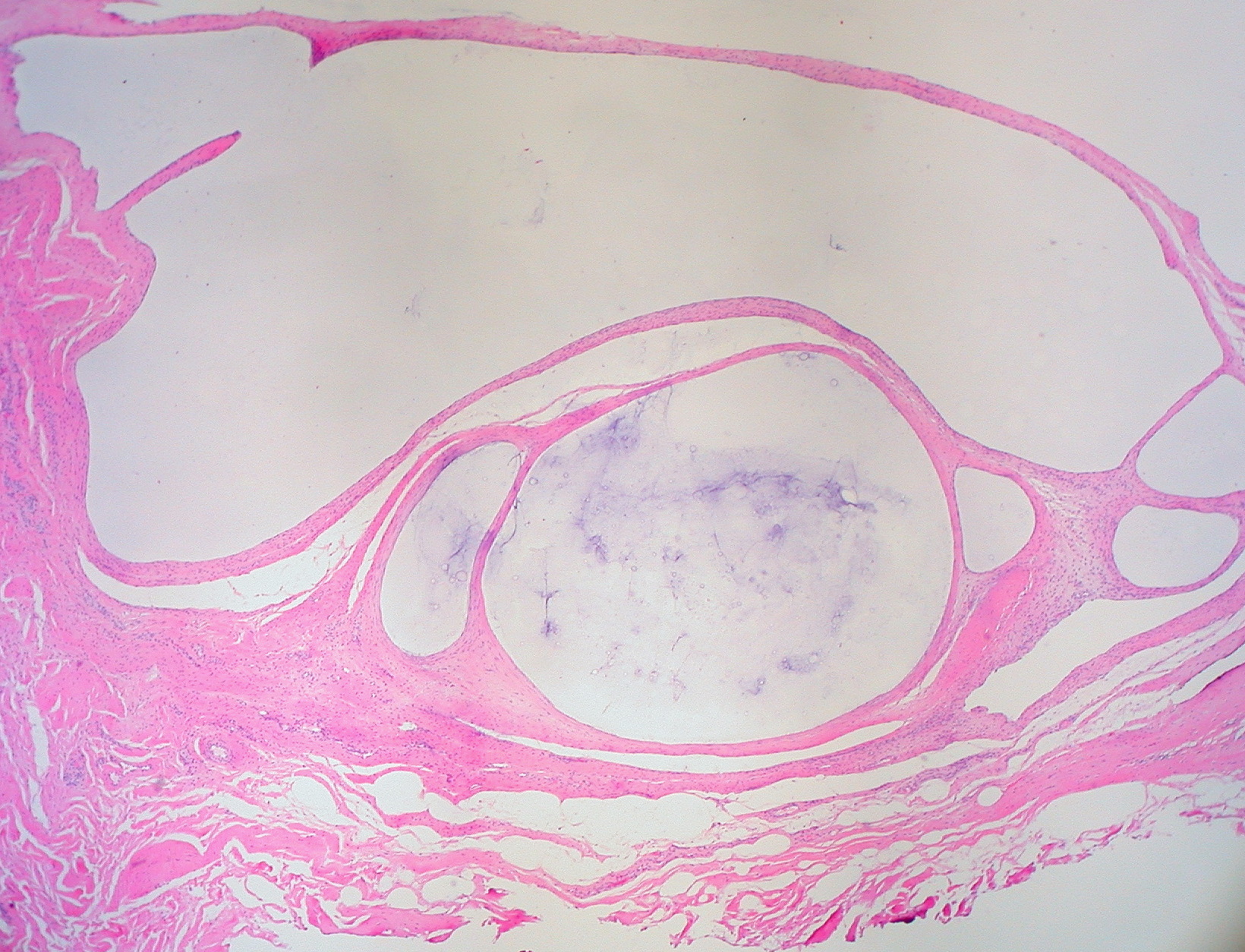
A ganglion cyst is a fluid-filled bump associated with a joint or tendon sheath. It most often occurs at the back of the wrist, followed by the front of the wrist. Onset is often over several months, typically with no further symptoms. Occasionally, pain or numbness may occur. Complications may include carpal tunnel syndrome. The cause is unknown. The underlying mechanism is believed to involve an outpouching of the synovial membrane. Risk factors include gymnastics activity. Diagnosis is typically based on examination with light shining through the lesion being supportive. Medical imaging may be done to rule out other potential causes. Treatment options include watchful waiting, splinting the affected joint, needle aspiration, or surgery. About half the time, they resolve on their own. About three per 10,000 people newly develop ganglion of the wrist or hand a year. They most commonly occur in young and middle-aged females. Presentation The average size of these cysts is 2.0 ... [...More Info...] [...Related Items...] OR: [Wikipedia] [Google] [Baidu] |
Calcaneal Spur
A calcaneal spur (also known as a heel spur) is a bony outgrowth from the calcaneal tuberosity (heel bone). Calcaneal spurs are typically detected by x-ray examination. It is a form of exostosis. When a foot is exposed to constant stress, calcium deposits build up on the bottom of the heel bone. Generally, this has no effect on a person's daily life. However, repeated damage can cause these deposits to pile up on each other, causing a spur-shaped deformity, called a calcaneal (or heel) spur. An inferior calcaneal spur is located on the inferior aspect of the calcaneus and is typically a response to plantar fasciitis over a period, but may also be associated with ankylosing spondylitis (typically in children). A posterior calcaneal spur develops on the back of the heel at the insertion of the Achilles tendon. An inferior calcaneal spur consists of a calcification of the calcaneus, which lies superior to the plantar fascia at the insertion of the plantar fascia. A posterior calcane ... [...More Info...] [...Related Items...] OR: [Wikipedia] [Google] [Baidu] |


