|
Eosinophilic Gastroenteritis
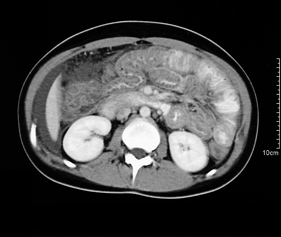
Eosinophilic gastroenteritis (EG or EGE) is a rare and heterogeneous condition characterized by patchy or diffuse eosinophilic infiltration of gastrointestinal (GI) tissue, first described by Kaijser in 1937.Kaijser R. Zur Kenntnis der allergischen Affektionen des Verdauugskanals vom Standpunkt des Chirurgen aus. Arch Klin Chir 1937; 188:36–64. Presentation may vary depending on location as well as depth and extent of bowel wall involvement and usually runs a chronic relapsing course. It can be classified into mucosal, muscular and serosal types based on the depth of involvement. Any part of the GI tract can be affected, and isolated biliary tract involvement has also been reported. The stomach is the organ most commonly affected, followed by the small intestine and the colon. Signs and symptoms EG typically presents with a combination of chronic nonspecific GI symptoms which include abdominal pain, diarrhea, occasional nausea and vomiting, weight loss and abdominal distens ... [...More Info...] [...Related Items...] OR: [Wikipedia] [Google] [Baidu] |
Eosinophilic
Eosinophilic (Greek suffix -phil-, meaning ''loves eosin'') is the staining of tissues, cells, or organelles after they have been washed with eosin, a dye. Eosin is an acidic dye for staining cell cytoplasm, collagen, and muscle fibers. ''Eosinophilic'' describes the appearance of cells and structures seen in histological sections that take up the staining dye eosin. Such eosinophilic structures are, in general, composed of protein. Eosin is usually combined with a stain called hematoxylin to produce a hematoxylin- and eosin-stained section (also called an H&E stain, HE or H+E section). It is the most widely used histological stain for a medical diagnosis. When a pathologist examines a biopsy of a suspected cancer, they will stain the biopsy with H&E. Some structures seen inside cells are described as being eosinophilic; for example, Lewy and Mallory bodies. [...More Info...] [...Related Items...] OR: [Wikipedia] [Google] [Baidu] |
Malabsorption
Malabsorption is a state arising from abnormality in absorption of food nutrients across the gastrointestinal (GI) tract. Impairment can be of single or multiple nutrients depending on the abnormality. This may lead to malnutrition and a variety of anaemias. Normally the human gastrointestinal tract digests and absorbs dietary nutrients with remarkable efficiency. A typical Western diet ingested by an adult in one day includes approximately 100 g of fat, 400 g of carbohydrate, 100 g of protein, 2 L of fluid, and the required sodium, potassium, chloride, calcium, vitamins, and other elements. Salivary, gastric, intestinal, hepatic, and pancreatic secretions add an additional 7–8 L of protein-, lipid-, and electrolyte-containing fluid to intestinal contents. This massive load is reduced by the small and large intestines to less than 200 g of stool that contains less than 8 g of fat, 1–2 g of nitrogen, and less than 20 mmol each of Na+, K+, Cl–, HCO3–, Ca2+, or Mg ... [...More Info...] [...Related Items...] OR: [Wikipedia] [Google] [Baidu] |
Eosinophilia
Eosinophilia is a condition in which the eosinophil count in the peripheral blood exceeds . Hypereosinophilia is an elevation in an individual's circulating blood eosinophil count above 1.5 x 10⁹/ L (i.e. 1,500/ μL). The hypereosinophilic syndrome is a sustained elevation in this count above 1.5 x 10⁹/L (i.e. 1,500/μL) that is also associated with evidence of eosinophil-based tissue injury. Eosinophils usually account for less than 7% of the circulating leukocytes. A marked increase in non-blood tissue eosinophil count noticed upon histopathologic examination is diagnostic for tissue eosinophilia. Several causes are known, with the most common being some form of allergic reaction or parasitic infection. Diagnosis of eosinophilia is via a complete blood count (CBC), but diagnostic procedures directed at the underlying cause vary depending on the suspected condition(s). An absolute eosinophil count is not generally needed if the CBC shows marked eosinophilia. The location of ... [...More Info...] [...Related Items...] OR: [Wikipedia] [Google] [Baidu] |
Duodenal Ulcer
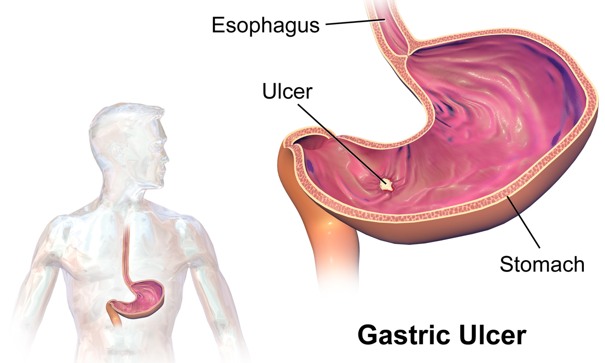
Peptic ulcer disease (PUD) is a break in the inner lining of the stomach, the first part of the small intestine, or sometimes the lower esophagus. An ulcer in the stomach is called a gastric ulcer, while one in the first part of the intestines is a duodenal ulcer. The most common symptoms of a duodenal ulcer are waking at night with upper abdominal pain and upper abdominal pain that improves with eating. With a gastric ulcer, the pain may worsen with eating. The pain is often described as a burning or dull ache. Other symptoms include belching, vomiting, weight loss, or poor appetite. About a third of older people have no symptoms. Complications may include bleeding, perforation, and blockage of the stomach. Bleeding occurs in as many as 15% of cases. Common causes include the bacteria '' Helicobacter pylori'' and non-steroidal anti-inflammatory drugs (NSAIDs). Other, less common causes include tobacco smoking, stress as a result of other serious health conditions, Behçet ... [...More Info...] [...Related Items...] OR: [Wikipedia] [Google] [Baidu] |
Appendicitis
Appendicitis is inflammation of the appendix. Symptoms commonly include right lower abdominal pain, nausea, vomiting, and decreased appetite. However, approximately 40% of people do not have these typical symptoms. Severe complications of a ruptured appendix include widespread, painful inflammation of the inner lining of the abdominal wall and sepsis. Appendicitis is caused by a blockage of the hollow portion of the appendix. This is most commonly due to a calcified "stone" made of feces. Inflamed lymphoid tissue from a viral infection, parasites, gallstone, or tumors may also cause the blockage. This blockage leads to increased pressures in the appendix, decreased blood flow to the tissues of the appendix, and bacterial growth inside the appendix causing inflammation. The combination of inflammation, reduced blood flow to the appendix and distention of the appendix causes tissue injury and tissue death. If this process is left untreated, the appendix may burst, releasing b ... [...More Info...] [...Related Items...] OR: [Wikipedia] [Google] [Baidu] |
Splenitis
Splenomegaly is an enlargement of the spleen. The spleen usually lies in the left upper quadrant (LUQ) of the human abdomen. Splenomegaly is one of the four cardinal signs of ''hypersplenism'' which include: some reduction in number of circulating blood cells affecting granulocytes, erythrocytes or platelets in any combination; a compensatory proliferative response in the bone marrow; and the potential for correction of these abnormalities by splenectomy. Splenomegaly is usually associated with increased workload (such as in hemolytic anemias), which suggests that it is a response to hyperfunction. It is therefore not surprising that splenomegaly is associated with any disease process that involves abnormal red blood cells being destroyed in the spleen. Other common causes include congestion due to portal hypertension and infiltration by leukemias and lymphomas. Thus, the finding of an enlarged spleen, along with caput medusae, is an important sign of portal hypertension. Definit ... [...More Info...] [...Related Items...] OR: [Wikipedia] [Google] [Baidu] |
Pancreatitis
Pancreatitis is a condition characterized by inflammation of the pancreas. The pancreas is a large organ behind the stomach that produces digestive enzymes and a number of hormones. There are two main types: acute pancreatitis, and chronic pancreatitis. Signs and symptoms of pancreatitis include pain in the upper abdomen, nausea and vomiting. The pain often goes into the back and is usually severe. In acute pancreatitis, a fever may occur, and symptoms typically resolve in a few days. In chronic pancreatitis weight loss, fatty stool, and diarrhea may occur. Complications may include infection, bleeding, diabetes mellitus, or problems with other organs. The two most common causes of acute pancreatitis are a gallstone blocking the common bile duct after the pancreatic duct has joined; and heavy alcohol use. Other causes include direct trauma, certain medications, infections such as mumps, and tumors. Chronic pancreatitis may develop as a result of acute pancreatitis. It is mos ... [...More Info...] [...Related Items...] OR: [Wikipedia] [Google] [Baidu] |
Corticosteroids
Corticosteroids are a class of steroid hormones that are produced in the adrenal cortex of vertebrates, as well as the synthetic analogues of these hormones. Two main classes of corticosteroids, glucocorticoids and mineralocorticoids, are involved in a wide range of physiological processes, including stress response, immune response, and regulation of inflammation, carbohydrate metabolism, protein catabolism, blood electrolyte levels, and behavior. Some common naturally occurring steroid hormones are cortisol (), corticosterone (), cortisone () and aldosterone (). (Note that cortisone and aldosterone are isomers.) The main corticosteroids produced by the adrenal cortex are cortisol and aldosterone. Classes * Glucocorticoids such as cortisol affect carbohydrate, fat, and protein metabolism, and have anti-inflammatory, immunosuppressive, anti-proliferative, and vasoconstrictive effects. Anti-inflammatory effects are mediated by blocking the action of inflammatory ... [...More Info...] [...Related Items...] OR: [Wikipedia] [Google] [Baidu] |
Serous Membrane
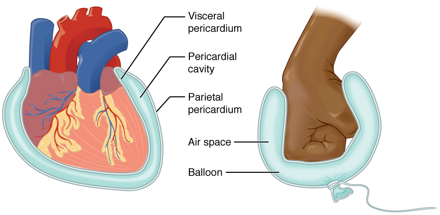
The serous membrane (or serosa) is a smooth tissue membrane of mesothelium lining the contents and inner walls of body cavities, which secrete serous fluid to allow lubricated sliding movements between opposing surfaces. The serous membrane that covers internal organs is called a ''visceral'' membrane; while the one that covers the cavity wall is called the ''parietal'' membrane. Between the two opposing serosal surfaces is often a potential space, mostly empty except for the small amount of serous fluid. The Latin anatomical name is '' tunica serosa''. Serous membranes line and enclose several body cavities, also known as serous cavities, where they secrete a lubricating fluid which reduces friction from movements. Serosa is entirely different from the adventitia, a connective tissue layer which binds together structures rather than reducing friction between them. The serous membrane covering the heart and lining the mediastinum is referred to as the pericardium, the se ... [...More Info...] [...Related Items...] OR: [Wikipedia] [Google] [Baidu] |
Intussusception (medical Disorder)
Intussusception is a medical condition in which a part of the intestine folds into the section immediately ahead of it. It typically involves the small bowel and less commonly the large bowel. Symptoms include abdominal pain which may come and go, vomiting, abdominal bloating, and bloody stool. It often results in a small bowel obstruction. Other complications may include peritonitis or bowel perforation. The cause in children is typically unknown; in adults a ''lead point'' is sometimes present. Risk factors in children include certain infections, diseases like cystic fibrosis, and intestinal polyps. Risk factors in adults include endometriosis, bowel adhesions, and intestinal tumors. Diagnosis is often supported by medical imaging. In children, ultrasound is preferred while in adults a CT scan is preferred. Intussusception is an emergency requiring rapid treatment. Treatment in children is typically by an enema with surgery used if this is not successful. Dexamethas ... [...More Info...] [...Related Items...] OR: [Wikipedia] [Google] [Baidu] |
Cecum
The cecum or caecum is a pouch within the peritoneum that is considered to be the beginning of the large intestine. It is typically located on the right side of the body (the same side of the body as the appendix, to which it is joined). The word cecum (, plural ceca ) stems from the Latin '' caecus'' meaning blind. It receives chyme from the ileum, and connects to the ascending colon of the large intestine. It is separated from the ileum by the ileocecal valve (ICV) or Bauhin's valve. It is also separated from the colon by the cecocolic junction. While the cecum is usually intraperitoneal, the ascending colon is retroperitoneal. In herbivores, the cecum stores food material where bacteria are able to break down the cellulose. In humans, the cecum is involved in absorption of salts and electrolytes and lubricates the solid waste that passes into the large intestine. Structure Development The cecum and appendix are formed by the enlargement of the postarterial segment of ... [...More Info...] [...Related Items...] OR: [Wikipedia] [Google] [Baidu] |


