|
External Anal Sphincter
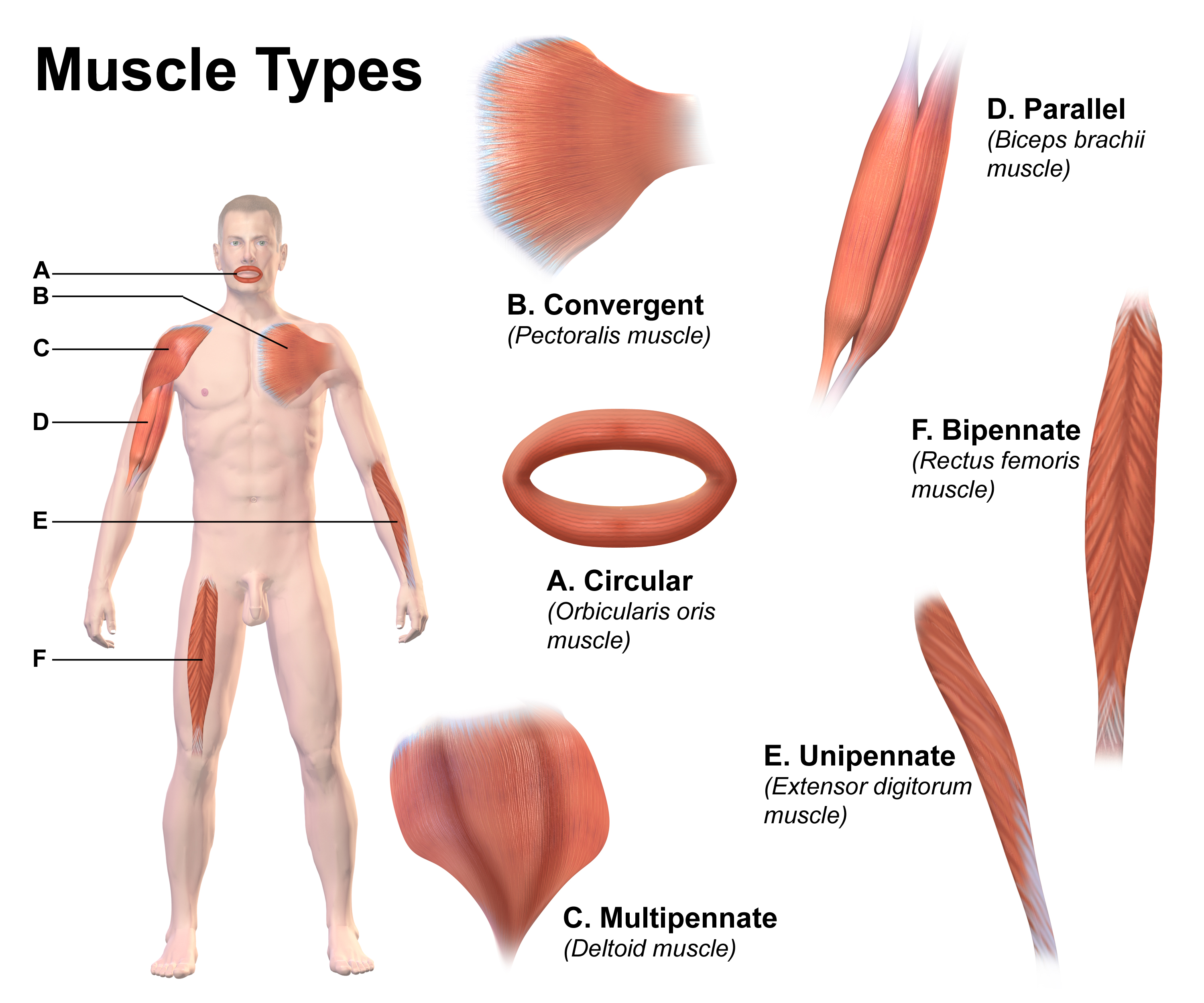
The external anal sphincter (or sphincter ani externus) is an oval tube of skeletal muscle fibers. Distally, it is adherent to the skin surrounding the margin of the anus. It exhibits a resting state of tonical contraction and also contracts during the bulbospongiosus reflex. Anatomy The external anal sphincter is far more substantial than the internal anal sphincter. The proximal portion of external anal sphincter overlaps the internal anal sphincter (which terminates distally a little distance proximal to the anal orifice) superficially; where the two overlap, they are separated by the intervening conjoint longitudinal muscle. Structure Historically, the sphincter was described as consisting of three parts (deep, superficial, and subcutaneous). This is not supported by current anatomical knowledge. Some sources still describe it in two layers, deep (or proximal) and superficial (or distal or subcutaneous). Some of the muscles fibres decussate at the anterior midline and po ... [...More Info...] [...Related Items...] OR: [Wikipedia] [Google] [Baidu] |
Anal Canal
The anal canal is the part that connects the rectum to the anus, located below the level of the pelvic diaphragm. It is located within the anal triangle of the perineum, between the right and left ischioanal fossa. As the final functional segment of the bowel, it functions to regulate release of excrement by two muscular sphincter complexes. The anus is the aperture at the terminal portion of the anal canal. Structure In humans, the anal canal is approximately long, from the anorectal junction to the anus. It is directed downwards and backwards. It is surrounded by inner involuntary and outer voluntary sphincters which keep the lumen closed in the form of an anteroposterior slit. The canal is differentiated from the rectum by a transition along the internal surface from endodermal to skin-like ectodermal tissue. The anal canal is traditionally divided into two segments, upper and lower, separated by the pectinate line (also known as the dentate line): * upper zo ... [...More Info...] [...Related Items...] OR: [Wikipedia] [Google] [Baidu] |
Anus
In mammals, invertebrates and most fish, the anus (: anuses or ani; from Latin, 'ring' or 'circle') is the external body orifice at the ''exit'' end of the digestive tract (bowel), i.e. the opposite end from the mouth. Its function is to facilitate the defecation, expulsion of wastes that remain after digestion. Bowel contents that pass through the anus include the gaseous flatus and the semi-solid feces, which (depending on the type of animal) include: indigestible matter such as bones, hair pellet (ornithology), pellets, endozoochory, endozoochorous seeds and gastrolith, digestive rocks; Summary at residual food material after the digestible nutrients have been extracted, for example cellulose or lignin; ingested matter which would be toxic if it remained in the digestive tract; excretion, excreted metabolites like bilirubin-containing bile; and dead mucosal epithelia or excess gut bacteria and other endosymbionts. Passage of feces through the anus is typically controlled by ... [...More Info...] [...Related Items...] OR: [Wikipedia] [Google] [Baidu] |
Perineum
The perineum (: perineums or perinea) in placentalia, placental mammals is the space between the anus and the genitals. The human perineum is between the anus and scrotum in the male or between the anus and vulva in the female. The perineum is the region of the body between the pubic symphysis (pubic arch) and the coccyx (tail bone), including the perineal body and surrounding structures. The perineal raphe is visible and pronounced to varying degrees. Etymology The word entered English from late Latin via Greek language, Greek περίναιος ~ περίνεος ''perinaios, perineos'', itself from περίνεος, περίνεοι 'male genitals' and earlier περίς ''perís'' 'penis' through influence from πηρίς ''pērís'' 'scrotum'. The term was originally understood as a purely male body-part with the perineal raphe seen as a continuation of the scrotal septum since Virilization, masculinization causes the development of a large anogenital distance in men, i ... [...More Info...] [...Related Items...] OR: [Wikipedia] [Google] [Baidu] |
Puborectalis Muscle
The levator ani is a broad, thin muscle group, situated on either side of the pelvis. It is formed from three muscle components: the pubococcygeus, the iliococcygeus, and the puborectalis. It is attached to the inner surface of each side of the lesser pelvis, and these unite to form the greater part of the pelvic floor. The coccygeus muscle completes the pelvic floor, which is also called the ''pelvic diaphragm''. It supports the viscera in the pelvic cavity, and surrounds the various structures that pass through it. The levator ani is the main pelvic floor muscle and contracts rhythmically during female orgasm, and painfully during vaginismus. Structure The levator ani is made up of 3 parts: * Iliococcygeus muscle * Pubococcygeus muscle * Puborectalis muscle The iliococcygeus arises from the inner side of the ischium (the lower and back part of the hip bone) and from the posterior part of the tendinous arch of the obturator fascia, and is attached to the coccyx and ... [...More Info...] [...Related Items...] OR: [Wikipedia] [Google] [Baidu] |
Internal Anal Sphincter
The internal anal sphincter, IAS, or sphincter ani internus is a ring of smooth muscle that surrounds about 2.5–4.0 cm of the anal canal. It is about 5 mm thick, and is formed by an aggregation of the smooth (involuntary) circular muscle fibers of the rectum. The internal anal sphincter aids the sphincter ani externus to occlude the anal aperture and aids in the expulsion of the feces. Its action is entirely involuntary. It is normally in a state of continuous maximal contraction to prevent leakage of faeces or gases. Sympathetic stimulation stimulates and maintains the sphincter's contraction, and parasympathetic stimulation inhibits it. It becomes relaxed in response to distention of the rectal ampulla, requiring voluntary contraction of the puborectalis and external anal sphincter to maintain continence, and also contracts during the bulbospongiosus reflex. Structure The internal anal sphincter is the specialised thickened terminal portion of the inner circular ... [...More Info...] [...Related Items...] OR: [Wikipedia] [Google] [Baidu] |
Slow Twitch Fiber
Skeletal muscle (commonly referred to as muscle) is one of the three types of vertebrate muscle tissue, the others being cardiac muscle and smooth muscle. They are part of the voluntary muscular system and typically are attached by tendons to bones of a skeleton. The skeletal muscle cells are much longer than in the other types of muscle tissue, and are also known as ''muscle fibers''. The tissue of a skeletal muscle is striated – having a striped appearance due to the arrangement of the sarcomeres. A skeletal muscle contains multiple fascicles – bundles of muscle fibers. Each individual fiber and each muscle is surrounded by a type of connective tissue layer of fascia. Muscle fibers are formed from the fusion of developmental myoblasts in a process known as myogenesis resulting in long multinucleated cells. In these cells, the nuclei, termed ''myonuclei'', are located along the inside of the cell membrane. Muscle fibers also have multiple mitochondria to meet energy needs ... [...More Info...] [...Related Items...] OR: [Wikipedia] [Google] [Baidu] |
Nerve To Levator Ani .Essential Clinical Anatomy. K.L. Moore & A.M. Agur. Lippincott, 2 ed. 2002. Page 217
The levator ani nerve is a nerve to the levator ani muscles. It originates from sacral spinal nerve 4 The sacral spinal nerve 4 (S4) is a spinal nerve of the sacral segment. References Nerves of the lower limb and lower torso {{neuroanatomy-stub ...[...More Info...] [...Related Items...] OR: [Wikipedia] [Google] [Baidu] |
Inferior Anal Nerves
The inferior rectal nerves (inferior anal nerves, inferior hemorrhoidal nerve) usually branch from the pudendal nerve but occasionally arises directly from the sacral plexus; they cross the ischiorectal fossa along with the inferior rectal artery and veins, toward the anal canal and the lower end of the rectum, and is distributed to the sphincter ani externus (external anal sphincter, EAS) and to the integument (skin) around the anus. Branches of this nerve communicate with the perineal branch of the posterior femoral cutaneous and with the posterior scrotal nerves at the forepart of the perineum. Supplies Cutaneous innervation below the pectinate line and external anal sphincter. See also * Inferior rectal artery Additional images File:Gray405.png, The perineum. The integument and superficial layer of superficial fascia reflected. File:Gray837.png, Sacral plexus of the right side. (Hemorrhoidal branch of pudic labeled at bottom right.) References External links Detai ... [...More Info...] [...Related Items...] OR: [Wikipedia] [Google] [Baidu] |
Perineal Body
The perineum (: perineums or perinea) in placental mammals is the space between the anus and the genitals. The human perineum is between the anus and scrotum in the male or between the anus and vulva in the female. The perineum is the region of the body between the pubic symphysis (pubic arch) and the coccyx (tail bone), including the perineal body and surrounding structures. The perineal raphe is visible and pronounced to varying degrees. Etymology The word entered English from late Latin via Greek περίναιος ~ περίνεος ''perinaios, perineos'', itself from περίνεος, περίνεοι 'male genitals' and earlier περίς ''perís'' 'penis' through influence from πηρίς ''pērís'' 'scrotum'. The term was originally understood as a purely male body-part with the perineal raphe seen as a continuation of the scrotal septum since masculinization causes the development of a large anogenital distance in men, in comparison to the corresponding ... [...More Info...] [...Related Items...] OR: [Wikipedia] [Google] [Baidu] |
Conjoint Longitudinal Muscle
The conjoint longitudinal muscle is a muscle layer in the wall of the anal canal between the internal anal sphincter (deep to CLM) and external anal sphincter (superficial to CLM). It is continuous proximally with the longitudinal (outer) smooth muscle layer of the rectum. It receives autonomic innervation in common with the internal anal sphincter The internal anal sphincter, IAS, or sphincter ani internus is a ring of smooth muscle that surrounds about 2.5–4.0 cm of the anal canal. It is about 5 mm thick, and is formed by an aggregation of the smooth (involuntary) circular musc .... Anatomy Distally, the muscle detaches and extends in three directions. The projections of the muscle represent pathways for the spread of infections. Development The CLM is especially prominent in the foetus, but is gradually replaced by connective tissue with age. Function Contraction of the CLM shortens and widens the anal canal, and everts the anal orifice. References Exte ... [...More Info...] [...Related Items...] OR: [Wikipedia] [Google] [Baidu] |
Bulbospongiosus Reflex
The bulbocavernosus reflex (BCR), bulbospongiosus reflex (BSR) or "Osinski reflex" is a polysynaptic reflex that is useful in testing for spinal shock and gaining information about the state of spinal cord injuries (SCI). ''Bulbocavernosus'' is an older term for ''bulbospongiosus'', thus this reflex may also be referred to as the bulbospongiosus reflex. Procedure The test involves monitoring internal/external anal sphincter contraction in response to squeezing the glans penis or clitoris, or tugging on an indwelling Foley catheter. This reflex can also be tested electrophysiologically, by stimulating the penis or vulva and recording from the anal sphincter. This testing modality is used in intraoperative neurophysiology monitoring to verify the function of sensory and motor sacral roots as well as the conus medullaris. Trauma The reflex is spinal mediated and involves S2– S4. The absence of the reflex in a person with acute paralysis from trauma indicates spinal shock wh ... [...More Info...] [...Related Items...] OR: [Wikipedia] [Google] [Baidu] |
Integument
In biology, an integument is the tissue surrounding an organism's body or an organ within, such as skin, a husk, Exoskeleton, shell, germ or Peel (fruit), rind. Etymology The term is derived from ''integumentum'', which is Latin for "a covering". In a transferred, or figurative sense, it could mean a cloak or a disguise. In English, "integument" is a fairly modern word, its origin having been traced back to the early seventeenth century; and refers to a material or layer with which anything is enclosed, clothed, or covered in the sense of "clad" or "coated", as with a skin or husk. Botanical usage In botany, the term "integument" may be used as it is in zoology, referring to the covering of an organ. When the context indicates nothing to the contrary, the word commonly refers to an envelope covering the nucellus of the Ovule#Integuments, chivai and chalaza, ovule. The integument may consist of one layer (unitegmic) or two layers (bitegmic), each of which consisting of two or ... [...More Info...] [...Related Items...] OR: [Wikipedia] [Google] [Baidu] |

