|
Extensor Digitorum Muscle
The extensor digitorum muscle (also known as extensor digitorum communis) is a muscle of the posterior forearm present in humans and other animals. It extends the medial four digits of the hand. Extensor digitorum is innervated by the posterior interosseous nerve, which is a branch of the radial nerve. Structure The extensor digitorum muscle arises from the lateral epicondyle of the humerus, by the common tendon; from the intermuscular septa between it and the adjacent muscles, and from the antebrachial fascia. It divides below into four tendons, which pass, together with that of the extensor indicis proprius, through a separate compartment of the dorsal carpal ligament, within a mucous sheath. The tendons then diverge on the back of the hand, and are inserted into the middle and distal phalanges of the fingers in the following manner.''Gray's anatomy'' (1918), see infobox Opposite the metacarpophalangeal articulation each tendon is bound by fasciculi to the collateral ligamen ... [...More Info...] [...Related Items...] OR: [Wikipedia] [Google] [Baidu] |
Lateral Epicondyle Of The Humerus
The lateral epicondyle of the humerus is a large, tuberculated eminence, curved a little forward, and giving attachment to the radial collateral ligament of the elbow joint, and to a tendon common to the origin of the supinator and some of the extensor muscles. Specifically, these extensor muscles include the anconeus muscle, the supinator, extensor carpi radialis brevis, extensor digitorum, extensor digiti minimi, and extensor carpi ulnaris. In birds, where the arm is somewhat rotated compared to other tetrapods, it is termed dorsal epicondyle of the humerus. In comparative anatomy, the term ''ectepicondyle'' is sometimes used. A common injury associated with the lateral epicondyle of the humerus is lateral epicondylitis also known as tennis elbow. Repetitive overuse of the forearm, as seen in tennis or other sports, can result in inflammation of "the tendons that join the forearm muscles on the outside of the elbow. The forearm muscles and tendons become damaged from overu ... [...More Info...] [...Related Items...] OR: [Wikipedia] [Google] [Baidu] |
Metacarpophalangeal
The metacarpophalangeal joints (MCP) are situated between the metacarpal bones and the proximal phalanges of the fingers. These joints are of the condyloid kind, formed by the reception of the rounded heads of the metacarpal bones into shallow cavities on the proximal ends of the proximal phalanges. Being condyloid, they allow the movements of flexion, extension, abduction, adduction and circumduction (see anatomical terms of motion) at the joint. Structure Ligaments Each joint has: * palmar ligaments of metacarpophalangeal articulations * collateral ligaments of metacarpophalangeal articulations Dorsal surfaces The dorsal surfaces of these joints are covered by the expansions of the Extensor tendons, together with some loose areolar tissue which connects the deep surfaces of the tendons to the bones. Function The movements which occur in these joints are flexion, extension, adduction, abduction, and circumduction; the movements of abduction and adduction are very ... [...More Info...] [...Related Items...] OR: [Wikipedia] [Google] [Baidu] |
Extensor Digitorum Longus Muscle
The extensor digitorum longus is a pennate muscle, situated at the lateral part of the front of the leg. Structure It arises from the lateral condyle of the tibia The tibia (; : tibiae or tibias), also known as the shinbone or shankbone, is the larger, stronger, and anterior (frontal) of the two Leg bones, bones in the leg below the knee in vertebrates (the other being the fibula, behind and to the outsi ...; from the upper three-quarters of the anterior surface of the body of the fibula; from the upper part of the interosseous membrane; from the deep surface of the fascia; and from the intermuscular septa between it and the tibialis anterior on the medial, and the peroneal muscles on the lateral side. Between it and the tibialis anterior are the upper portions of the anterior tibial vessels and deep peroneal nerve. The muscle passes under the superior and inferior extensor retinaculum of foot in company with the fibularis tertius, and divides into four slips, which ... [...More Info...] [...Related Items...] OR: [Wikipedia] [Google] [Baidu] |
Extensor Digitorum Brevis Muscle
The extensor digitorum brevis muscle (sometimes EDB) is a muscle Muscle is a soft tissue, one of the four basic types of animal tissue. There are three types of muscle tissue in vertebrates: skeletal muscle, cardiac muscle, and smooth muscle. Muscle tissue gives skeletal muscles the ability to muscle contra ... on the upper surface of the foot that helps extend digits 2 through 4. Structure The muscle originates from the forepart of the upper and lateral surface of the calcaneus (in front of the groove for the peroneus brevis tendon), from the interosseous talocalcaneal ligament and the stem of the inferior extensor retinaculum. The fibres pass obliquely forwards and medially across the dorsum of the foot and end in four tendons. The medial part of the muscle, also known as extensor hallucis brevis, ends in a tendon which crosses the dorsalis pedis artery and inserts into the dorsal surface of the base of the proximal phalanx of the great toe. The other three tendons ins ... [...More Info...] [...Related Items...] OR: [Wikipedia] [Google] [Baidu] |
Palmar Interossei Muscles
In human anatomy, the palmar or volar interossei (interossei volares in older literature) are four muscles, one on the thumb that is occasionally missing, and three small, unipennate, central muscles in the hand that lie between the Metacarpus, metacarpal bones and are attached to the Index finger, index, Ring finger, ring, and Little finger, little fingers. They are smaller than the dorsal interossei of the hand. Structure All palmar interossei originate along the shaft of the metacarpal bone of the digit on which they act. They are inserted into the base of the Phalanx bone, proximal phalanx and the extensor expansion of the Extensor digitorum muscle, extensor digitorum of the same digit. Pollical palmar interosseous The first palmar interosseous is located at the thumb's medial side. Passing between the first dorsal interosseous and the oblique head of Adductor pollicis muscle, adductor pollicis, it is inserted on the base of the thumb's proximal phalanx together with Adductor ... [...More Info...] [...Related Items...] OR: [Wikipedia] [Google] [Baidu] |
Dorsal Interossei Of The Hand
In human anatomy, the dorsal interossei (DI) are four muscles in the back of the hand that act to abduct (spread) the index, middle, and ring fingers away from the hand's midline (ray of middle finger) and assist in flexion at the metacarpophalangeal The metacarpophalangeal joints (MCP) are situated between the metacarpal bones and the proximal phalanges of the fingers. These joints are of the condyloid kind, formed by the reception of the rounded heads of the metacarpal bones into shallow ... joints and extension at the interphalangeal joints of the index, middle and ring fingers. Structure There are four dorsal interossei in each hand. They are specified as 'dorsal' to contrast them with the palmar interossei, which are located on the anterior side of the metacarpals. The dorsal interosseous muscles are bipennate, with each muscle arising by two heads from the adjacent sides of the metacarpal bones, but more extensively from the metacarpal bone of the finger into whic ... [...More Info...] [...Related Items...] OR: [Wikipedia] [Google] [Baidu] |
Interphalangeal Articulations Of Hand
The interphalangeal joints of the hand are the hinge joints between the phalanges of the fingers that provide flexion towards the palm of the hand. There are two sets in each finger (except in the thumb, which has only one joint): * "proximal interphalangeal joints" (PIJ or PIP), those between the first (also called proximal) and second (intermediate) phalanges * "distal interphalangeal joints" (DIJ or DIP), those between the second (intermediate) and third (distal) phalanges Anatomically, the proximal and distal interphalangeal joints are very similar. There are some minor differences in how the palmar plates are attached proximally and in the segmentation of the flexor tendon sheath, but the major differences are the smaller dimension and reduced mobility of the distal joint. Joint structure The PIP joint exhibits great lateral stability. Its transverse diameter is greater than its antero-posterior diameter and its thick collateral ligaments are tight in all positions dur ... [...More Info...] [...Related Items...] OR: [Wikipedia] [Google] [Baidu] |
Metacarpophalangeal Joint
The metacarpophalangeal joints (MCP) are situated between the metacarpal bones and the proximal phalanges of the fingers. These joints are of the condyloid kind, formed by the reception of the rounded heads of the metacarpal bones into shallow cavities on the proximal ends of the proximal phalanges. Being condyloid, they allow the movements of flexion, extension, abduction, adduction and circumduction (see anatomical terms of motion) at the joint. Structure Ligaments Each joint has: * palmar ligaments of metacarpophalangeal articulations * collateral ligaments of metacarpophalangeal articulations Dorsal surfaces The dorsal surfaces of these joints are covered by the expansions of the Extensor tendons, together with some loose areolar tissue which connects the deep surfaces of the tendons to the bones. Function The movements which occur in these joints are flexion, extension, adduction, abduction, and circumduction; the movements of abduction and adduction are very ... [...More Info...] [...Related Items...] OR: [Wikipedia] [Google] [Baidu] |
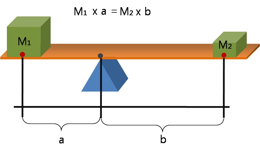
Lever
A lever is a simple machine consisting of a beam (structure), beam or rigid rod pivoted at a fixed hinge, or '':wikt:fulcrum, fulcrum''. A lever is a rigid body capable of rotating on a point on itself. On the basis of the locations of fulcrum, load, and effort, the lever is divided into Lever#Types of levers, three types. It is one of the six simple machines identified by Renaissance scientists. A lever amplifies an input force to provide a greater output force, which is said to provide leverage, which is mechanical advantage gained in the system, equal to the ratio of the output force to the input force. As such, the lever is a mechanical advantage device, trading off force against movement. Etymology The word "lever" entered English language, English around 1300 from . This sprang from the stem of the verb ''lever'', meaning "to raise". The verb, in turn, goes back to , itself from the adjective ''levis'', meaning "light" (as in "not heavy"). The word's primary origin is the ... [...More Info...] [...Related Items...] OR: [Wikipedia] [Google] [Baidu] |
Juncturae Tendinum
In human anatomy, juncturae tendinum or ''connexus intertendinei'' refers to the connective tissues that link the tendons of the extensor digitorum communis, and sometimes, to the tendon of the extensor digiti minimi. Juncturae tendinum are located on the dorsal aspect of the hand in the first, second and third inter-metacarpal spaces proximal to the metacarpophalangeal joint. Structure Juncturae tendinum are narrow bands of connective tissues that extend between the tendons of the extensor digitorum communis and the extensor digiti minimi. It is classified into three distinct types (Type 1, 2 and 3) depending on morphology. * Type 1: This is a thin and filamentous juncturae tendinum. Its shape can either be square, rhomboidal or triangular. * Type 2: This type is more tendinous and thicker than type 1 juncturae, and it is also located more distal than the type 1. * Type 3: Type 3 juncturae refers to the slips from the extensor digitorum communis. Type 3 juncture is furth ... [...More Info...] [...Related Items...] OR: [Wikipedia] [Google] [Baidu] |
Ulna
The ulna or ulnar bone (: ulnae or ulnas) is a long bone in the forearm stretching from the elbow to the wrist. It is on the same side of the forearm as the little finger, running parallel to the Radius (bone), radius, the forearm's other long bone. Longer and thinner than the radius, the ulna is considered to be the smaller long bone of the lower arm. The corresponding bone in the Human leg#Structure, lower leg is the fibula. Structure The ulna is a long bone found in the forearm that stretches from the elbow to the wrist, and when in standard anatomical position, is found on the Medial (anatomy), medial side of the forearm. It is broader close to the elbow, and narrows as it approaches the wrist. Close to the elbow, the ulna has a bony Process (anatomy), process, the olecranon process, a hook-like structure that fits into the olecranon fossa of the humerus. This prevents hyperextension and forms a hinge joint with the trochlea of the humerus. There is also a radial notch for ... [...More Info...] [...Related Items...] OR: [Wikipedia] [Google] [Baidu] |
Lumbricals Of The Hand
The lumbricals are intrinsic muscles of the hand that flexion, flex the metacarpophalangeal joints, and extension (kinesiology), extend the Interphalangeal articulations of hand, interphalangeal joints. p. 97 The lumbrical muscle of the foot, lumbrical muscles of the foot also have a similar action, though they are of less clinical concern. Structure The lumbricals are four, small, worm-like muscles on each hand. These muscles are unusual in that they do not attach to bone. Instead, they attach proximally to the tendons of flexor digitorum profundus, and distally to the extensor expansions. The first and second lumbricals are Anatomical terms of muscle, unipennate, while the third and fourth lumbricals are Anatomical terms of muscle, bipennate. Nerve supply The first and second lumbricals (the most radial two) are Nerve, innervated by the median nerve. The third and fourth lumbricals (most ulnar two) are innervated by the deep branch of ulnar nerve. This is the usual inne ... [...More Info...] [...Related Items...] OR: [Wikipedia] [Google] [Baidu] |


