|
Ethmoid Sinus
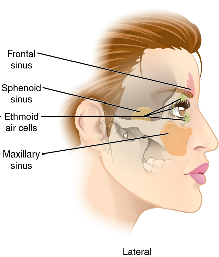
The ethmoid sinuses or ethmoid air cells of the ethmoid bone are one of the four paired paranasal sinuses. Unlike the other three pairs of paranasal sinuses which consist of one or two large cavities, the ethmoidal sinuses entail a number of small air-filled cavities ("air cells"). The cells are located within the lateral mass (labyrinth) of each ethmoid bone and are variable in both size and number.Illustrated Anatomy of the Head and Neck, Fehrenbach and Herring, Elsevier, 2012, page 64 The cells are grouped into anterior, middle, and posterior groups; the groups differ in their drainage modalities, though all ultimately drain into either the superior or the middle nasal meatus of the lateral wall of the nasal cavity. Structure The ethmoid air cells consist of numerous thin-walled cavities in the ethmoidal labyrinthOtorhinolaryngology, Head and Neck Surgery, Anniko, Springer, 2010, page 188 that represent invaginations of the mucous membrane of the nasal wall into the ethmoid bo ... [...More Info...] [...Related Items...] OR: [Wikipedia] [Google] [Baidu] |
Paranasal Sinus
Paranasal sinuses are a group of four paired skeletal pneumaticity, air-filled spaces that surround the nasal cavity. The maxillary sinuses are located under the eyes; the frontal sinuses are above the eyes; the Ethmoid sinus, ethmoidal sinuses are between the eyes and the sphenoidal sinuses are behind the eyes. The sinus (anatomy), sinuses are named for the Facial skeleton, facial bones and sphenoid bone in which they are located. Their role is disputed. Structure Humans possess four pairs of paranasal sinuses, divided into subgroups that are named according to the bones within which the sinuses lie. They are all innervated by branches of the trigeminal nerve (CN V). * The maxillary sinuses, the largest of the paranasal sinuses, are under the Human eye, eyes, in the maxillary bones (open in the back of the semilunar hiatus of the nose). They are innervated by the maxillary nerve (CN V2). * The frontal sinuses, superior to the eyes, in the frontal bone, which forms the hard part o ... [...More Info...] [...Related Items...] OR: [Wikipedia] [Google] [Baidu] |
Superior Nasal Concha
The superior nasal concha is a small, curved plate of bone representing a medial bony process of the labyrinth of the ethmoid bone. The superior nasal concha forms the roof of the superior nasal meatus. Anatomy Anatomical relations The superior nasal concha is situated posterosuperiorly to the middle nasal concha. It forms the superior boundary of the superior nasal meatus. Superior to the superior nasal concha is the sphenoethmoidal recess where the sphenoid sinus communicates with the nasal cavity; the sphenoethmoidal recess is interposed between the superior nasal concha, and (the anterior aspect of) the body of sphenoid bone. The sphenoid sinus ostium exists medial to the superior turbinate. See also * Nasal concha In anatomy, a nasal concha (; : conchae; ; Latin for 'shell'), also called a nasal turbinate or turbinal, is a long, narrow, curled shelf of bone tissue, bone that protrudes into the breathing passage of the nose in humans and various other anim ... Add ... [...More Info...] [...Related Items...] OR: [Wikipedia] [Google] [Baidu] |
Optic Nerve
In neuroanatomy, the optic nerve, also known as the second cranial nerve, cranial nerve II, or simply CN II, is a paired cranial nerve that transmits visual system, visual information from the retina to the brain. In humans, the optic nerve is derived from optic stalks during the seventh week of development and is composed of retinal ganglion cell axons and glial cells; it extends from the optic disc to the optic chiasma and continues as the optic tract to the lateral geniculate nucleus, Pretectal area, pretectal nuclei, and superior colliculus. Structure The optic nerve has been classified as the second of twelve paired cranial nerves, but it is technically a myelinated tract of the central nervous system, rather than a classical nerve of the peripheral nervous system because it is derived from an out-pouching of the diencephalon (optic stalks) during embryonic development. As a consequence, the fibers of the optic nerve are covered with myelin produced by oligodendrocytes, r ... [...More Info...] [...Related Items...] OR: [Wikipedia] [Google] [Baidu] |
Anterior Clinoid Process
The anterior clinoid process is a posterior projection of the sphenoid bone at the junction of the medial end of either lesser wing of sphenoid bone with the body of sphenoid bone. The bilateral processes flank the sella turcica anteriorly. The ACP is an important structure for cranial and endovascular surgical operations for several structures, including the pituitary gland and the internal carotid artery. Anatomy The anterior clinoid process is a pyramid-shaped bony projection of the lesser wing of the sphenoid bone and forms part of the lateral wall of the optic canal. Between each ACP lies the sella turcica, which holds the pituitary gland. Additionally, the ACP is part of the anterior roof of the cavernous sinus. The posterior and inferior portions of the ACP border the internal carotid artery. Attachments The free border of the tentorium cerebelli extends anteriorly on either side beyond the attached border of the same side (which ends anteriorly at the posterior clin ... [...More Info...] [...Related Items...] OR: [Wikipedia] [Google] [Baidu] |
Sphenoid Sinus
The sphenoid sinus is a paired paranasal sinus in the body of the sphenoid bone. It is one pair of the four paired paranasal sinuses.Illustrated Anatomy of the Head and Neck, Fehrenbach and Herring, Elsevier, 2012, page 64 The two sphenoid sinuses are separated from each other by a septum. Each sphenoid sinus communicates with the nasal cavity via the opening of sphenoidal sinus. The two sphenoid sinuses vary in size and shape, and are usually asymmetrical. Structure On average, a sphenoid sinus measures 2.2 cm vertical height, 2 cm in transverse breadth; and 2.2 cm antero-posterior depth. Each spehoid sinus is in the body of sphenoid bone, just under the sella turcica. The sphenoid sinuses are separated from each other medially by the septum of sphenoidal sinuses, which is usually asymmetrical. An opening of sphenoidal sinus forms a passage between each sphenoidal sinus and the nasal cavity. Posteriorly, an opening of sphenoidal sinus opens into the sphenoidal ... [...More Info...] [...Related Items...] OR: [Wikipedia] [Google] [Baidu] |
Ethmoidal Infundibulum
The ethmoidal infundibulum is a funnel-shaped/slit-like/curved opening/passage/space/cleft upon the anterosuperior portion of the middle nasal meatus (and thus of the lateral wall of the nasal cavity) at the hiatus semilunaris (which represents the medial extremity of the infundibulum). The anterior ethmoidal air cells, and (usually) the frontonasal duct (which drains the frontal sinus) open into the ethmoidal infundibulum. The ethmoidal infundibulum extends anterosuperiorly from its opening into the nasal cavity. Anatomy The ethmoidal infundibulum is bordered medially by the uncinate process of the ethmoid bone, and laterally by the orbital plate of the ethmoid bone. The ethmoid infundibulum leads towards the maxillary hiatus.The anterior ethmoidal cells The ethmoid sinuses or ethmoid air cells of the ethmoid bone are one of the four paired paranasal sinuses. Unlike the other three pairs of paranasal sinuses which consist of one or two large cavities, the ethmoidal sinus ... [...More Info...] [...Related Items...] OR: [Wikipedia] [Google] [Baidu] |
Lamina Papyracea
The orbital lamina of ethmoid bone (or lamina papyracea or orbital lamina) is a smooth, oblong, paper-thin bone plate which forms the lateral wall of the labyrinth of the ethmoid bone. It covers the middle and posterior ethmoidal cells, and forms a large part of the medial wall of the orbit. It articulates above with the orbital plate of the frontal bone, below with the maxilla In vertebrates, the maxilla (: maxillae ) is the upper fixed (not fixed in Neopterygii) bone of the jaw formed from the fusion of two maxillary bones. In humans, the upper jaw includes the hard palate in the front of the mouth. The two maxil ... and the orbital process of palatine bone, in front with the lacrimal, and behind with the sphenoid. Its name lamina papyracea is an appropriate description, as this part of the ethmoid bone is paper-thin and fractures easily. A fracture here could cause entrapment of the medial rectus muscle. Additional images File:Slide4fen.JPG, Orbital lamina of et ... [...More Info...] [...Related Items...] OR: [Wikipedia] [Google] [Baidu] |
Facial Nerve
The facial nerve, also known as the seventh cranial nerve, cranial nerve VII, or simply CN VII, is a cranial nerve that emerges from the pons of the brainstem, controls the muscles of facial expression, and functions in the conveyance of taste sensations from the anterior two-thirds of the tongue. The nerve typically travels from the pons through the facial canal in the temporal bone and exits the skull at the stylomastoid foramen. It arises from the brainstem from an area posterior to the cranial nerve VI (abducens nerve) and anterior to cranial nerve VIII (vestibulocochlear nerve). The facial nerve also supplies preganglionic parasympathetic fibers to several head and neck ganglia. The facial and intermediate nerves can be collectively referred to as the nervus intermediofacialis. The path of the facial nerve can be divided into six segments: # intracranial (cisternal) segment (from brainstem pons to internal auditory canal) # meatal (canalicular) segment (with ... [...More Info...] [...Related Items...] OR: [Wikipedia] [Google] [Baidu] |
Pterygopalatine Ganglion
The pterygopalatine ganglion (aka Meckel's ganglion, nasal ganglion, or sphenopalatine ganglion) is a parasympathetic ganglion in the pterygopalatine fossa. It is one of four parasympathetic ganglia of the head and neck, (the others being the submandibular, otic, and ciliary ganglion). It is innervated by the Vidian nerve (formed by the greater superficial petrosal nerve branch of the facial nerve and deep petrosal nerve) and maxillary division of the trigeminal nerve. Its postsynaptic axons project to the lacrimal glands and nasal mucosa. The flow of blood to the nasal mucosa, in particular the venous plexus of the conchae, is regulated by the pterygopalatine ganglion and heats or cools the air in the nose. Structure The pterygopalatine ganglion (of Meckel), the largest of the parasympathetic ganglia associated with the branches of the maxillary nerve, is deeply placed in the pterygopalatine fossa, close to the sphenopalatine foramen. It is triangular or heart-shaped ... [...More Info...] [...Related Items...] OR: [Wikipedia] [Google] [Baidu] |
Trigeminal Nerve
In neuroanatomy, the trigeminal nerve (literal translation, lit. ''triplet'' nerve), also known as the fifth cranial nerve, cranial nerve V, or simply CN V, is a cranial nerve responsible for Sense, sensation in the face and motor functions such as biting and chewing; it is the most complex of the cranial nerves. Its name (''trigeminal'', ) derives from each of the two nerves (one on each side of the pons) having three major branches: the ophthalmic nerve (V), the maxillary nerve (V), and the mandibular nerve (V). The ophthalmic and maxillary nerves are purely sensory, whereas the mandibular nerve supplies motor as well as sensory (or "cutaneous") functions. Adding to the complexity of this nerve is that Autonomic nervous system, autonomic nerve fibers as well as special sensory fibers (taste) are contained within it. The motor division of the trigeminal nerve derives from the Basal plate (neural tube), basal plate of the embryonic pons, and the sensory division originates in ... [...More Info...] [...Related Items...] OR: [Wikipedia] [Google] [Baidu] |
Ophthalmic Nerve
The ophthalmic nerve (CN V1) is a sensory nerve of the head. It is one of three divisions of the trigeminal nerve (CN V), a cranial nerve. It has three major branches which provide sensory innervation to the eye, and the skin of the upper face and anterior scalp, as well as other structures of the head. Structure Origin The ophthalmic nerve is the first branch of the trigeminal nerve (CN V), the first and smallest of its three divisions. It arises from the superior part of the trigeminal ganglion. Course It passes anterior-ward along the lateral wall of the cavernous sinus inferior to the oculomotor nerve (CN III) and trochlear nerve (N IV). It exits the skull into the orbit through the superior orbital fissure. Branches Within the skull, the ophthalmic nerve produces: * meningeal branch (tentorial nerve) The ophthalmic nerve divides into three major branches which pass through the superior orbital fissure: * frontal nerve ** supraorbital nerve ** supratrochlea ... [...More Info...] [...Related Items...] OR: [Wikipedia] [Google] [Baidu] |
Anterior Ethmoidal Nerve
The anterior ethmoidal nerve is a nerve of the head. It is a branch of the nasociliary nerve (itself a branch of the ophthalmic nerve (V1)). It arises in the orbit, and enters first the cranial cavity and then the nasal cavity. It provides sensory innervation to part of the meninges, parts of the nasal cavity, and part of the skin of the nose. Structure Origin The anterior ethmoidal nerve is a terminal branch of the nasociliary nerve, a branch of the ophthalmic nerve (CN V1), itself a branch of the trigeminal nerve (CN V). It branches near the medial wall of the orbit. Course It passes through the anterior ethmoidal canal alongside the anterior ethmoidal artery and vein to emerge in anterior cranial fossa through the anterior ethmoidal foramen (at the junction of the cribiform plate of ethmoid bone and orbital part of frontal bone). Within the cranial cavity, it passes anterior-ward (external to the dura mater) along a groove upon the superior surface of the cribrifor ... [...More Info...] [...Related Items...] OR: [Wikipedia] [Google] [Baidu] |


