|
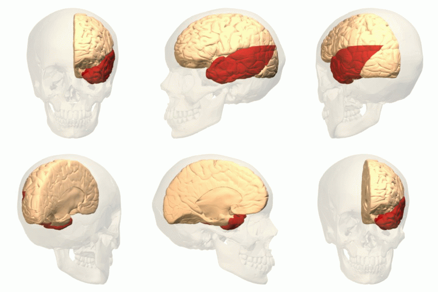
Entorhinal Cortex
The entorhinal cortex (EC) is an area of the brain's allocortex, located in the medial temporal lobe, whose functions include being a widespread network hub for memory, navigation, and the perception of time.Integrating time from experience in the lateral entorhinal cortex Albert Tsao, Jørgen Sugar, Li Lu, Cheng Wang, James J. Knierim, May-Britt Moser & Edvard I. Moser Naturevolume 561, pages57–62 (2018) The EC is the main interface between the hippocampus and neocortex. The EC-hippocampus system plays an important role in declarative (autobiographical/episodic/semantic) memories and in particular spatial memories including memory formation, memory consolidation, and memory optimization in sleep. The EC is also responsible for the pre-processing (familiarity) of the input signals in the reflex nictitating membrane response of classical trace conditioning; the association of impulses from the eye and the ear occurs in the entorhinal cortex. Anatomy The entorhina ... [...More Info...] [...Related Items...] OR: [Wikipedia] [Google] [Baidu] |
Temporal Lobe
The temporal lobe is one of the four major lobes of the cerebral cortex in the brain of mammals. The temporal lobe is located beneath the lateral fissure on both cerebral hemispheres of the mammalian brain. The temporal lobe is involved in processing sensory input into derived meanings for the appropriate retention of visual memory, language comprehension, and emotion association. ''Temporal'' refers to the head's temples. Structure The temporal lobe consists of structures that are vital for declarative or long-term memory. Declarative (denotative) or explicit memory is conscious memory divided into semantic memory (facts) and episodic memory (events). The medial temporal lobe structures are critical for long-term memory, and include the hippocampal formation, perirhinal cortex, parahippocampal, and entorhinal neocortical regions. The hippocampus is critical for memory formation, and the surrounding medial temporal cortex is currently theorized to be critical f ... [...More Info...] [...Related Items...] OR: [Wikipedia] [Google] [Baidu] |
Medial (anatomy)
Standard anatomical terms of location are used to describe unambiguously the anatomy of humans and other animals. The terms, typically derived from Latin or Greek roots, describe something in its standard anatomical position. This position provides a definition of what is at the front ("anterior"), behind ("posterior") and so on. As part of defining and describing terms, the body is described through the use of anatomical planes and axes. The meaning of terms that are used can change depending on whether a vertebrate is a biped or a quadruped, due to the difference in the neuraxis, or if an invertebrate is a non-bilaterian. A non-bilaterian has no anterior or posterior surface for example but can still have a descriptor used such as proximal or distal in relation to a body part that is nearest to, or furthest from its middle. International organisations have determined vocabularies that are often used as standards for subdisciplines of anatomy. For example, '' Terminolog ... [...More Info...] [...Related Items...] OR: [Wikipedia] [Google] [Baidu] |
Reelin
Reelin, encoded by the ''RELN'' gene, is a large secreted extracellular matrix glycoprotein that helps regulate processes of neuronal migration and positioning in the developing brain by controlling cell–cell interactions. Besides this important role in early Developmental biology, development, reelin continues to work in the adult brain. It modulates synaptic plasticity by enhancing the induction and maintenance of long-term potentiation. It also stimulates dendrite and dendritic spine development in the hippocampus, and regulates the continuing migration of neuroblasts generated in adult neurogenesis sites of the subventricular zone, subventricular and subgranular zones. It is found not only in the brain but also in the liver, Thyroid, thyroid gland, adrenal gland, fallopian tube, breast and in comparatively lower levels across a range of anatomical regions. Reelin has been suggested to be implicated in pathogenesis of several brain diseases. The expression of the protein has ... [...More Info...] [...Related Items...] OR: [Wikipedia] [Google] [Baidu] |
Grid Cells
A grid cell is a type of neuron within the entorhinal cortex that fires at regular intervals as an animal navigates an open area, allowing it to understand its position in space by storing and integrating information about location, distance, and direction. Grid cells have been found in many animals, including rats, mice, bats, monkeys, and humans. Grid cells were discovered in 2005 by Edvard Moser, May-Britt Moser, and their students Torkel Hafting, Marianne Fyhn, and Sturla Molden at the Centre for the Biology of Memory (CBM) in Norway. They were awarded the 2014 Nobel Prize in Physiology or Medicine together with John O'Keefe for their discoveries of cells that constitute a positioning system in the brain. The arrangement of spatial firing fields, all at equal distances from their neighbors, led to a hypothesis that these cells encode a neural representation of Euclidean space. The discovery also suggested a mechanism for dynamic computation of self-position based on continu ... [...More Info...] [...Related Items...] OR: [Wikipedia] [Google] [Baidu] |
Nobel Prize In Physiology Or Medicine
The Nobel Prize in Physiology or Medicine () is awarded yearly by the Nobel Assembly at the Karolinska Institute for outstanding discoveries in physiology or medicine. The Nobel Prize is not a single prize, but five separate prizes that, according to Alfred Nobel's 1895 will, are awarded "to those who, during the preceding year, have conferred the greatest benefit to humankind". Nobel Prizes are awarded in the fields of Physics, Medicine or Physiology, Chemistry, Literature, and Peace. The Nobel Prize is presented annually on the anniversary of Alfred Nobel's death, 10 December. As of 2024, 115 Nobel Prizes in Physiology or Medicine have been awarded to 229 laureates, 216 men and 13 women. The first one was awarded in 1901 to the German physiologist, Emil von Behring, for his work on serum therapy and the development of a vaccine against diphtheria. The first woman to receive the Nobel Prize in Physiology or Medicine, Gerty Cori, received it in 1947 for her role in elucida ... [...More Info...] [...Related Items...] OR: [Wikipedia] [Google] [Baidu] |
Cognitive Map
A cognitive map is a type of mental representation used by an individual to order their personal store of information about their everyday or metaphorical spatial environment, and the relationship of its component parts. The concept was introduced by Edward Tolman in 1948. He tried to explain the behavior of rats that appeared to learn the spatial layout of a maze, and subsequently the concept was applied to other animals, including humans. The term was later generalized by some researchers, especially in the field of operations research, to refer to a kind of semantic network representing an individual's personal knowledge or schemas. Overview Cognitive maps have been studied in various fields, such as psychology, education, archaeology, planning, geography, cartography, architecture, landscape architecture, urban planning, management and history. Because of the broad use and study of cognitive maps, it has become a colloquialism for almost any mental representation or model ... [...More Info...] [...Related Items...] OR: [Wikipedia] [Google] [Baidu] |
Brodmann Area 34
Brodmann area 34 is a region of the brain. It has been described as part of the entorhinal area and the superior temporal gyrus. The entorhinal area is the main interface between the hippocampus and neocortex and involved in memory, navigation and the perception of time. Destruction of Brodmann area 34 results in ipsilateral anosmia Anosmia, also known as smell blindness, is the lack of ability to detect one or more smells. Anosmia may be temporary or permanent. It differs from hyposmia, which is a decreased sensitivity to some or all smells. Anosmia can be categorized int .... See also * Brodmann area References 34 Olfactory system Temporal lobe Medial surface of cerebral hemisphere {{Neuroanatomy-stub ... [...More Info...] [...Related Items...] OR: [Wikipedia] [Google] [Baidu] |
Brodmann Area 28
Brodmann area 28 is a subdivision of the cerebral cortex defined on the basis of cytoarchitecture. It is located on the medial aspect of the temporal lobe and is part of the entorhinal cortex Human In humans, Brodmann area 28, and Brodmann area 34 together constitute approximately the entorhinal cortex. Guenon Brodmann regarded the location of area 28 adjacent to the hippocampus as imprecisely represented in the illustration of the cortex of the guenon brain in Brodmann-1909. It is located on the medial aspect of the temporal lobe. Distinctive features The molecular layer (I) is unusually wide; the external granular layer (II) contains nests of, for the most part, multipolar cells: the external pyramidal layer (III) contains medium-sized pyramidal cells which merge with cells of the internal pyramidal layer (V); a clear cell free zone represents sublayer 5b of layer V; the multiform layer is wide and has a less clear two sublayer structure; the internal granular layer (I ... [...More Info...] [...Related Items...] OR: [Wikipedia] [Google] [Baidu] |
Prefrontal Cortex
In mammalian brain anatomy, the prefrontal cortex (PFC) covers the front part of the frontal lobe of the cerebral cortex. It is the association cortex in the frontal lobe. The PFC contains the Brodmann areas BA8, BA9, BA10, BA11, BA12, BA13, BA14, BA24, BA25, BA32, BA44, BA45, BA46, and BA47. This brain region is involved in a wide range of higher-order cognitive functions, including speech formation (Broca's area), gaze ( frontal eye fields), working memory ( dorsolateral prefrontal cortex), and risk processing (e.g. ventromedial prefrontal cortex). The basic activity of this brain region is considered to be orchestration of thoughts and actions in accordance with internal goals. Many authors have indicated an integral link between a person's will to live, personality, and the functions of the prefrontal cortex. This brain region has been implicated in executive functions, such as planning, decision making, working memory, personality expression, moderating ... [...More Info...] [...Related Items...] OR: [Wikipedia] [Google] [Baidu] |
Parahippocampal Gyrus
The parahippocampal gyrus (or hippocampal gyrus') is a grey matter cortical region, a gyrus of the brain that surrounds the hippocampus and is part of the limbic system. The region plays an important role in memory encoding and retrieval. It has been involved in some cases of hippocampal sclerosis. Asymmetry has been observed in schizophrenia. Structure The anterior part of the gyrus includes the perirhinal and entorhinal cortices. The term parahippocampal cortex is used to refer to an area that encompasses both the posterior parahippocampal gyrus and the medial portion of the fusiform gyrus. Function Scene recognition The parahippocampal place area (PPA) is a sub-region of the parahippocampal cortex that lies medially in the inferior temporo-occipital cortex. PPA plays an important role in the encoding and recognition of environmental scenes (rather than faces). fMRI studies indicate that this region of the brain becomes highly active when human subjects view topograp ... [...More Info...] [...Related Items...] OR: [Wikipedia] [Google] [Baidu] |
Perirhinal Cortex
The perirhinal cortex is a brain cortex, cortical region in the medial temporal lobe that is made up of Brodmann areas Brodmann area 35, 35 and Brodmann area 36, 36. It receives highly processed sensory information from all sensory regions, and is generally accepted to be an important region for memory. It is bordered caudally by postrhinal cortex or parahippocampal gyrus, parahippocampal cortex (homologous regions in rodents and primates, respectively) and ventrally and Anatomical terms of location#Left and right (lateral), and medial, medially by entorhinal cortex. Structure The perirhinal cortex is composed of two regions: areas 36 and 35. Area 36 is sometimes divided into three subdivisions: 36d is the most rostral and dorsal, 36r ventral and caudal, and 36c the most caudal. Area 35 can be divided in the same manner, into 35d and 35v (for dorsal and ventral, respectively). Area 36 is six-layered, dysgranular, meaning that its layer IV is relatively sparse. Area 35 is agran ... [...More Info...] [...Related Items...] OR: [Wikipedia] [Google] [Baidu] |
Subiculum
The subiculum (Latin for "support") also known as the subicular complex, or subicular cortex, is the most inferior component of the hippocampal formation. It lies between the entorhinal cortex and the CA1 hippocampal subfield. The subicular complex comprises a set of four related structures including the prosubiculum, presubiculum, postsubiculum and parasubiculum. Name The subiculum got its name from Karl Friedrich Burdach in his three-volume work ''Vom Bau und Leben des Gehirns'' (Vol. 2, §199). He originally named it subiculum cornu ammonis and so associated it with the rest of the hippocampal subfields. Structure The subicular complex receives input from CA1 and entorhinal cortical layer III pyramidal neurons and is the main output of the hippocampus proper. The pyramidal neurons send projections to the nucleus accumbens, septal nuclei, prefrontal cortex, lateral hypothalamus, nucleus reuniens, mammillary nuclei, entorhinal cortex and amygdala. The pyramidal neuro ... [...More Info...] [...Related Items...] OR: [Wikipedia] [Google] [Baidu] |


