|
ECG
Electrocardiography is the process of producing an electrocardiogram (ECG or EKG), a recording of the heart's electrical activity through repeated cardiac cycles. It is an electrogram of the heart which is a graph of voltage versus time of the electrical activity of the heart using electrodes placed on the skin. These electrodes detect the small electrical changes that are a consequence of cardiac muscle depolarization followed by repolarization during each cardiac cycle (heartbeat). Changes in the normal ECG pattern occur in numerous cardiac abnormalities, including: * Cardiac rhythm disturbances, such as atrial fibrillation and ventricular tachycardia; * Inadequate coronary artery blood flow, such as myocardial ischemia and myocardial infarction; * and electrolyte disturbances, such as hypokalemia. Traditionally, "ECG" usually means a 12-lead ECG taken while lying down as discussed below. However, other devices can record the electrical activity of the heart ... [...More Info...] [...Related Items...] OR: [Wikipedia] [Google] [Baidu] |
Myocardial Infarction
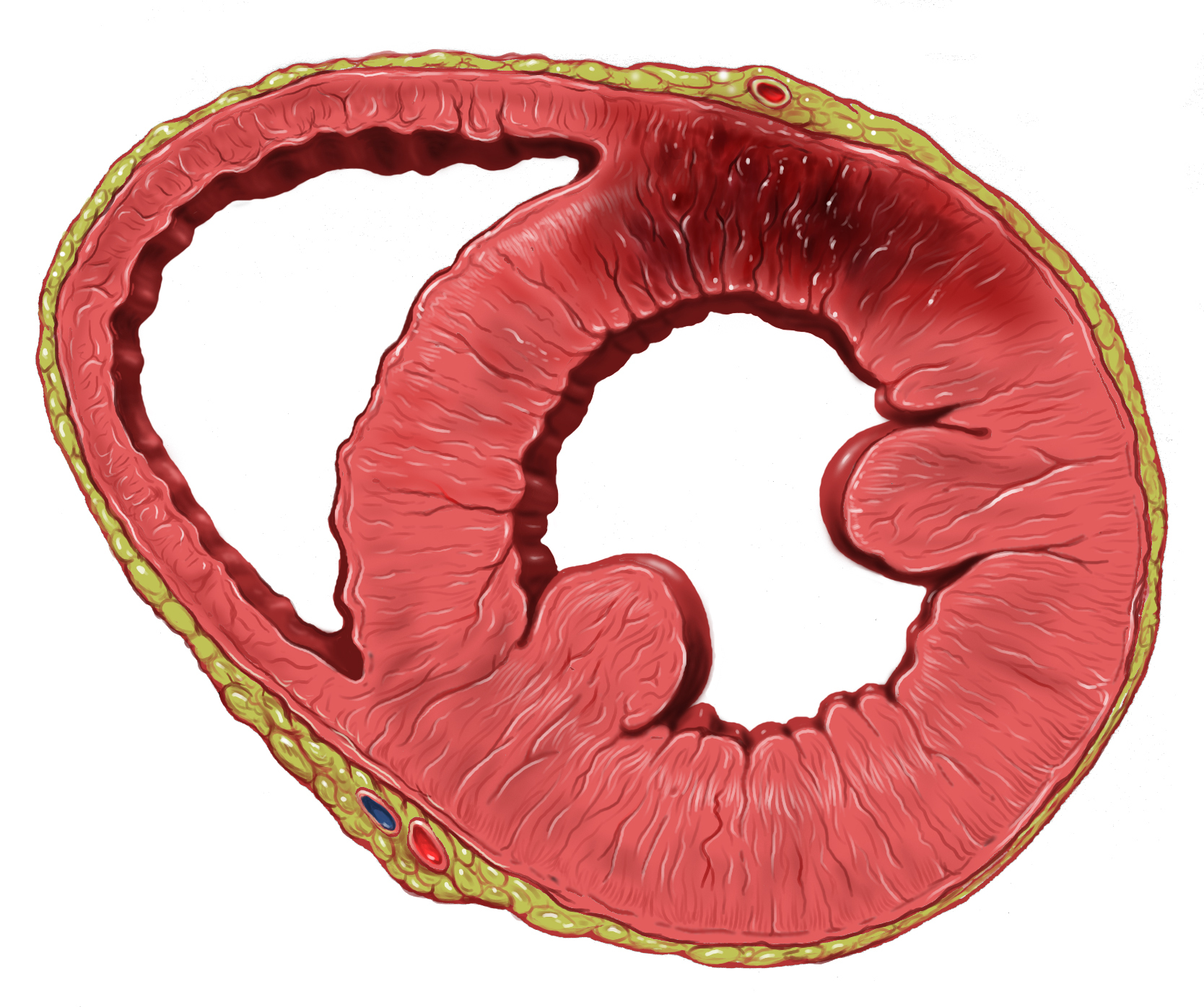
A myocardial infarction (MI), commonly known as a heart attack, occurs when Ischemia, blood flow decreases or stops in one of the coronary arteries of the heart, causing infarction (tissue death) to the heart muscle. The most common symptom is retrosternal Angina, chest pain or discomfort that classically radiates to the left shoulder, arm, or jaw. The pain may occasionally feel like heartburn. This is the dangerous type of acute coronary syndrome. Other symptoms may include shortness of breath, nausea, presyncope, feeling faint, a diaphoresis, cold sweat, Fatigue, feeling tired, and decreased level of consciousness. About 30% of people have atypical symptoms. Women more often present without chest pain and instead have neck pain, arm pain or feel tired. Among those over 75 years old, about 5% have had an MI with little or no history of symptoms. An MI may cause heart failure, an Cardiac arrhythmia, irregular heartbeat, cardiogenic shock or cardiac arrest. Most MIs occur d ... [...More Info...] [...Related Items...] OR: [Wikipedia] [Google] [Baidu] |
Sinus Rhythm
A sinus rhythm is any cardiac rhythm in which depolarisation of the cardiac muscle begins at the sinus node. It is necessary, but not sufficient, for normal electrical activity within the heart. On the electrocardiogram (ECG), a sinus rhythm is characterised by the presence of P waves that are normal in morphology. The term normal sinus rhythm (NSR) is sometimes used to denote a specific type of sinus rhythm where all other measurements on the ECG also fall within designated normal limits, giving rise to the characteristic appearance of the ECG when the electrical conduction system of the heart is functioning normally; however, other sinus rhythms can be entirely normal in particular patient groups and clinical contexts, so the term is sometimes considered a misnomer and its use is sometimes discouraged. Other types of sinus rhythm that can be normal include sinus tachycardia, sinus bradycardia, and sinus arrhythmia. Sinus rhythms may be present together with various othe ... [...More Info...] [...Related Items...] OR: [Wikipedia] [Google] [Baidu] |
Atrial Fibrillation
Atrial fibrillation (AF, AFib or A-fib) is an Heart arrhythmia, abnormal heart rhythm (arrhythmia) characterized by fibrillation, rapid and irregular beating of the Atrium (heart), atrial chambers of the heart. It often begins as short periods of abnormal cardiac cycle, beating, which become longer or continuous over time. It may also start as other forms of arrhythmia such as atrial flutter that then transform into AF. Episodes can be asymptomatic. Symptomatic episodes may involve heart palpitations, syncope (medicine), fainting, Presyncope, lightheadedness, Unconsciousness, loss of consciousness, or shortness of breath. Atrial fibrillation is associated with an increased risk of heart failure, dementia, and stroke. It is a type of supraventricular tachycardia. Atrial fibrillation frequently results from bursts of tachycardia that originate in muscle bundles extending from the Atrium (heart), atrium to the pulmonary veins. Pulmonary vein isolation by catheter ablation, trans ... [...More Info...] [...Related Items...] OR: [Wikipedia] [Google] [Baidu] |
Heart
The heart is a muscular Organ (biology), organ found in humans and other animals. This organ pumps blood through the blood vessels. The heart and blood vessels together make the circulatory system. The pumped blood carries oxygen and nutrients to the tissue, while carrying metabolic waste such as carbon dioxide to the lungs. In humans, the heart is approximately the size of a closed fist and is located between the lungs, in the middle compartment of the thorax, chest, called the mediastinum. In humans, the heart is divided into four chambers: upper left and right Atrium (heart), atria and lower left and right Ventricle (heart), ventricles. Commonly, the right atrium and ventricle are referred together as the right heart and their left counterparts as the left heart. In a healthy heart, blood flows one way through the heart due to heart valves, which prevent cardiac regurgitation, backflow. The heart is enclosed in a protective sac, the pericardium, which also contains a sma ... [...More Info...] [...Related Items...] OR: [Wikipedia] [Google] [Baidu] |
Ventricular Tachycardia
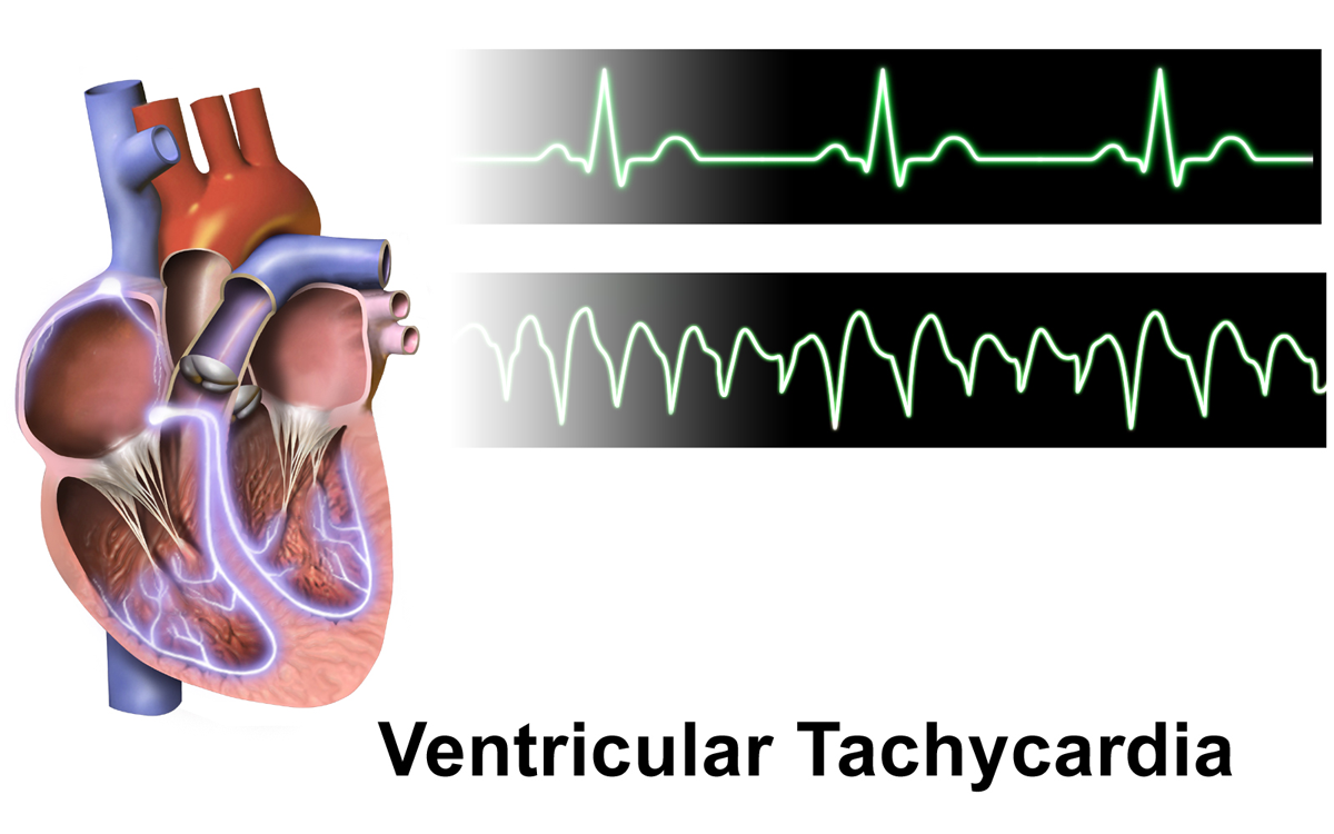
Ventricular tachycardia (V-tach or VT) is a cardiovascular disorder in which fast heart rate occurs in the ventricles of the heart. Although a few seconds of VT may not result in permanent problems, longer periods are dangerous; and multiple episodes over a short period of time are referred to as an electrical storm. Short periods may occur without symptoms, or present with lightheadedness, palpitations, shortness of breath, chest pain, and decreased level of consciousness. Ventricular tachycardia may lead to coma and persistent vegetative state due to lack of blood and oxygen to the brain. Ventricular tachycardia may result in ventricular fibrillation (VF) and turn into cardiac arrest. This conversion of the VT into VF is called the degeneration of the VT. It is found initially in about 7% of people in cardiac arrest. Ventricular tachycardia can occur due to coronary heart disease, aortic stenosis, cardiomyopathy, electrolyte imbalance, or a heart attack. Diagnosis is ... [...More Info...] [...Related Items...] OR: [Wikipedia] [Google] [Baidu] |
Holter Monitor
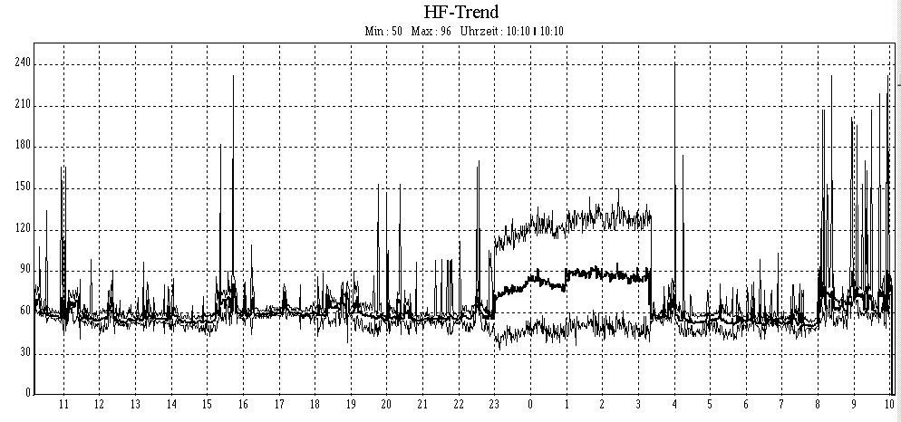
In medicine, a Holter monitor (often simply Holter) is a type of ambulatory electrocardiography device, a portable device for cardiac monitoring (the monitoring of the electrical activity of the cardiovascular system) for at least 24 hours. The Holter's most common use is for monitoring ECG heart activity (electrocardiography or ECG). Its extended recording period is sometimes useful for observing occasional cardiac arrhythmias which would be difficult to identify in a shorter period. For patients having more transient symptoms, a cardiac event monitor which can be worn for a month or more can be used. When used to study the heart, much like standard electrocardiography, the Holter monitor records electrical signals from the heart via a series of electrodes attached to the chest. Electrodes are placed over bones to minimize artifacts from muscular activity. The number and position of electrodes varies by model, but most Holter monitors employ between three and eight. These e ... [...More Info...] [...Related Items...] OR: [Wikipedia] [Google] [Baidu] |
Electrogram
An electrogram (EGM) is a recording of electrical activity of organs such as the brain and heart, measured by monitoring changes in electric potential. Historically, it also referred to an instrument to measure atmospheric electrical potential. Brain Electroencephalography (EEG) An electroencephalogram (EEG) is an electrical recording of the activity of the brain taken from the scalp. An EEG can be used to diagnose seizures, sleep disorders, and for monitoring of level of anesthesia during surgery. Electrocorticography (ECoG or iEEG) An electrocorticogram is an electrical recording of the brain measured intracranially, that is, from within the brain. Eye Electrooculography (EOG) An electrooculogram (EOG) is an electrical recording of the potential between the cornea and the retina, and does not change with visual stimuli. An EOG can measure movements of the eyes and can help in diagnosis of nystagmus. Electroretinography (ERG) An electroretinogram (ERG) is an elec ... [...More Info...] [...Related Items...] OR: [Wikipedia] [Google] [Baidu] |
Repolarization
In neuroscience, repolarization refers to the change in membrane potential that returns it to a negative value just after the depolarization phase of an action potential which has changed the membrane potential to a positive value. The repolarization phase usually returns the membrane potential back to the resting membrane potential. The efflux of potassium (K+) ions results in the falling phase of an action potential. The ions pass through the selectivity filter of the K+ channel pore. Repolarization typically results from the movement of positively charged K+ ions out of the cell. The repolarization phase of an action potential initially results in hyperpolarization, attainment of a membrane potential, termed the afterhyperpolarization, that is more negative than the resting potential. Repolarization usually takes several milliseconds. Repolarization is a stage of an action potential in which the cell experiences a decrease of voltage due to the efflux of potassium (K+) io ... [...More Info...] [...Related Items...] OR: [Wikipedia] [Google] [Baidu] |
Hypokalemia
Hypokalemia is a low level of potassium (K+) in the blood serum. Mild low potassium does not typically cause symptoms. Symptoms may include feeling tired, leg cramps, weakness, and constipation. Low potassium also increases the risk of an abnormal heart rhythm, which is often too slow and can cause cardiac arrest. Causes of hypokalemia include vomiting, diarrhea, medications like furosemide and steroids, dialysis, diabetes insipidus, hyperaldosteronism, hypomagnesemia, and not enough intake in the diet. Normal potassium levels in humans are between 3.5 and 5.0 mmol/L (3.5 and 5.0 mEq/L) with levels below 3.5 mmol/L defined as hypokalemia. It is classified as severe when levels are less than 2.5 mmol/L. Low levels may also be suspected based on an electrocardiogram (ECG). The opposite state is called hyperkalemia that means high level of potassium in the blood serum. The speed at which potassium should be replaced depends on whether or not there are symptom ... [...More Info...] [...Related Items...] OR: [Wikipedia] [Google] [Baidu] |
Myocardial Ischemia
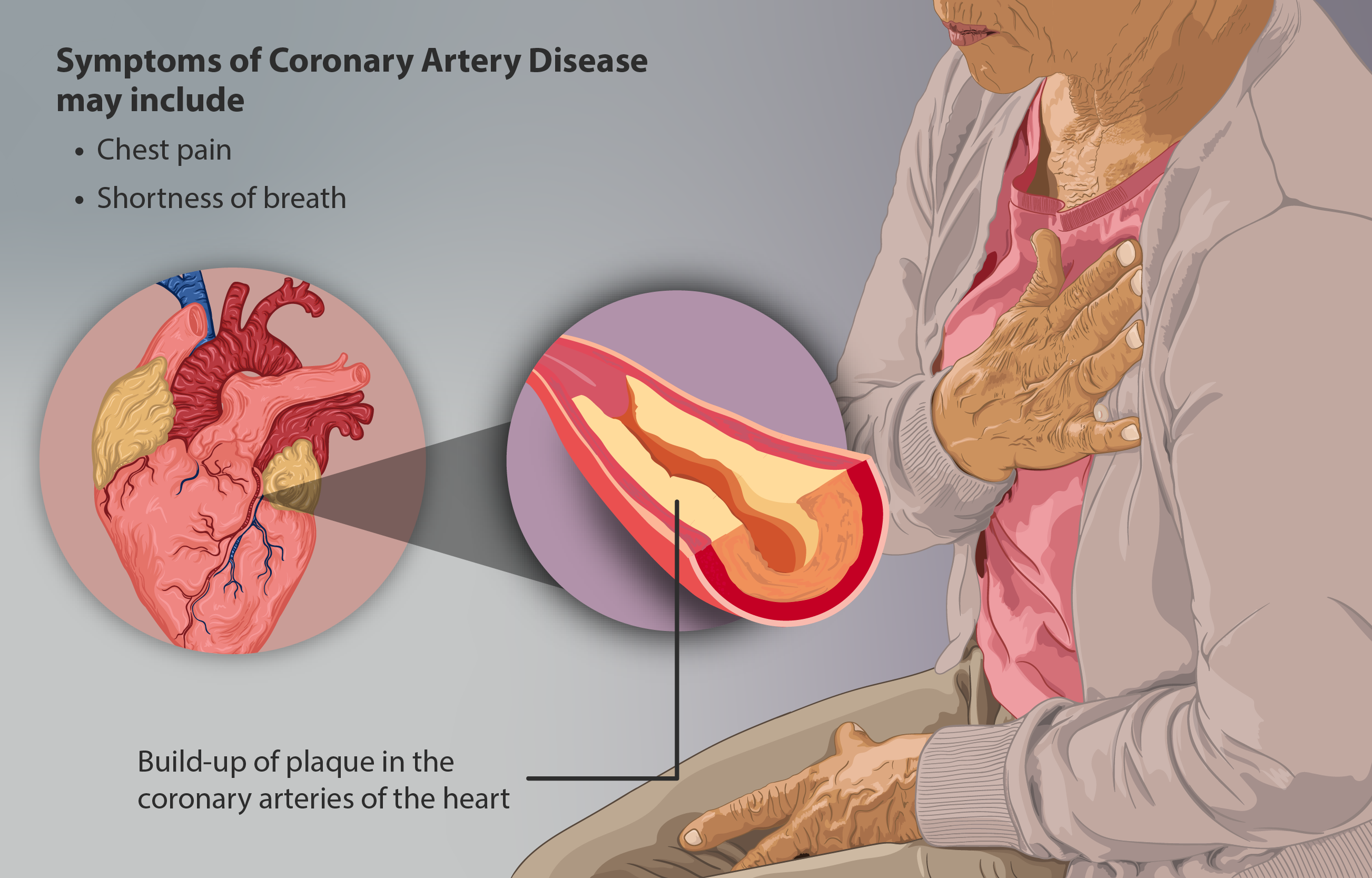
Coronary artery disease (CAD), also called coronary heart disease (CHD), or ischemic heart disease (IHD), is a type of heart disease involving the reduction of blood flow to the cardiac muscle due to a build-up of atheromatous plaque in the arteries of the heart. It is the most common of the cardiovascular diseases. CAD can cause stable angina, unstable angina, myocardial ischemia, and myocardial infarction. A common symptom is angina, which is chest pain or discomfort that may travel into the shoulder, arm, back, neck, or jaw. Occasionally it may feel like heartburn. In stable angina, symptoms occur with exercise or emotional stress, last less than a few minutes, and improve with rest. Shortness of breath may also occur and sometimes no symptoms are present. In many cases, the first sign is a heart attack. Other complications include heart failure or an abnormal heartbeat. Risk factors include high blood pressure, smoking, diabetes mellitus, lack of exercise, obesity, h ... [...More Info...] [...Related Items...] OR: [Wikipedia] [Google] [Baidu] |
Electroencephalography
Electroencephalography (EEG) is a method to record an electrogram of the spontaneous electrical activity of the brain. The biosignal, bio signals detected by EEG have been shown to represent the postsynaptic potentials of pyramidal neurons in the neocortex and allocortex. It is typically non-invasive, with the EEG electrodes placed along the scalp (commonly called "scalp EEG") using the 10–20 system (EEG), International 10–20 system, or variations of it. Electrocorticography, involving surgical placement of electrodes, is sometimes called Electrocorticography, "intracranial EEG". Clinical interpretation of EEG recordings is most often performed by visual inspection of the tracing or quantitative EEG, quantitative EEG analysis. Voltage fluctuations measured by the EEG bioamplifier, bio amplifier and electrodes allow the evaluation of normal Brain activity and meditation, brain activity. As the electrical activity monitored by EEG originates in neurons in the underlying Huma ... [...More Info...] [...Related Items...] OR: [Wikipedia] [Google] [Baidu] |
Smartwatch
A smartwatch is a portable wearable computer that resembles a wristwatch. Most modern smartwatches are operated via a touchscreen, and rely on mobile apps that run on a connected device (such as a smartphone) in order to provide core functions. Early smartwatches were capable of performing basic functions like calculator watch, calculating, displaying digital time, translating text, and playing games. More recent models often offer features comparable to smartphones, including apps, a mobile operating system, Bluetooth and Wi-Fi connectivity, and the ability to function as portable media players or FM broadcasting, FM radios. Some high-end models have cellular capabilities, allowing users to make and receive phone calls. While internal hardware varies, most smartwatches have a backlit Liquid-crystal display, LCD or OLED electronic visual display and are powered by a rechargeable lithium-ion battery. They may also incorporate GPS navigation device, GPS receivers, digital cameras ... [...More Info...] [...Related Items...] OR: [Wikipedia] [Google] [Baidu] |


