|
Diverticulum
In medicine or biology, a diverticulum is an outpouching of a hollow (or a fluid-filled) structure in the body. Depending upon which layers of the structure are involved, diverticula are described as being either true or false. In medicine, the term usually implies the structure is not normally present, but in embryology, the term is used for some normal structures arising from others, as for instance the thyroid diverticulum, which arises from the tongue. The word comes from Latin ''dīverticulum'', "bypath" or "byway". Classification Diverticula are described as being true or false depending upon the layers involved: *False diverticula (also known as "pseudodiverticula") do not involve muscular layers or adventitia. False diverticula, in the gastrointestinal tract for instance, involve only the submucosa and mucosa. *True diverticula involve all layers of the structure, including muscularis propria and adventitia, such as Meckel's diverticulum. Embryology *The kidne ... [...More Info...] [...Related Items...] OR: [Wikipedia] [Google] [Baidu] |
Meckel's Diverticulum
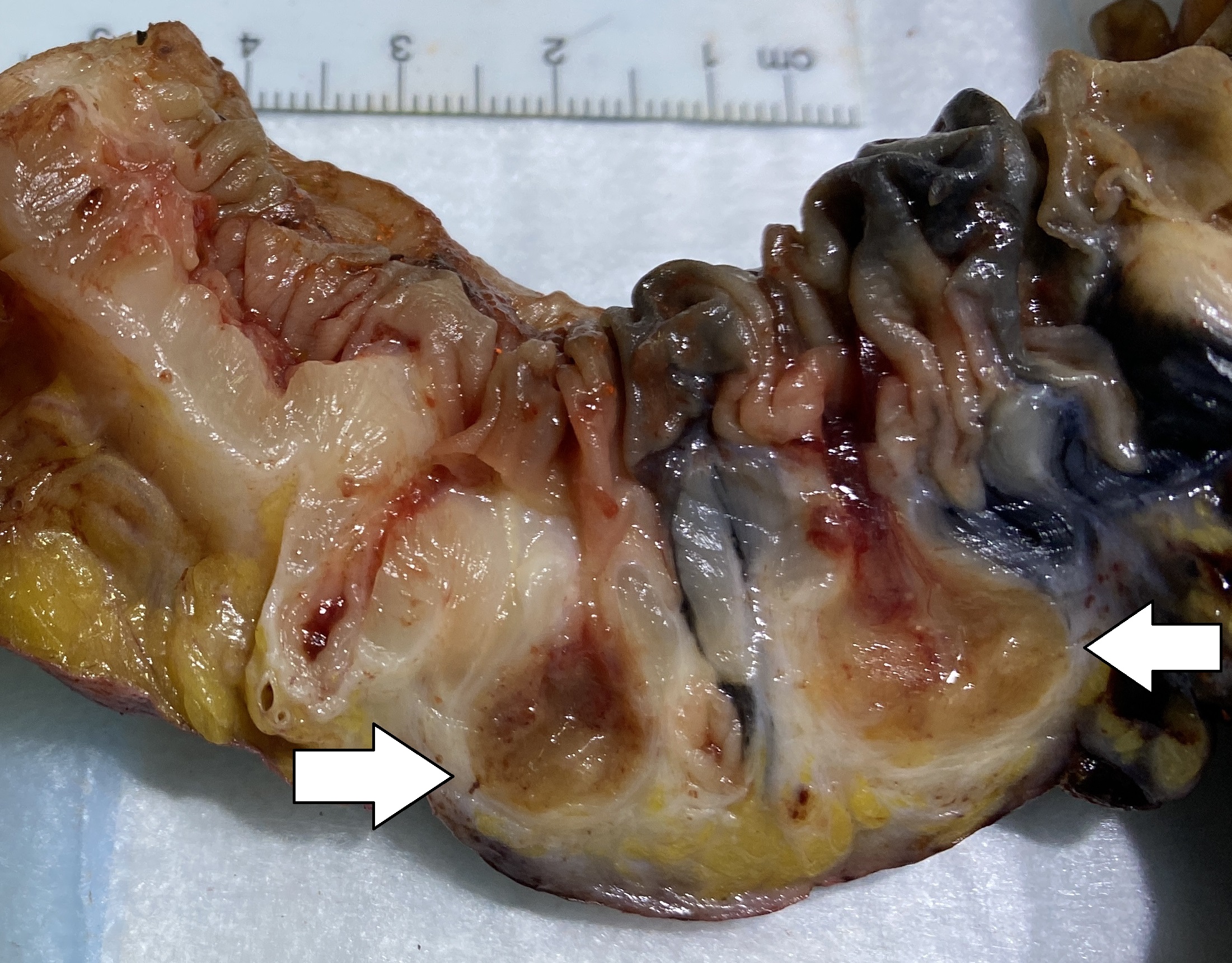
A Meckel's diverticulum, a true congenital diverticulum, is a slight bulge in the small intestine present at birth and a vestigial remnant of the omphalomesenteric duct (also called the vitelline duct or yolk stalk). It is the most common malformation of the Human gastrointestinal tract, gastrointestinal tract and is present in approximately 2% of the population, with males more frequently experiencing symptoms. Meckel's diverticulum was first explained by Fabricius Hildanus in the sixteenth century and later named after Johann Friedrich Meckel, who described the embryological origin of this type of diverticulum in 1809. Signs and symptoms The majority of people with a Meckel's diverticulum are asymptomatic. An asymptomatic Meckel's diverticulum is called a ''silent'' Meckel's diverticulum. If symptoms do occur, they typically appear before the age of two years. The most common presenting symptom is painless rectal bleeding such as melaena-like black offensive stools, followed by i ... [...More Info...] [...Related Items...] OR: [Wikipedia] [Google] [Baidu] |
Zenker's Diverticulum
A Zenker's diverticulum, also pharyngeal pouch, is a diverticulum of the mucosa of the human pharynx, just above the cricopharyngeal muscle (i.e. above the upper sphincter of the esophagus). It is a pseudo diverticulum (not involving all layers of the esophageal wall). It was named in 1877 after German pathologist Friedrich Albert von Zenker. Signs and symptoms In simple words, when there is excessive pressure within the lower pharynx, the weakest portion of the pharyngeal wall balloons out, forming a diverticulum which may reach several centimetres in diameter. More precisely, while traction and pulsion mechanisms have long been deemed the main factors promoting development of a Zenker's diverticulum, current consensus considers occlusive mechanisms to be most important: uncoordinated swallowing, impaired relaxation and spasm of the cricopharyngeus muscle lead to an increase in pressure within the distal pharynx, so that its wall herniates through the point of least resistan ... [...More Info...] [...Related Items...] OR: [Wikipedia] [Google] [Baidu] |
Diverticulosis
Diverticulosis is the condition of having multiple pouches ( diverticula) in the colon that are not inflamed. These are outpockets of the colonic mucosa and submucosa through weaknesses of muscle layers in the colon wall. Diverticula do not cause symptoms in most people. Diverticular disease occurs when diverticula become clinically inflamed, a condition known as diverticulitis. Diverticula typically occur in the sigmoid colon, which is commonplace for increased pressure. The left side of the colon is more commonly affected in the United States while the right side is more commonly affected in Asia. Diagnosis is often during routine colonoscopy or as an incidental finding during CT scan. It is common in Western countries with about half of those over the age of 60 affected in Canada and the United States. Diverticula are uncommon before the age of 40, and increase in incidence beyond that age. Rates are lower in Africa; the reasons for this remain unclear but may invo ... [...More Info...] [...Related Items...] OR: [Wikipedia] [Google] [Baidu] |
Vitelline Duct
In the human embryo, the vitelline duct, also known as the vitellointestinal duct, the yolk stalk, the omphaloenteric duct, or the omphalomesenteric duct, is a long narrow tube that joins the yolk sac to the midgut lumen of the developing fetus. It appears at the end of the fourth week, when the yolk sac presents the appearance of a small pear-shaped vesicle (the umbilical vesicle). Function Obliteration Generally, the duct fully obliterates (narrows and disappears) during the 5–6th week of fertilization age (9th week of gestational age), but a failure of the duct to close is termed a vitelline fistula. This results in discharge of meconium from the umbilicus. About two percent of fetuses exhibit a type of vitelline fistula characterized by persistence of the proximal part of the vitelline duct as a diverticulum protruding from the small intestine, Meckel's diverticulum, which is typically situated within two feet of the ileocecal junction and may be attached by a fibrous ... [...More Info...] [...Related Items...] OR: [Wikipedia] [Google] [Baidu] |
Cricopharyngeal Muscle
The inferior pharyngeal constrictor muscle is a skeletal muscle of the neck. It is the thickest of the three outer pharyngeal muscles. It arises from the sides of the cricoid cartilage and the thyroid cartilage. It is supplied by the vagus nerve (CN X). It is active during swallowing, and partially during breathing and speech. It may be affected by Zenker's diverticulum. Structure The inferior pharyngeal constrictor muscle is composed of two parts. The first part (and more superior) arises from the thyroid cartilage (thyropharyngeal part), and the second part arises from the cricoid cartilage (cricopharyngeal part). * On the ''thyroid cartilage'', it arises from the oblique line on the side of the lamina, from the surface behind this nearly as far as the posterior border and from the inferior horn of the thyroid cartilage. * From the ''cricoid cartilage'', it arises in the interval between the cricothyroid muscle in front, and the articular facet for the inferior horn of the t ... [...More Info...] [...Related Items...] OR: [Wikipedia] [Google] [Baidu] |
Killian's Triangle
Killian's dehiscence (also known as Killian's triangle) is a triangular area in the wall of the pharynx between the cricopharyngeus and thyropharyngeus which are the two parts of the inferior constrictors(also see Pharyngeal pouch). It can be seen as a locus minoris resistentiae. Similar triangular area between circular fibres of cricopharyngeus and longitudinal fibres of esophagus is Lamier's triangle or Lamier-hackermann's area. Clinical significance It represents a potentially weak spot where a pharyngoesophageal diverticulum (Zenker's diverticulum) is more likely to occur. Eponym It is named after the German ENT surgeon Gustav Killian Gustav Killian (2 June 1860 – 24 February 1921) was a German laryngologist and founder of the bronchoscopy. Life and death His father Johann Baptist Caesar Killian (1820–1889), the son of a ''städtischen Wegeaufsehers'' an urban way oversee .... References Human head and neck Otorhinolaryngology {{anatomy-stub ... [...More Info...] [...Related Items...] OR: [Wikipedia] [Google] [Baidu] |
Thyroid
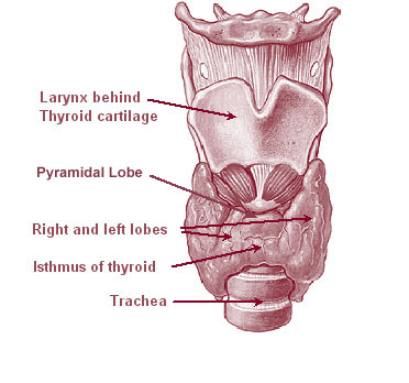
The thyroid, or thyroid gland, is an endocrine gland in vertebrates. In humans it is in the neck and consists of two connected lobes. The lower two thirds of the lobes are connected by a thin band of tissue called the thyroid isthmus. The thyroid is located at the front of the neck, below the Adam's apple. Microscopically, the functional unit of the thyroid gland is the spherical thyroid follicle, lined with follicular cells (thyrocytes), and occasional parafollicular cells that surround a lumen containing colloid. The thyroid gland secretes three hormones: the two thyroid hormones triiodothyronine (T3) and thyroxine (T4)and a peptide hormone, calcitonin. The thyroid hormones influence the metabolic rate and protein synthesis, and in children, growth and development. Calcitonin plays a role in calcium homeostasis. Secretion of the two thyroid hormones is regulated by thyroid-stimulating hormone (TSH), which is secreted from the anterior pituitary gland. TSH is reg ... [...More Info...] [...Related Items...] OR: [Wikipedia] [Google] [Baidu] |
Thyroid Diverticulum
The thyroid pouch or thyroid diverticulum is the embryological structure of the second pharyngeal arch from which thyroid follicular cells derive. It grows from the floor of the pharynx. See also * Diverticulum In medicine or biology, a diverticulum is an outpouching of a hollow (or a fluid-filled) structure in the body. Depending upon which layers of the structure are involved, diverticula are described as being either true or false. In medicine, t ... References Human head and neck Embryology Thyroid {{developmental-biology-stub ... [...More Info...] [...Related Items...] OR: [Wikipedia] [Google] [Baidu] |
Diverticulitis
Diverticulitis, specifically colonic diverticulitis, is a gastrointestinal disease characterized by inflammation of abnormal pouches— diverticula—which can develop in the wall of the large intestine. Symptoms typically include lower abdominal pain of sudden onset, but the onset may also occur over a few days. There may also be nausea; and diarrhea or constipation. Fever or blood in the stool suggests a complication. Repeated attacks may occur. The causes of diverticulitis are unclear. Risk factors may include obesity, lack of exercise, smoking, a family history of the disease, and use of nonsteroidal anti-inflammatory drugs (NSAIDs). The role of a low fiber diet as a risk factor is unclear. Having pouches in the large intestine that are not inflamed is known as diverticulosis. Inflammation occurs in between 10% and 25% at some point in time, and is due to a bacterial infection. Diagnosis is typically by CT scan, though blood tests, colonoscopy, or a lower gastrointes ... [...More Info...] [...Related Items...] OR: [Wikipedia] [Google] [Baidu] |
Tongue
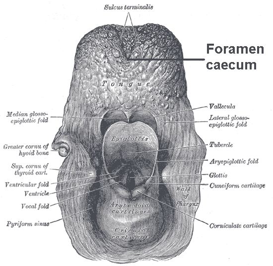
The tongue is a muscular organ in the mouth of a typical tetrapod. It manipulates food for mastication and swallowing as part of the digestive process, and is the primary organ of taste. The tongue's upper surface (dorsum) is covered by taste buds housed in numerous lingual papillae. It is sensitive and kept moist by saliva and is richly supplied with nerves and blood vessels. The tongue also serves as a natural means of cleaning the teeth. A major function of the tongue is the enabling of speech in humans and vocalization in other animals. The human tongue is divided into two parts, an oral part at the front and a pharyngeal part at the back. The left and right sides are also separated along most of its length by a vertical section of fibrous tissue (the lingual septum) that results in a groove, the median sulcus, on the tongue's surface. There are two groups of muscles of the tongue. The four intrinsic muscles alter the shape of the tongue and are not attached to bone. ... [...More Info...] [...Related Items...] OR: [Wikipedia] [Google] [Baidu] |
Foramen Caecum (tongue)
The tongue is a muscular organ in the mouth of a typical tetrapod. It manipulates food for mastication and swallowing as part of the digestive process, and is the primary organ of taste. The tongue's upper surface (dorsum) is covered by taste buds housed in numerous lingual papillae. It is sensitive and kept moist by saliva and is richly supplied with nerves and blood vessels. The tongue also serves as a natural means of cleaning the teeth. A major function of the tongue is the enabling of speech in humans and vocalization in other animals. The human tongue is divided into two parts, an oral part at the front and a pharyngeal part at the back. The left and right sides are also separated along most of its length by a vertical section of fibrous tissue (the lingual septum) that results in a groove, the median sulcus, on the tongue's surface. There are two groups of muscles of the tongue. The four intrinsic muscles alter the shape of the tongue and are not attached to bone. Th ... [...More Info...] [...Related Items...] OR: [Wikipedia] [Google] [Baidu] |
Development Of The Urinary And Reproductive Organs
The development of the urinary system begins during prenatal development, and relates to the development of the urogenital system – both the organs of the urinary system and the sex organs of the reproductive system. The development continues as a part of sexual differentiation. The urinary and reproductive organs are developed from the intermediate mesoderm. The permanent organs of the adult are preceded by a set of structures which are purely embryonic, and which with the exception of the ducts disappear almost entirely before birth. These embryonic structures are on either side; the pronephros, the mesonephros and the metanephros of the kidney, and the Wolffian and Müllerian ducts of the sex organ. The pronephros disappears very early; the structural elements of the mesonephros mostly degenerate, but the gonad is developed in their place, with which the Wolffian duct remains as the duct in males, and the Müllerian as that of the female. Some of the tubules of the mesone ... [...More Info...] [...Related Items...] OR: [Wikipedia] [Google] [Baidu] |


