|
Decussation
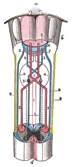
Decussation is used in biological contexts to describe a crossing (due to the shape of the Roman numeral for ten, an uppercase 'X' (), ). In Latin anatomical terms, the form is used, e.g. . Similarly, the anatomical term Chiasm (anatomy), chiasma is named after the Greek uppercase 'Χ' (Chi (letter), chi). Whereas a decussation refers to a crossing within the central nervous system, various kinds of crossings in the peripheral nervous system are called chiasma. Examples include: * In the brain, where nerve fibers obliquely cross from one lateral side of the brain to the other, that is to say they cross at a level other than their origin. See for examples decussation of pyramids and sensory decussation. In neuroanatomy, the term ''chiasma'' is reserved for crossing of- or within nerves such as in the optic chiasm. * In Botany, botanical phyllotaxis, leaf taxology, the word ''decussate'' describes an opposite leaves, opposite pattern of leaves which has successive pairs at right ... [...More Info...] [...Related Items...] OR: [Wikipedia] [Google] [Baidu] |
Sensory Decussation
The sensory decussation or decussation of the lemnisci is a decussation (a crossing over) of axons from the gracile nucleus and cuneate nucleus, known together as the dorsal column nuclei. The dorsal column nuclei are responsible for fine touch, vibration, proprioception and two-point discrimination. The fibers of this decussation are called the internal arcuate fibers and are found at the superior aspect of the closed medulla oblongata, superior to the motor decussation. Neurons of these nuclei are second-order neurons in the dorsal column–medial lemniscus pathway. Structure At the level of the closed medulla in the posterior white column, two large nuclei namely the gracile nucleus and the cuneate nucleus can be found. The two nuclei receive the impulse from the two ascending tracts: fasciculus gracilis and fasciculus cuneatus. After the two tracts terminate upon these nuclei, the heavily myelinated fibres arise and ascend anteromedially around the periaqueducta ... [...More Info...] [...Related Items...] OR: [Wikipedia] [Google] [Baidu] |
Contralateral Brain
The contralateral organization of the forebrain (Latin: contra‚ against; latus‚ side; lateral‚ sided) is the property that the Cerebral hemisphere, hemispheres of the cerebrum and the thalamus represent mainly the contralateral side of the body. Consequently, the left side of the forebrain mostly represents the right side of the body, and the right side of the brain primarily represents the left side of the body. The contralateral organization involves both executive and sensory functions (e.g., a left-sided brain lesion may cause a right-sided hemiplegia). The contralateral organization is only present in vertebrates. A number of theories have been put forward to explain this phenomenon, but none are generally accepted. These include, among others, Santiago Ramón y Cajal, Cajal's #Visual map theory, visual map theory, different topological approaches, the #Comparing inversion, somatic twist and axial twist, somatic twist theory and the axial twist theory. Anatomy Ana ... [...More Info...] [...Related Items...] OR: [Wikipedia] [Google] [Baidu] |
Decussation Of Pyramids
In neuroanatomy, the medullary pyramids are paired white matter structures of the brainstem's medulla oblongata that contain motor fibers of the corticospinal and corticobulbar tracts – known together as the pyramidal tracts. The lower limit of the pyramids is marked when the fibers cross (decussate). Structure The ventral portion of the medulla oblongata contains the medullary pyramids. These two ridge-like structures travel along the length of the medulla oblongata and are bordered medially by the anterior median fissure. They each have an anterolateral sulcus along their lateral borders, where the hypoglossal nerve emerges from. Also at the side of each pyramid there is a pronounced bulge known as an olive. Fibers of the posterior column, which transmit sensory and proprioceptive information, are located behind the pyramids on the medulla oblongata. The medullary pyramids contain motor fibers that are known as the corticobulbar and corticospinal tracts. The cortico ... [...More Info...] [...Related Items...] OR: [Wikipedia] [Google] [Baidu] |
Axial Twist Theory
The axial twist theory (wiktionary:also known as, a.k.a. axial twist ''hypothesis'') is a proposed scientific theory that has been proposed to explain a range of unusual aspects of the body plan of vertebrates (including humans). It states that the Rostral (anatomical term), rostral part of the head is "turned around" regarding the rest of the body. This end-part consists of the face (eyes, nose, and mouth) as well as part of the brain (cerebrum and thalamus). According to the theory, the vertebrate body has a left-handed chirality#Biology, chirality. The axial twist theory competes with a Axial twist theory#Relation to other theories and hypotheses, number of other proposals that focus on more limited, specific aspects, most of which explain Contralateral brain, contralateral forebrain organization, the phenomenon that the left side of the brain mainly controls the right side of the body and vice versa. None of the proposed theories explaining this phenomenon, including axial twis ... [...More Info...] [...Related Items...] OR: [Wikipedia] [Google] [Baidu] |
Decussation Of Pyramids
In neuroanatomy, the medullary pyramids are paired white matter structures of the brainstem's medulla oblongata that contain motor fibers of the corticospinal and corticobulbar tracts – known together as the pyramidal tracts. The lower limit of the pyramids is marked when the fibers cross (decussate). Structure The ventral portion of the medulla oblongata contains the medullary pyramids. These two ridge-like structures travel along the length of the medulla oblongata and are bordered medially by the anterior median fissure. They each have an anterolateral sulcus along their lateral borders, where the hypoglossal nerve emerges from. Also at the side of each pyramid there is a pronounced bulge known as an olive. Fibers of the posterior column, which transmit sensory and proprioceptive information, are located behind the pyramids on the medulla oblongata. The medullary pyramids contain motor fibers that are known as the corticobulbar and corticospinal tracts. The cortico ... [...More Info...] [...Related Items...] OR: [Wikipedia] [Google] [Baidu] |
Anatomical Terms Of Neuroanatomy
This article describes anatomical terminology that is used to describe the central and peripheral nervous systems - including the brain, brainstem, spinal cord, and nerves. Anatomical terminology in neuroanatomy Neuroanatomy, like other aspects of anatomy, uses specific terminology to describe anatomical structures. This terminology helps ensure that a structure is described accurately, with minimal ambiguity. Terms also help ensure that structures are described consistently, depending on their structure or function. Terms are often derived from Latin and Greek, and like other areas of anatomy are generally standardised based on internationally accepted lexicons such as Terminologia Anatomica. To help with consistency, humans and other species are assumed when described to be in ''standard anatomical position'', with the body standing erect and facing observer, arms at sides, palms forward. Location Anatomical terms of location depend on the location and species that is be ... [...More Info...] [...Related Items...] OR: [Wikipedia] [Google] [Baidu] |
Chiasm (anatomy)
In anatomy a chiasm is the spot where two structures cross, forming an X-shape (). Examples of chiasms are: * A tendinous chiasm, the spot where two tendons cross. For example, the tendon of the flexor digitorum superficialis muscle, and the tendon of the flexor digitorum longus muscle which even forms two chiasms. * In neuroanatomy, the crossing of fibres of a nerve or the crossing of two nerves. Neural chiasms Different types of crossings of nerves are referred to as chiasm: * Type I: Two nerves can ''cross one over the other'' (sagittal plane) without fusing, e.g., the trochlear nerve (see figure). * Type II: Two nerves can ''merge'' while at least part of the fibres cross the midline (see figure 2). * Type III: The fibres ''within'' a single nerve cross, such that the order of the functional map is reversed, e.g., the optic chiasms of various invertebrates such as insects and cephalopods. * Type IV: A torsion or loop by 180 degrees of a nerve can also reverse the order of ... [...More Info...] [...Related Items...] OR: [Wikipedia] [Google] [Baidu] |
Optic Chiasm
In neuroanatomy, the optic chiasm, or optic chiasma (; , ), is the part of the brain where the optic nerves cross. It is located at the bottom of the brain immediately inferior to the hypothalamus. The optic chiasm is found in all vertebrates, although in cyclostomes (lampreys and hagfishes), it is located within the brain. This article is about the optic chiasm of vertebrates, which is the best known nerve chiasm, but not every chiasm denotes a crossing of the body midline (e.g., in some invertebrates, see Chiasm (anatomy)). A midline crossing of nerves inside the brain is called a decussation (see Definition of types of crossings). Structure In all vertebrates, the optic nerves of the left and the right eye meet in the body midline, ventral to the brain. In many vertebrates the left optic nerve crosses over the right one without fusing with it. In vertebrates with a large overlap of the visual fields of the two eyes, i.e., most mammals and birds, but also amphibians, ... [...More Info...] [...Related Items...] OR: [Wikipedia] [Google] [Baidu] |
Tooth Enamel
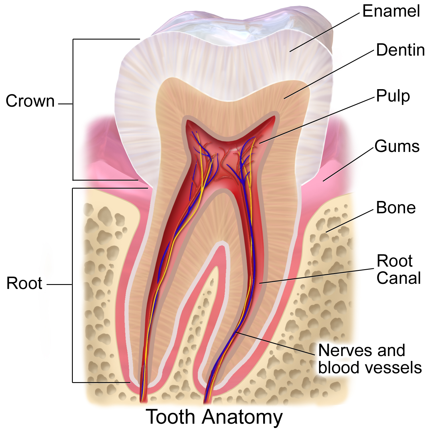
Tooth enamel is one of the four major Tissue (biology), tissues that make up the tooth in humans and many animals, including some species of fish. It makes up the normally visible part of the tooth, covering the Crown (tooth), crown. The other major tissues are dentin, cementum, and Pulp (tooth), dental pulp. It is a very hard, white to off-white, highly mineralised substance that acts as a barrier to protect the tooth but can become susceptible to degradation, especially by acids from food and drink. In rare circumstances enamel fails to form, leaving the underlying dentin exposed on the surface. Features Enamel is the hardest substance in the human body and contains the highest percentage of minerals (at 96%),Ross ''et al.'', p. 485 with water and organic material composing the rest.Ten Cate's Oral Histology, Nancy, Elsevier, pp. 70–94 The primary mineral is hydroxyapatite, which is a crystalline calcium phosphate. Enamel is formed on the tooth while the tooth develops wit ... [...More Info...] [...Related Items...] OR: [Wikipedia] [Google] [Baidu] |
Dysdercus Decussatus (13476425975)
''Dysdercus'' is a widespread genus of true bugs in the family Pyrrhocoridae; a number of species attacking cotton bolls may be called "cotton stainers." Description Species may be confused with bugs in the family Lygaeidae, but can be distinguished by the lack of ocelli on the head. They can be readily distinguished from most other genera of Pyrrhocoridae by the strong white markings at the junction of the head and thorax, and along the sides of the thorax, and often abdomen. Ecology Some members of the genus attack cotton bolls and are known as "cotton stainers." There are several species of tachinid flies that are parasitoids of ''Dysdercus'' nymphs and have been used as biocontrol agents. Species ; subgenus ''Dysdercus'' Guérin-Méneville, 1831 * '' Dysdercus albofasciatus'' Berg, 1878 * '' Dysdercus albomaculatus'' Doesburg, 1968 * '' Dysdercus andreae'' (Linnaeus, 1758) * '' Dysdercus basialbus'' Schmidt, 1932 * '' Dysdercus bimaculatus'' (Stål, 1854) * '' Dysdercus ... [...More Info...] [...Related Items...] OR: [Wikipedia] [Google] [Baidu] |
Taxonomy (biology)
In biology, taxonomy () is the science, scientific study of naming, defining (Circumscription (taxonomy), circumscribing) and classifying groups of biological organisms based on shared characteristics. Organisms are grouped into taxon, taxa (singular: taxon), and these groups are given a taxonomic rank; groups of a given rank can be aggregated to form a more inclusive group of higher rank, thus creating a taxonomic hierarchy. The principal ranks in modern use are domain (biology), domain, kingdom (biology), kingdom, phylum (''division'' is sometimes used in botany in place of ''phylum''), class (biology), class, order (biology), order, family (biology), family, genus, and species. The Swedish botanist Carl Linnaeus is regarded as the founder of the current system of taxonomy, having developed a ranked system known as Linnaean taxonomy for categorizing organisms. With advances in the theory, data and analytical technology of biological systematics, the Linnaean system has transfo ... [...More Info...] [...Related Items...] OR: [Wikipedia] [Google] [Baidu] |


