|
Cystinosis
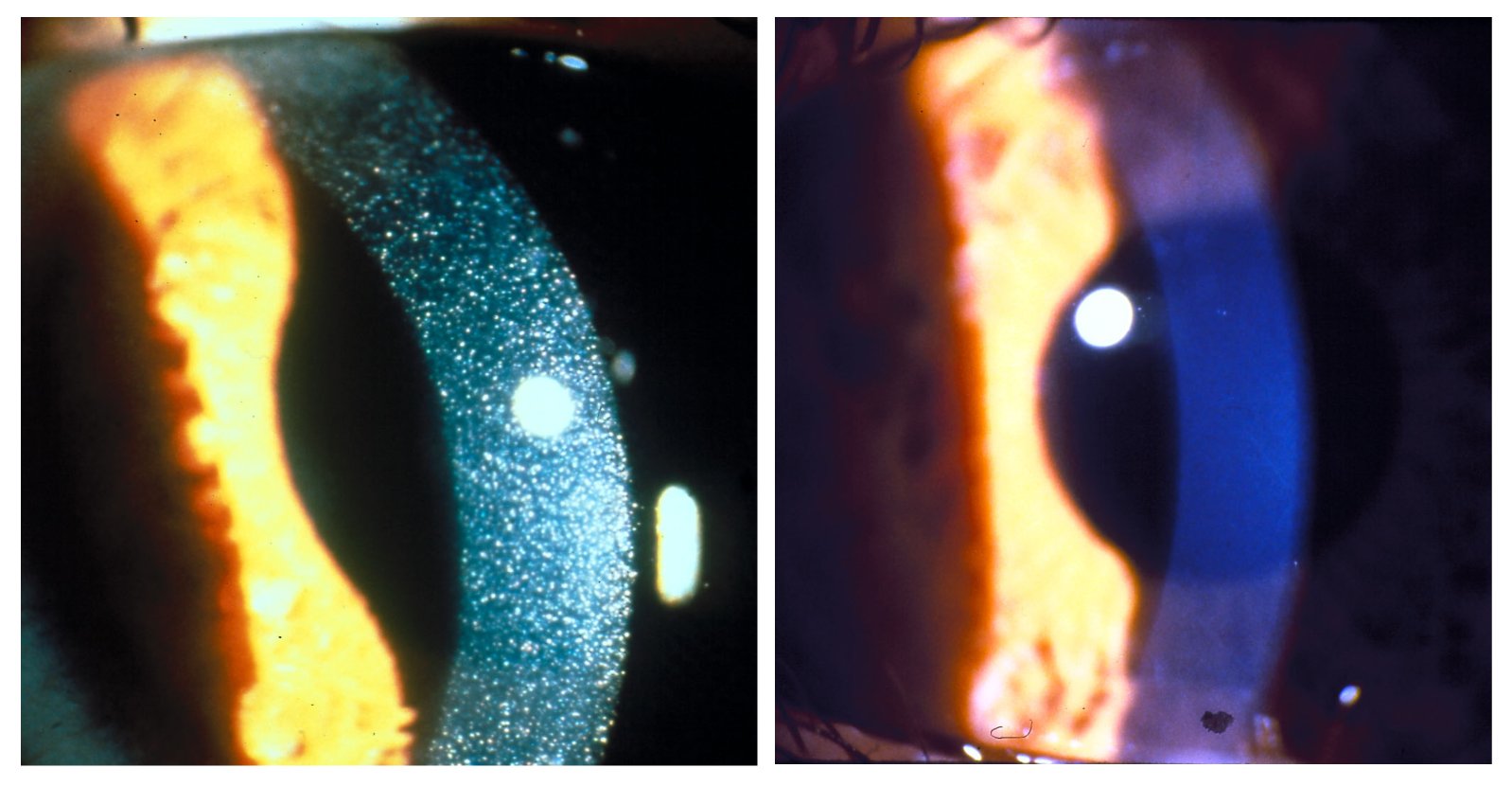
Cystinosis is a lysosomal storage disease characterized by the abnormal accumulation of cystine, the oxidized dimer of the amino acid cysteine. It is a genetic disorder that follows an autosomal recessive inheritance pattern. It is a rare autosomal recessive disorder resulting from accumulation of free cystine in lysosomes, eventually leading to intracellular crystal formation throughout the body. Cystinosis is the most common cause of Fanconi syndrome in the pediatric age group. Fanconi syndrome occurs when the function of cells in renal tubules is impaired, leading to abnormal amounts of carbohydrates and amino acids in the urine, excessive urination, and low blood levels of potassium and phosphates. Cystinosis was the first documented genetic disease belonging to the group of lysosomal storage disease disorders.Nesterova G, Gahl WA. Cystinosis: the evolution of a treatable disease. Pediatr Nephrol 2012;28:51–9. Cystinosis is caused by mutations in the '' CTNS'' gene that code ... [...More Info...] [...Related Items...] OR: [Wikipedia] [Google] [Baidu] |
CTNS (gene)
''CTNS may also refer to the Center for Theology and the Natural Sciences.'' ''CTNS'' is the gene that encodes the protein cystinosin in humans. Cystinosin is a lysosomal seven-transmembrane protein that functions as an active transporter for the export of cystine molecules out of the lysosome. Mutations in ''CTNS'' are responsible for cystinosis, an autosomal recessive lysosomal storage disease. Gene ''The CTNS'' gene is located on the p arm of human chromosome 17, at position 13.2. It spans base pairs 3,636,468 and 3,661,542, and comprises 12 exons. In 1995, the gene was localized to the short arm of chromosome 17. An international collaborative effort finally succeeded in isolating ''CTNS'' by positional cloning in 1998. The CTNSN323K, CTNSK280R, and CTNSN288K mutations completely stop the movement of CySS out of the lysosome via cystinosin. /sup> interestingly, CTNSN323K and CTNSK280R are related to juvenile nephropathic cystinosis while CTNSN288K mutations are found i ... [...More Info...] [...Related Items...] OR: [Wikipedia] [Google] [Baidu] |
Cysteamine
Cysteamine is a chemical compound that can be biosynthesized in mammals, including humans, by the degradation of coenzyme A. The intermediate pantetheine is broken down into cysteamine and pantothenic acid. It is the biosynthetic precursor to the neurotransmitter hypotaurine. It is a stable aminothiol, i.e., an organic compound containing both an amine and a thiol functional groups. Cysteamine is a white, water-soluble solid. It is often used as salts of the ammonium derivative SCH2CH2NH3sup>+ including the hydrochloride, phosphocysteamine, and bitartrate. As a medication, cysteamine, sold under the brand name Cystagon among others, is indicated to treat cystinosis. Medical uses Cysteamine is used to treat cystinosis. It is available by mouth (capsule and extended release capsule) and in eye drops. When applied topically it can scavenge free radicals and lighten skin that's been darkened as a result of post-inflammatory hyperpigmentation, sun exposure and Melasma ... [...More Info...] [...Related Items...] OR: [Wikipedia] [Google] [Baidu] |
Cystine
Cystine is the oxidized derivative of the amino acid cysteine and has the formula (SCH2CH(NH2)CO2H)2. It is a white solid that is poorly soluble in water. As a residue in proteins, cystine serves two functions: a site of redox reactions and a mechanical linkage that allows proteins to retain their three-dimensional structure. Formation and reactions Structure Cystine is the disulfide derived from the amino acid cysteine. The conversion can be viewed as an oxidation: : Cystine contains a disulfide bond, two amine groups, and two carboxylic acid groups. As for other amino acids, the amine and carboxylic acid groups exist is rapid equilibrium with the ammonium-carboxylate tautomer. The great majority of the literature concerns the ''l,l-''cystine, derived from ''l''-cysteine. Other isomers include ''d,d''-cystine and the meso isomer d,l-cystine, neither of which is biologically significant. Occurrence Cystine is common in many foods such as eggs, meat, dairy products, and whole ... [...More Info...] [...Related Items...] OR: [Wikipedia] [Google] [Baidu] |
Metabolism
Metabolism (, from el, μεταβολή ''metabolē'', "change") is the set of life-sustaining chemical reactions in organisms. The three main functions of metabolism are: the conversion of the energy in food to energy available to run cellular processes; the conversion of food to building blocks for proteins, lipids, nucleic acids, and some carbohydrates; and the elimination of metabolic wastes. These enzyme-catalyzed reactions allow organisms to grow and reproduce, maintain their structures, and respond to their environments. The word metabolism can also refer to the sum of all chemical reactions that occur in living organisms, including digestion and the transportation of substances into and between different cells, in which case the above described set of reactions within the cells is called intermediary (or intermediate) metabolism. Metabolic reactions may be categorized as '' catabolic'' – the ''breaking down'' of compounds (for example, of glucose to pyruv ... [...More Info...] [...Related Items...] OR: [Wikipedia] [Google] [Baidu] |
Thyroid
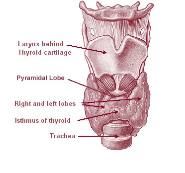
The thyroid, or thyroid gland, is an endocrine gland in vertebrates. In humans it is in the neck and consists of two connected lobes. The lower two thirds of the lobes are connected by a thin band of tissue called the thyroid isthmus. The thyroid is located at the front of the neck, below the Adam's apple. Microscopically, the functional unit of the thyroid gland is the spherical thyroid follicle, lined with follicular cells (thyrocytes), and occasional parafollicular cells that surround a lumen containing colloid. The thyroid gland secretes three hormones: the two thyroid hormones triiodothyronine (T3) and thyroxine (T4)and a peptide hormone, calcitonin. The thyroid hormones influence the metabolic rate and protein synthesis, and in children, growth and development. Calcitonin plays a role in calcium homeostasis. Secretion of the two thyroid hormones is regulated by thyroid-stimulating hormone (TSH), which is secreted from the anterior pituitary gland. TSH is reg ... [...More Info...] [...Related Items...] OR: [Wikipedia] [Google] [Baidu] |
Diabetes
Diabetes, also known as diabetes mellitus, is a group of metabolic disorders characterized by a high blood sugar level ( hyperglycemia) over a prolonged period of time. Symptoms often include frequent urination, increased thirst and increased appetite. If left untreated, diabetes can cause many health complications. Acute complications can include diabetic ketoacidosis, hyperosmolar hyperglycemic state, or death. Serious long-term complications include cardiovascular disease, stroke, chronic kidney disease, foot ulcers, damage to the nerves, damage to the eyes, and cognitive impairment. Diabetes is due to either the pancreas not producing enough insulin, or the cells of the body not responding properly to the insulin produced. Insulin is a hormone which is responsible for helping glucose from food get into cells to be used for energy. There are three main types of diabetes mellitus: * Type 1 diabetes results from failure of the pancreas to produce enough insulin du ... [...More Info...] [...Related Items...] OR: [Wikipedia] [Google] [Baidu] |
Photophobia
Photophobia is a medical symptom of abnormal intolerance to visual perception of light. As a medical symptom photophobia is not a morbid fear or phobia, but an experience of discomfort or pain to the eyes due to light exposure or by presence of actual physical sensitivity of the eyes, though the term is sometimes additionally applied to abnormal or irrational fear of light such as heliophobia. The term ''photophobia'' comes from the Greek φῶς (''phōs''), meaning "light", and φόβος (''phóbos''), meaning "fear". Causes Patients may develop photophobia as a result of several different medical conditions, related to the eye, the nervous system, genetic, or other causes. Photophobia may manifest itself in an increased response to light starting at any step in the visual system, such as: *Too much light entering the eye. Too much light can enter the eye if it is damaged, such as with corneal abrasion and retinal damage, or if its pupil(s) is unable to normally constrict ... [...More Info...] [...Related Items...] OR: [Wikipedia] [Google] [Baidu] |
Cornea
The cornea is the transparent front part of the eye that covers the iris, pupil, and anterior chamber. Along with the anterior chamber and lens, the cornea refracts light, accounting for approximately two-thirds of the eye's total optical power. In humans, the refractive power of the cornea is approximately 43 dioptres. The cornea can be reshaped by surgical procedures such as LASIK. While the cornea contributes most of the eye's focusing power, its focus is fixed. Accommodation (the refocusing of light to better view near objects) is accomplished by changing the geometry of the lens. Medical terms related to the cornea often start with the prefix "'' kerat-''" from the Greek word κέρας, ''horn''. Structure The cornea has unmyelinated nerve endings sensitive to touch, temperature and chemicals; a touch of the cornea causes an involuntary reflex to close the eyelid. Because transparency is of prime importance, the healthy cornea does not have or need blood ve ... [...More Info...] [...Related Items...] OR: [Wikipedia] [Google] [Baidu] |
Acidosis
Acidosis is a process causing increased acidity in the blood and other body tissues (i.e., an increase in hydrogen ion concentration). If not further qualified, it usually refers to acidity of the blood plasma. The term ''acidemia'' describes the state of low blood pH, while ''acidosis'' is used to describe the processes leading to these states. Nevertheless, the terms are sometimes used interchangeably. The distinction may be relevant where a patient has factors causing both acidosis and alkalosis, wherein the relative severity of both determines whether the result is a high, low, or normal pH. Acidemia is said to occur when arterial pH falls below 7.35 (except in the fetus – see below), while its counterpart (alkalemia) occurs at a pH over 7.45. Arterial blood gas analysis and other tests are required to separate the main causes. The rate of cellular metabolic activity affects and, at the same time, is affected by the pH of the body fluids. In mammals, the normal pH of ... [...More Info...] [...Related Items...] OR: [Wikipedia] [Google] [Baidu] |
Hypophosphatemic Rickets
X-linked hypophosphatemia (XLH) is an X-linked dominant form of rickets (or osteomalacia) that differs from most cases of dietary deficiency rickets in that vitamin D supplementation does not cure it. It can cause bone deformity including short stature and genu varum (bow-leggedness). It is associated with a mutation in the '' PHEX'' gene sequence (Xp.22) and subsequent inactivity of the PHEX protein. ''PHEX'' mutations lead to an elevated circulating (systemic) level of the hormone FGF23 which results in renal phosphate wasting, and locally in the extracellular matrix of bones and teeth an elevated level of the mineralization/calcification-inhibiting protein osteopontin. An inactivating mutation in the PHEX gene results in an increase in systemic circulating FGF23, and a decrease in the enzymatic activity of the PHEX enzyme which normally removes (degrades) mineralization-inhibiting osteopontin protein; in XLH, the decreased PHEX enzyme activity leads to an accumulation of inhibit ... [...More Info...] [...Related Items...] OR: [Wikipedia] [Google] [Baidu] |
Kidney
The kidneys are two reddish-brown bean-shaped organs found in vertebrates. They are located on the left and right in the retroperitoneal space, and in adult humans are about in length. They receive blood from the paired renal arteries; blood exits into the paired renal veins. Each kidney is attached to a ureter, a tube that carries excreted urine to the bladder. The kidney participates in the control of the volume of various body fluids, fluid osmolality, acid–base balance, various electrolyte concentrations, and removal of toxins. Filtration occurs in the glomerulus: one-fifth of the blood volume that enters the kidneys is filtered. Examples of substances reabsorbed are solute-free water, sodium, bicarbonate, glucose, and amino acids. Examples of substances secreted are hydrogen, ammonium, potassium and uric acid. The nephron is the structural and functional unit of the kidney. Each adult human kidney contains around 1 million nephrons, while a mouse kidney co ... [...More Info...] [...Related Items...] OR: [Wikipedia] [Google] [Baidu] |


