|
Corneal Collagen Cross-linking
Corneal cross-linking (CXL) with riboflavin (vitamin B2) and UV-A light is a surgical treatment for corneal ectasia such as keratoconus, PMD, and post-LASIK ectasia. It is used in an attempt to make the cornea stronger. According to a 2015 Cochrane review, there is insufficient evidence to determine if it is useful in keratoconus. In 2016, the US Food and Drug Administration approved riboflavin ophthalmic solution crosslinking based on three 12-month clinical trials. Medical uses A 2015 Cochrane review found that the evidence on corneal cross-linking was insufficient to determine if it is an effective procedure for the treatment of keratoconus. Adverse effects Among those with keratoconus who worsen CXL may be used. In this group, the most common side effects are haziness of the cornea, punctate keratitis, corneal striae, corneal epithelium defect, and eye pain. In those who use it after post-LASIK ectasia, the most common side effects are haziness of the cornea, corn ... [...More Info...] [...Related Items...] OR: [Wikipedia] [Google] [Baidu] |
Riboflavin
Riboflavin, also known as vitamin B2, is a vitamin found in food and sold as a dietary supplement. It is essential to the formation of two major coenzymes, flavin mononucleotide and flavin adenine dinucleotide. These coenzymes are involved in energy metabolism, cellular respiration, and antibody production, as well as normal growth and development. The coenzymes are also required for the metabolism of niacin, vitamin B6, and folate. Riboflavin is prescribed to treat corneal thinning, and taken orally, may reduce the incidence of migraine headaches in adults. Riboflavin deficiency is rare and is usually accompanied by deficiencies of other vitamins and nutrients. It may be prevented or treated by oral supplements or by injections. As a water-soluble vitamin, any riboflavin consumed in excess of nutritional requirements is not stored; it is either not absorbed or is absorbed and quickly excreted in urine, causing the urine to have a bright yellow tint. Natural sources of rib ... [...More Info...] [...Related Items...] OR: [Wikipedia] [Google] [Baidu] |
Microscopy
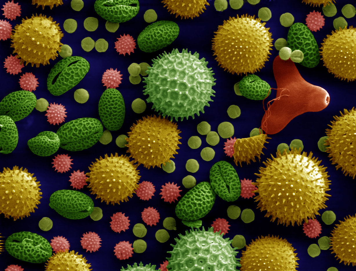
Microscopy is the technical field of using microscopes to view objects and areas of objects that cannot be seen with the naked eye (objects that are not within the resolution range of the normal eye). There are three well-known branches of microscopy: optical, electron, and scanning probe microscopy, along with the emerging field of X-ray microscopy. Optical microscopy and electron microscopy involve the diffraction, reflection, or refraction of electromagnetic radiation/electron beams interacting with the specimen, and the collection of the scattered radiation or another signal in order to create an image. This process may be carried out by wide-field irradiation of the sample (for example standard light microscopy and transmission electron microscopy) or by scanning a fine beam over the sample (for example confocal laser scanning microscopy and scanning electron microscopy). Scanning probe microscopy involves the interaction of a scanning probe with the surface of the objec ... [...More Info...] [...Related Items...] OR: [Wikipedia] [Google] [Baidu] |
Moorfields Eye Hospital NHS Foundation Trust
Moorfields Eye Hospital NHS Foundation Trust is an NHS foundation trust which runs Moorfields Eye Hospital. The Trust employs over 1,700 people. Over 24,000 ophthalmic operations are carried out and over 300,000 patients are seen by the hospital each year. The trust delivers its services from its main site on City Road and through its distributed network of over 20 other 'satellite' clinics located in other parts of London and the South East including Ealing, Teddington, Tooting, Mile End, Harrow and Tottenham. Backing up NHS Direct, Moorfields has a specialised ophthalmic telephone advice line, Moorfields Direct. Moorfields was one of the first NHS foundation trusts, and is a founder member of the UCLPartners, an academic health science centre. It plans to move its main hospital from the current City Road site to St Pancras Hospital in Camden. This will cost £352 million and it is planned to be operational in 2025–26. Commercial activities In addition to its NHS cl ... [...More Info...] [...Related Items...] OR: [Wikipedia] [Google] [Baidu] |
Soft Contact Lens
Contact lenses, or simply contacts, are thin lenses placed directly on the surface of the eyes. Contact lenses are ocular prosthetic devices used by over 150 million people worldwide, and they can be worn to correct vision or for cosmetic or therapeutic reasons. In 2010, the worldwide market for contact lenses was estimated at $6.1 billion, while the US soft lens market was estimated at $2.1 billion.Nichols, Jason J., et a"ANNUAL REPORT: Contact Lenses 2010" January 2011. Multiple analysts estimated that the global market for contact lenses would reach $11.7 billion by 2015. , the average age of contact lens wearers globally was 31 years old, and two-thirds of wearers were female.Morgan, Philip B., et al"International Contact Lens Prescribing in 2010" ''Contact Lens Spectrum''. October 2011. People choose to wear contact lenses for many reasons. Aesthetics and cosmetics are main motivating factors for people who want to avoid wearing glasses or to change the appearance or ... [...More Info...] [...Related Items...] OR: [Wikipedia] [Google] [Baidu] |
Epithelium
Epithelium or epithelial tissue is one of the four basic types of animal tissue, along with connective tissue, muscle tissue and nervous tissue. It is a thin, continuous, protective layer of compactly packed cells with a little intercellular matrix. Epithelial tissues line the outer surfaces of organs and blood vessels throughout the body, as well as the inner surfaces of cavities in many internal organs. An example is the epidermis, the outermost layer of the skin. There are three principal shapes of epithelial cell: squamous (scaly), columnar, and cuboidal. These can be arranged in a singular layer of cells as simple epithelium, either squamous, columnar, or cuboidal, or in layers of two or more cells deep as stratified (layered), or ''compound'', either squamous, columnar or cuboidal. In some tissues, a layer of columnar cells may appear to be stratified due to the placement of the nuclei. This sort of tissue is called pseudostratified. All glands are made up of epithe ... [...More Info...] [...Related Items...] OR: [Wikipedia] [Google] [Baidu] |
Dresden University Of Technology
TU Dresden (for german: Technische Universität Dresden, abbreviated as TUD and often wrongly translated as "Dresden University of Technology") is a public research university, the largest institute of higher education in the city of Dresden, the largest university in Saxony and one of the 10 largest universities in Germany with 32,389 students . The name Technische Universität Dresden has only been used since 1961; the history of the university, however, goes back nearly 200 years to 1828. This makes it one of the oldest colleges of technology in Germany, and one of the country’s oldest universities, which in German today refers to institutes of higher education that cover the entire curriculum. The university is a member of TU9, a consortium of the nine leading German Institutes of Technology. The university is one of eleven German universities which succeeded in the Excellence Initiative in 2012, thus getting the title of a "University of Excellence". The TU Dresden succee ... [...More Info...] [...Related Items...] OR: [Wikipedia] [Google] [Baidu] |
Keratometry
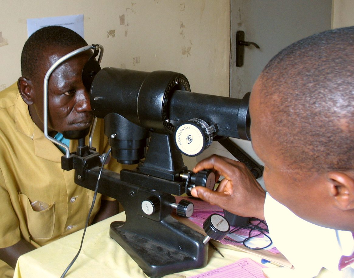
A keratometer, also known as an ophthalmometer, is a diagnostic instrument for measuring the curvature of the anterior surface of the cornea, particularly for assessing the extent and axis of astigmatism. It was invented by the German physiologist Hermann von Helmholtz in 1851, although an earlier model was developed in 1796 by Jesse Ramsden and Everard Home Sir Everard Home, 1st Baronet, FRS (6 May 1756, in Kingston upon Hull – 31 August 1832, in London) was a British surgeon. Home was born in Kingston-upon-Hull and educated at Westminster School. He gained a scholarship to Trinity College, Ca .... A keratometer uses the relationship between object size (O), image size (I), the distance between the reflective surface and the object (d), and the radius of the reflective surface (R). If three of these variables are known (or fixed), the fourth can be calculated using the formula :R = 2d \frac{O} There are two distinct variants of determining R; Javal-Schiotz type kerato ... [...More Info...] [...Related Items...] OR: [Wikipedia] [Google] [Baidu] |
Sonography
Medical ultrasound includes diagnostic techniques (mainly medical imaging, imaging techniques) using ultrasound, as well as therapeutic ultrasound, therapeutic applications of ultrasound. In diagnosis, it is used to create an image of internal body structures such as tendons, muscles, joints, blood vessels, and internal organs, to measure some characteristics (e.g. distances and velocities) or to generate an informative audible sound. Its aim is usually to find a source of disease or to exclude pathology. The usage of ultrasound to produce visual images for medicine is called medical ultrasonography or simply sonography. The practice of examining pregnant women using ultrasound is called obstetric ultrasonography, and was an early development of clinical ultrasonography. Ultrasound is composed of sound waves with frequency, frequencies which are significantly higher than the range of human hearing (>20,000 Hz). Ultrasonic images, also known as sonograms, are created by se ... [...More Info...] [...Related Items...] OR: [Wikipedia] [Google] [Baidu] |
B-scan
Medical ultrasound includes diagnostic techniques (mainly imaging techniques) using ultrasound, as well as therapeutic applications of ultrasound. In diagnosis, it is used to create an image of internal body structures such as tendons, muscles, joints, blood vessels, and internal organs, to measure some characteristics (e.g. distances and velocities) or to generate an informative audible sound. Its aim is usually to find a source of disease or to exclude pathology. The usage of ultrasound to produce visual images for medicine is called medical ultrasonography or simply sonography. The practice of examining pregnant women using ultrasound is called obstetric ultrasonography, and was an early development of clinical ultrasonography. Ultrasound is composed of sound waves with frequencies which are significantly higher than the range of human hearing (>20,000 Hz). Ultrasonic images, also known as sonograms, are created by sending pulses of ultrasound into tissue using a pro ... [...More Info...] [...Related Items...] OR: [Wikipedia] [Google] [Baidu] |
Pachymetry
Corneal pachymetry is the process of measuring the thickness of the cornea. A pachymeter is a medical device used to measure the thickness of the eye's cornea. It is used to perform corneal pachymetry prior to refractive surgery, for Keratoconus screening, LRI surgery and is useful in screening for patients suspected of developing glaucoma among other uses. Process It can be done using either ultrasonic or optical methods . The contact methods, such as ultrasound and optical such as confocal microscopy (CONFOSCAN), or noncontact methods such as optical biometry with a single Scheimpflug camera (such as SIRIUS or PENTACAM), or a Dual Scheimpflug camera (such as GALILEI), or Optical Coherence Tomography (OCT, such as Visante) and online Optical Coherence Pachymetry (OCP, such as ORBSCAN). Corneal Pachymetry is essential prior to a refractive surgery procedure for ensuring sufficient corneal thickness to prevent abnormal bulging of the cornea, a side effect known as ectasia. P ... [...More Info...] [...Related Items...] OR: [Wikipedia] [Google] [Baidu] |
Corneal Topography
Corneal topography, also known as photokeratoscopy or videokeratography, is a non-invasive medical imaging technique for mapping the anterior curvature of the cornea, the outer structure of the eye. Since the cornea is normally responsible for some 70% of the eye's refractive power, its topography is of critical importance in determining the quality of vision and corneal health. The three-dimensional map is therefore a valuable aid to the examining ophthalmologist or optometrist and can assist in the diagnosis and treatment of a number of conditions; in planning cataract surgery and intraocular lens implantation; in planning refractive surgery such as LASIK, and evaluating its results; or in assessing the fit of contact lenses. A development of keratoscopy, corneal topography extends the measurement range from the four points a few millimeters apart that is offered by keratometry to a grid of thousands of points covering the entire cornea. The procedure is carried out in seconds ... [...More Info...] [...Related Items...] OR: [Wikipedia] [Google] [Baidu] |
UV-A
Ultraviolet (UV) is a form of electromagnetic radiation with wavelength from 10 nm (with a corresponding frequency around 30 PHz) to 400 nm (750 THz), shorter than that of visible light, but longer than X-rays. UV radiation is present in sunlight, and constitutes about 10% of the total electromagnetic radiation output from the Sun. It is also produced by electric arcs and specialized lights, such as mercury-vapor lamps, tanning lamps, and black lights. Although long-wavelength ultraviolet is not considered an ionizing radiation because its photons lack the energy to ionize atoms, it can cause chemical reactions and causes many substances to glow or fluoresce. Consequently, the chemical and biological effects of UV are greater than simple heating effects, and many practical applications of UV radiation derive from its interactions with organic molecules. Short-wave ultraviolet light damages DNA and sterilizes surfaces with which it comes into contact. For huma ... [...More Info...] [...Related Items...] OR: [Wikipedia] [Google] [Baidu] |


