|
Cystic Fibrosis–related Diabetes
A cyst is a closed sac, having a distinct envelope and division compared with the nearby tissue. Hence, it is a cluster of cells that have grouped together to form a sac (like the manner in which water molecules group together to form a bubble); however, the distinguishing aspect of a cyst is that the cells forming the "shell" of such a sac are distinctly abnormal (in both appearance and behaviour) when compared with all surrounding cells for that given location. A cyst may contain air, fluids, or semi-solid material. A collection of pus is called an abscess, not a cyst. Once formed, a cyst may resolve on its own. When a cyst fails to resolve, it may need to be removed surgically, but that would depend upon its type and location. Cancer-related cysts are formed as a defense mechanism for the body following the development of mutations that lead to an uncontrolled cellular division. Once that mutation has occurred, the affected cells divide incessantly and become cancerous, for ... [...More Info...] [...Related Items...] OR: [Wikipedia] [Google] [Baidu] |
H&E Stain
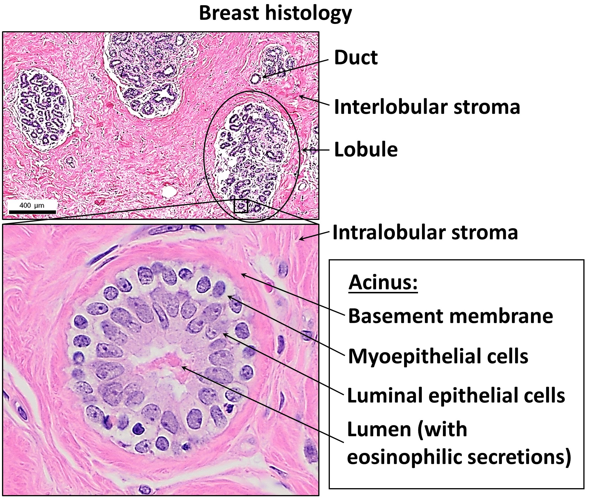
Hematoxylin and eosin stain ( or haematoxylin and eosin stain or hematoxylin–eosin stain; often abbreviated as H&E stain or HE stain) is one of the principal tissue stains used in histology. It is the most widely used stain in medical diagnosis and is often the ''gold standard.'' For example, when a pathologist looks at a biopsy of a suspected cancer, the histological section is likely to be stained with H&E. H&E is the combination of two histological stains: hematoxylin and eosin. The hematoxylin stains cell nuclei a purplish blue, and eosin stains the extracellular matrix and cytoplasm pink, with other structures taking on different shades, hues, and combinations of these colors. Hence a pathologist can easily differentiate between the nuclear and cytoplasmic parts of a cell, and additionally, the overall patterns of coloration from the stain show the general layout and distribution of cells and provides a general overview of a tissue sample's structure. Thus, patte ... [...More Info...] [...Related Items...] OR: [Wikipedia] [Google] [Baidu] |
Blood Vessel
Blood vessels are the tubular structures of a circulatory system that transport blood throughout many Animal, animals’ bodies. Blood vessels transport blood cells, nutrients, and oxygen to most of the Tissue (biology), tissues of a Body (biology), body. They also take waste and carbon dioxide away from the tissues. Some tissues such as cartilage, epithelium, and the lens (anatomy), lens and cornea of the eye are not supplied with blood vessels and are termed ''avascular''. There are five types of blood vessels: the arteries, which carry the blood away from the heart; the arterioles; the capillaries, where the exchange of water and chemicals between the blood and the tissues occurs; the venules; and the veins, which carry blood from the capillaries back towards the heart. The word ''vascular'', is derived from the Latin ''vas'', meaning ''vessel'', and is mostly used in relation to blood vessels. Etymology * artery – late Middle English; from Latin ''arteria'', from Gree ... [...More Info...] [...Related Items...] OR: [Wikipedia] [Google] [Baidu] |
Nabothian Cyst
A nabothian cyst (or nabothian follicle) is a mucus-filled cyst on the surface of the cervix. They are most often caused when stratified squamous epithelium of the ectocervix (portion nearest to the vagina) grows over the simple columnar epithelium of the endocervix (portion nearest to the uterus). This tissue growth can block the cervical crypts (subdermal pockets usually 2–10 mm in diameter), trapping cervical mucus inside the crypts. Presentation Nabothian cysts appear most often as firm bumps on the cervix's surface. A woman may notice the cyst when inserting a diaphragm or cervical cap, or when checking the cervix as part of fertility awareness. A health care provider may notice the cysts during a pelvic exam. Nabothian cysts are also incidentally found during MRI imaging. During the healing process of chronic cervicitis, squamous epithelium of ectocervix proliferates and enter the cervical canal (endocervix), covering and obstructing the columnar epithelium of end ... [...More Info...] [...Related Items...] OR: [Wikipedia] [Google] [Baidu] |
Brainstem
The brainstem (or brain stem) is the posterior stalk-like part of the brain that connects the cerebrum with the spinal cord. In the human brain the brainstem is composed of the midbrain, the pons, and the medulla oblongata. The midbrain is continuous with the thalamus of the diencephalon through the tentorial notch, and sometimes the diencephalon is included in the brainstem. The brainstem is very small, making up around only 2.6 percent of the brain's total weight. It has the critical roles of regulating heart and respiratory system, respiratory function, helping to control heart rate and breathing rate. It also provides the main motor and sensory nerve supply to the face and neck via the cranial nerves. Ten pairs of cranial nerves come from the brainstem. Other roles include the regulation of the central nervous system and the body's sleep cycle. It is also of prime importance in the conveyance of motor and sensory pathways from the rest of the brain to the body, and from the b ... [...More Info...] [...Related Items...] OR: [Wikipedia] [Google] [Baidu] |
Spinal Cord
The spinal cord is a long, thin, tubular structure made up of nervous tissue that extends from the medulla oblongata in the lower brainstem to the lumbar region of the vertebral column (backbone) of vertebrate animals. The center of the spinal cord is hollow and contains a structure called the central canal, which contains cerebrospinal fluid. The spinal cord is also covered by meninges and enclosed by the neural arches. Together, the brain and spinal cord make up the central nervous system. In humans, the spinal cord is a continuation of the brainstem and anatomically begins at the occipital bone, passing out of the foramen magnum and then enters the spinal canal at the beginning of the cervical vertebrae. The spinal cord extends down to between the first and second lumbar vertebrae, where it tapers to become the cauda equina. The enclosing bony vertebral column protects the relatively shorter spinal cord. It is around long in adult men and around long in adult women. The diam ... [...More Info...] [...Related Items...] OR: [Wikipedia] [Google] [Baidu] |
Syrinx (medicine)
A syrinx is a rare, fluid-filled neuroglial cavity within the spinal cord ( syringomyelia), in the brain stem ( syringobulbia), or in the nerves of the elbow, usually in a young age. Presentation Symptoms usually begin insidiously between adolescence and age 45. Syringomyelia develops in the center of the spinal cord, causing a central cord syndrome. Pain and temperature sensory deficits occur early but may not be recognized for years. The first abnormality recognized may be a painless burn or cut. Syringomyelia typically causes weakness, atrophy, and often fasciculations and hyperreflexia of the hands and arms; a deficit in pain and temperature sensation in a capelike distribution over the shoulders, arms and back is characteristic. Light touch and position and vibration sensation are not affected. Later, spastic leg weakness develops. Deficits may be asymmetric. Syringobulbia may cause vertigo, nystagmus, unilateral or bilateral loss of facial sensation, lingual atrophy an ... [...More Info...] [...Related Items...] OR: [Wikipedia] [Google] [Baidu] |
Endothelial
The endothelium (: endothelia) is a single layer of squamous endothelial cells that line the interior surface of blood vessels and lymphatic vessels. The endothelium forms an interface between circulating blood or lymph in the lumen and the rest of the vessel wall. Endothelial cells in direct contact with blood are called vascular endothelial cells whereas those in direct contact with lymph are known as lymphatic endothelial cells. Vascular endothelial cells line the entire circulatory system, from the heart to the smallest capillaries. These cells have unique functions that include fluid filtration, such as in the glomerulus of the kidney, blood vessel tone, hemostasis, neutrophil recruitment, and hormone trafficking. Endothelium of the interior surfaces of the heart chambers is called endocardium. An impaired function can lead to serious health issues throughout the body. Structure The endothelium is a thin layer of single flat (squamous) cells that line the interior su ... [...More Info...] [...Related Items...] OR: [Wikipedia] [Google] [Baidu] |
Epithelial
Epithelium or epithelial tissue is a thin, continuous, protective layer of cells with little extracellular matrix. An example is the epidermis, the outermost layer of the skin. Epithelial ( mesothelial) tissues line the outer surfaces of many internal organs, the corresponding inner surfaces of body cavities, and the inner surfaces of blood vessels. Epithelial tissue is one of the four basic types of animal tissue, along with connective tissue, muscle tissue and nervous tissue. These tissues also lack blood or lymph supply. The tissue is supplied by nerves. There are three principal shapes of epithelial cell: squamous (scaly), columnar, and cuboidal. These can be arranged in a singular layer of cells as simple epithelium, either simple squamous, simple columnar, or simple cuboidal, or in layers of two or more cells deep as stratified (layered), or ''compound'', either squamous, columnar or cuboidal. In some tissues, a layer of columnar cells may appear to be stratified due ... [...More Info...] [...Related Items...] OR: [Wikipedia] [Google] [Baidu] |
Pseudocyst
Pseudocysts are like cysts, but lack epithelial or endothelial cells. Initial management consists of general supportive care. Symptoms and complications caused by pseudocysts require surgery. Computed tomography (CT) scans are used for initial imaging of cysts, and endoscopic ultrasounds are used in differentiating between cysts and pseudocysts. Endoscopic drainage is a popular and effective method of treating pseudocysts. This is not to be confused with the so-called "pseudocystic appearance", mainly radiographically, of other lesions, such as Stafne static bone cyst and aneurysmal bone cyst of the jaws. Signs and symptoms Pseudocysts are often asymptomatic. Symptoms are more common in larger pseudocysts, though the size and time present usually are poor indicators of potential complications. Pancreatic pseudocysts may cause abdominal pain, nausea and vomiting, a bloated feeling, and trouble eating or digesting food. They also may become infected, rupture, or block part of ... [...More Info...] [...Related Items...] OR: [Wikipedia] [Google] [Baidu] |
Epithelium
Epithelium or epithelial tissue is a thin, continuous, protective layer of cells with little extracellular matrix. An example is the epidermis, the outermost layer of the skin. Epithelial ( mesothelial) tissues line the outer surfaces of many internal organs, the corresponding inner surfaces of body cavities, and the inner surfaces of blood vessels. Epithelial tissue is one of the four basic types of animal tissue, along with connective tissue, muscle tissue and nervous tissue. These tissues also lack blood or lymph supply. The tissue is supplied by nerves. There are three principal shapes of epithelial cell: squamous (scaly), columnar, and cuboidal. These can be arranged in a singular layer of cells as simple epithelium, either simple squamous, simple columnar, or simple cuboidal, or in layers of two or more cells deep as stratified (layered), or ''compound'', either squamous, columnar or cuboidal. In some tissues, a layer of columnar cells may appear to be stratified d ... [...More Info...] [...Related Items...] OR: [Wikipedia] [Google] [Baidu] |
Pseudocyst
Pseudocysts are like cysts, but lack epithelial or endothelial cells. Initial management consists of general supportive care. Symptoms and complications caused by pseudocysts require surgery. Computed tomography (CT) scans are used for initial imaging of cysts, and endoscopic ultrasounds are used in differentiating between cysts and pseudocysts. Endoscopic drainage is a popular and effective method of treating pseudocysts. This is not to be confused with the so-called "pseudocystic appearance", mainly radiographically, of other lesions, such as Stafne static bone cyst and aneurysmal bone cyst of the jaws. Signs and symptoms Pseudocysts are often asymptomatic. Symptoms are more common in larger pseudocysts, though the size and time present usually are poor indicators of potential complications. Pancreatic pseudocysts may cause abdominal pain, nausea and vomiting, a bloated feeling, and trouble eating or digesting food. They also may become infected, rupture, or block part of ... [...More Info...] [...Related Items...] OR: [Wikipedia] [Google] [Baidu] |
Metaplasia
Metaplasia () is the transformation of a cell type to another cell type. The change from one type of cell to another may be part of a normal maturation process, or caused by some sort of abnormal stimulus. In simplistic terms, it is as if the original cells are not robust enough to withstand their environment, so they transform into another cell type better suited to their environment. If the stimulus causing metaplasia is removed or ceases, tissues return to their normal pattern of differentiation. Metaplasia is not synonymous with dysplasia, and is not considered to be an actual cancer. It is also contrasted with heteroplasia, which is the spontaneous abnormal growth of cytologic and histologic elements. Today, metaplastic changes are usually considered to be an early phase of carcinogenesis, specifically for those with a history of cancers or who are known to be susceptible to carcinogenic changes. Metaplastic change is thus often viewed as a premalignant condition th ... [...More Info...] [...Related Items...] OR: [Wikipedia] [Google] [Baidu] |


