|
Corynebacterium Freneyi
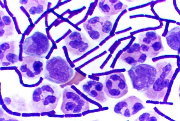
''Corynebacterium freneyi'' is a species of Gram-positive, non-motile, non-spore-forming, rod-shaped bacterium in the genus ''Corynebacterium''. It was first described in 2001 by Renaud et al. based on α-glucosidase-positive strains related to ''Corynebacterium xerosis''. Taxonomy ''C. freneyi'' was initially identified from clinical isolates that exhibited α-glucosidase activity and biochemical profiles similar to ''C. xerosis''. However, molecular analyses, including 16S rRNA gene sequencing and restriction fragment length polymorphism (RFLP) of the 16S-23S rRNA intergenic spacer region, demonstrated that ''C. freneyi'' is a distinct species. Clinical significance Although initially considered a rare isolate, ''C. freneyi'' has been implicated in various clinical specimens, including those from the female genital tract. Its pathogenic potential remains under investigation, but it has been associated with bacteremia Bloodstream infections (BSIs) are infections of blood c ... [...More Info...] [...Related Items...] OR: [Wikipedia] [Google] [Baidu] |
Gram-positive
In bacteriology, gram-positive bacteria are bacteria that give a positive result in the Gram stain test, which is traditionally used to quickly classify bacteria into two broad categories according to their type of cell wall. The Gram stain is used by microbiologists to place bacteria into two main categories, gram-positive (+) and gram-negative bacteria, gram-negative (−). Gram-positive bacteria have a thick layer of peptidoglycan within the cell wall, and gram-negative bacteria have a thin layer of peptidoglycan. Gram-positive bacteria retain the crystal violet stain used in the test, resulting in a purple color when observed through an optical microscope. The thick layer of peptidoglycan in the bacterial cell wall retains the Stain (biology), stain after it has been fixed in place by iodine. During the decolorization step, the decolorizer removes crystal violet from all other cells. Conversely, gram-negative bacteria cannot retain the violet stain after the decolorization ... [...More Info...] [...Related Items...] OR: [Wikipedia] [Google] [Baidu] |
Spore
In biology, a spore is a unit of sexual reproduction, sexual (in fungi) or asexual reproduction that may be adapted for biological dispersal, dispersal and for survival, often for extended periods of time, in unfavourable conditions. Spores form part of the Biological life cycle, life cycles of many plants, algae, fungus, fungi and protozoa. They were thought to have appeared as early as the mid-late Ordovician period as an adaptation of early land plants. Bacterial spores are not part of a sexual cycle, but are resistant structures used for survival under unfavourable conditions. Myxozoan spores release amoeboid infectious germs ("amoebulae") into their hosts for parasitic infection, but also reproduce within the hosts through the pairing of two nuclei within the plasmodium, which develops from the amoebula. In plants, spores are usually haploid and unicellular and are produced by meiosis in the sporangium of a diploid sporophyte. In some rare cases, a diploid spore is also p ... [...More Info...] [...Related Items...] OR: [Wikipedia] [Google] [Baidu] |
Corynebacterium
''Corynebacterium'' () is a genus of Gram-positive bacteria and most are aerobic. They are bacilli (rod-shaped), and in some phases of life they are, more specifically, club-shaped, which inspired the genus name ('' coryneform'' means "club-shaped"). They are widely distributed in nature in the microbiota of animals (including the human microbiota) and are mostly innocuous, most commonly existing in commensal relationships with their hosts. Some, such as '' C. glutamicum'', are commercially and industrially useful. Others can cause human disease, including, most notably, diphtheria, which is caused by '' C. diphtheriae''. Like various species of microbiota (including their relatives in the genera '' Arcanobacterium'' and '' Trueperella''), they are usually not pathogenic, but can occasionally capitalize opportunistically on atypical access to tissues (via wounds) or weakened host defenses. Taxonomy The genus ''Corynebacterium'' was created by Lehmann and Neumann in 18 ... [...More Info...] [...Related Items...] OR: [Wikipedia] [Google] [Baidu] |
Corynebacterium Xerosis
''Corynebacterium xerosis'' is a gram-positive, rod-shaped bacterium in the genus ''Corynebacterium''. Although it is frequently a harmless Commensalism, commensal organism living on the skin and in the mucous membranes, ''C. xerosis'' is also a clinically relevant Opportunistic infection, opportunistic pathogen that has been attributed to many different infections in animals and humans. However, its actual prominence in human medicine is up for debate due to early difficulties distinguishing it from other ''Corynebacterium'' species in clinical isolates. Characteristics The genome of ''C. xerosis'' is approximately 2.7 million base pairs long with over 2,000 genes encoding proteins and a high GC-content, G+C content. C''. xerosis'' was found to contain a series of plasmids, one of which confers resistance to common antibiotics such as chloramphenicol, kanamycin, streptomycin, and tetracycline and was named pTP10. This plasmid has since been introduced into ''Bacillus subtilis ... [...More Info...] [...Related Items...] OR: [Wikipedia] [Google] [Baidu] |
16S RRNA Gene
16S ribosomal RNA (or 16 S rRNA) is the RNA component of the 30S subunit of a prokaryotic ribosome (SSU rRNA). It binds to the Shine-Dalgarno sequence and provides most of the SSU structure. The genes coding for it are referred to as 16S rRNA genes and are used in reconstructing phylogenies, due to the slow rates of evolution of this region of the gene. Carl Woese and George E. Fox were two of the people who pioneered the use of 16S rRNA in phylogenetics in 1977. Multiple sequences of the 16S rRNA gene can exist within a single bacterium. Terminology The descriptor ''16S'' refers to the size of these ribosomal subunits as reflected indirectly by the speed at which they sediment when samples are centrifuged. Thus ''16S'' means 16 Svedburg units. Functions * Like the large (23S) ribosomal RNA, it has a structural role, acting as a scaffold defining the positions of the ribosomal proteins. * The 3-end contains the anti- Shine-Dalgarno sequence, which binds upstream to th ... [...More Info...] [...Related Items...] OR: [Wikipedia] [Google] [Baidu] |
Restriction Fragment Length Polymorphism
In molecular biology, restriction fragment length polymorphism (RFLP) is a technique that exploits variations in homologous DNA sequences, known as polymorphisms, populations, or species or to pinpoint the locations of genes within a sequence. The term may refer to a polymorphism itself, as detected through the differing locations of restriction enzyme sites, or to a related laboratory technique by which such differences can be illustrated. In RFLP analysis, a DNA sample is digested into fragments by one or more restriction enzymes, and the resulting ''restriction fragments'' are then separated by gel electrophoresis according to their size. RFLP analysis is now largely obsolete due to the emergence of inexpensive DNA sequencing technologies, but it was the first DNA profiling technique inexpensive enough to see widespread application. RFLP analysis was an important early tool in genome mapping, localization of genes for genetic disorders, determination of risk for disease, an ... [...More Info...] [...Related Items...] OR: [Wikipedia] [Google] [Baidu] |
Genital Tract
The reproductive system of an organism, also known as the genital system, is the biological system made up of all the anatomical organs involved in sexual reproduction. Many non-living substances such as fluids, hormones, and pheromones are also important accessories to the reproductive system. Unlike most organ systems, the sexes of differentiated species often have significant differences. These differences allow for a combination of genetic material between two individuals, which allows for the possibility of greater genetic fitness of the offspring.Reproductive System 2001 Body Guide powered by Adam Animals In mammals, the major organs of the reproductive system include the external gen ...[...More Info...] [...Related Items...] OR: [Wikipedia] [Google] [Baidu] |
Bacteremia
Bloodstream infections (BSIs) are infections of blood caused by blood-borne pathogens. The detection of microbes in the blood (most commonly accomplished by blood cultures) is always abnormal. A bloodstream infection is different from sepsis, which is characterized by severe Inflammation, inflammatory or immune responses of the host organism to pathogens. Bacteria can enter the bloodstream as a severe complication of infections (like pneumonia or meningitis), during surgery (especially when involving mucous membranes such as the gastrointestinal tract), or due to catheters and other foreign bodies entering the arteries or veins (including during intravenous drug abuse). Transient bacteremia can result after dental procedures or brushing of teeth. Bacteremia can have several important health consequences. Immune responses to the bacteria can cause sepsis and septic shock, which, particularly if severe sepsis and then septic shock occurs, have high mortality rates, especially if n ... [...More Info...] [...Related Items...] OR: [Wikipedia] [Google] [Baidu] |
Endocarditis
Endocarditis is an inflammation of the inner layer of the heart, the endocardium. It usually involves the heart valves. Other structures that may be involved include the interventricular septum, the chordae tendineae, the mural endocardium, or the surfaces of intracardiac devices. Endocarditis is characterized by lesions, known as '' vegetations'', which are masses of platelets, fibrin, microcolonies of microorganisms, and scant inflammatory cells. In the subacute form of infective endocarditis, a vegetation may also include a center of granulomatous tissue, which may fibrose or calcify. There are several ways to classify endocarditis. The simplest classification is based on cause: either ''infective'' or ''non-infective'', depending on whether a microorganism is the source of the inflammation or not. Regardless, the diagnosis of endocarditis is based on clinical features, investigations such as an echocardiogram, and blood cultures demonstrating the presence of endocar ... [...More Info...] [...Related Items...] OR: [Wikipedia] [Google] [Baidu] |
CCUG
Culture Collection University of Gothenburg (CCUG) is a Swedish microbial culture repository located in Gothenburg (Sweden) established by Enevold Falsen in 1968 and affiliated with the University of Gothenburg. The current curator is Prof. Dr. Edward R. B. Moore and it maintains bacterial, Filamentous fungi, filamentous fungal and yeasts cultures, but it does not hold extremophiles and does not dispatch the most hazardous organisms classified in biosafety level 3. More than 73,000 strains of more than 4,500 species have so far been examined, whereof more than 21,000 are displayed on Internet. It represents the largest public collection of bacteria in Europe. The CCUG has been devoted to the identification of bacteria. The search engine is sophisticated and useful for clinical microbiologists who may check their diagnosis of an unusual species or order on-line a reference strain. The CCUG also performs research and development of novel methods for rapid diagnosis of infectious dise ... [...More Info...] [...Related Items...] OR: [Wikipedia] [Google] [Baidu] |
Gram-positive Bacteria
In bacteriology, gram-positive bacteria are bacteria that give a positive result in the Gram stain test, which is traditionally used to quickly classify bacteria into two broad categories according to their type of cell wall. The Gram stain is used by microbiologists to place bacteria into two main categories, gram-positive (+) and gram-negative (−). Gram-positive bacteria have a thick layer of peptidoglycan within the cell wall, and gram-negative bacteria have a thin layer of peptidoglycan. Gram-positive bacteria retain the crystal violet stain used in the test, resulting in a purple color when observed through an optical microscope. The thick layer of peptidoglycan in the bacterial cell wall retains the stain after it has been fixed in place by iodine. During the decolorization step, the decolorizer removes crystal violet from all other cells. Conversely, gram-negative bacteria cannot retain the violet stain after the decolorization step; alcohol used in this stage ... [...More Info...] [...Related Items...] OR: [Wikipedia] [Google] [Baidu] |


