|
Cleidocranial2
Cleidocranial dysostosis (CCD), also called cleidocranial dysplasia, is a congenital disorder, birth defect that mostly affects the bones and teeth. The collarbones are typically either poorly developed or absent, which allows the shoulders to be brought close together. The front of the skull often does not close until later, and those affected are often shorter than average. Other symptoms may include a prominent forehead, wide set eyes, abnormal teeth, and a flat nose. Symptoms vary among people; however, cognitive function is typically unaffected. The condition is either heredity, inherited or occurs as a new mutation. It is inherited in an autosomal dominant manner. It is due to a defect in the RUNX2 gene which is involved in bone formation. Diagnosis is suspected based on symptoms and X-rays with confirmation by genetic testing. Other conditions that can produce similar symptoms include mandibuloacral dysplasia, pyknodysostosis, osteogenesis imperfecta, and Hajdu-Cheney synd ... [...More Info...] [...Related Items...] OR: [Wikipedia] [Google] [Baidu] |
Medical Genetics
Medical genetics is the branch of medicine that involves the diagnosis and management of hereditary disorders. Medical genetics differs from human genetics in that human genetics is a field of scientific research that may or may not apply to medicine, while medical genetics refers to the application of genetics to medical care. For example, research on the causes and inheritance of genetic disorders would be considered within both human genetics and medical genetics, while the diagnosis, management, and counselling people with genetic disorders would be considered part of medical genetics. In contrast, the study of typically non-medical phenotypes such as the genetics of eye color would be considered part of human genetics, but not necessarily relevant to medical genetics (except in situations such as albinism). ''Genetic medicine'' is a newer term for medical genetics and incorporates areas such as gene therapy, personalized medicine, and the rapidly emerging new medical specia ... [...More Info...] [...Related Items...] OR: [Wikipedia] [Google] [Baidu] |
Mandibuloacral Dysplasia
Mandibuloacral dysplasia (MAD) is a rare autosomal recessive syndrome characterized by mandibular hypoplasia, delayed cranial suture closure, dysplastic clavicles, abbreviated and club-shaped terminal phalanges, acroosteolysis, atrophy of the skin of the hands and feet, and typical facial changes.James, William; Berger, Timothy; Elston, Dirk (2005). ''Andrews' Diseases of the Skin: Clinical Dermatology''. (10th ed.). Saunders. . Types See also * Hereditary sclerosing poikiloderma * Skin lesion A skin condition, also known as cutaneous condition, is any medical condition that affects the integumentary system—the organ system that encloses the body and includes skin, nails, and related muscle and glands. The major function of this ... References External links Genodermatoses {{Genodermatoses-stub ... [...More Info...] [...Related Items...] OR: [Wikipedia] [Google] [Baidu] |
Forehead
In human anatomy, the forehead is an area of the head bounded by three features, two of the skull and one of the scalp. The top of the forehead is marked by the hairline, the edge of the area where hair on the scalp grows. The bottom of the forehead is marked by the supraorbital ridge, the bone feature of the skull above the eyes. The two sides of the forehead are marked by the temporal ridge, a bone feature that links the supraorbital ridge to the coronal suture line and beyond. However, the eyebrows do not form part of the forehead. In '' Terminologia Anatomica'', ''sinciput'' is given as the Latin equivalent to "forehead" (etymology of ''sinciput'': from ''semi-'' "half" and ''caput'' "head".). Structure The bone of the forehead is the squamous part of the frontal bone. The overlying muscles are the occipitofrontalis, procerus, and corrugator supercilii muscles, all of which are controlled by the temporal branch of the facial nerve. The sensory nerves of the forehea ... [...More Info...] [...Related Items...] OR: [Wikipedia] [Google] [Baidu] |
Hyperdontia
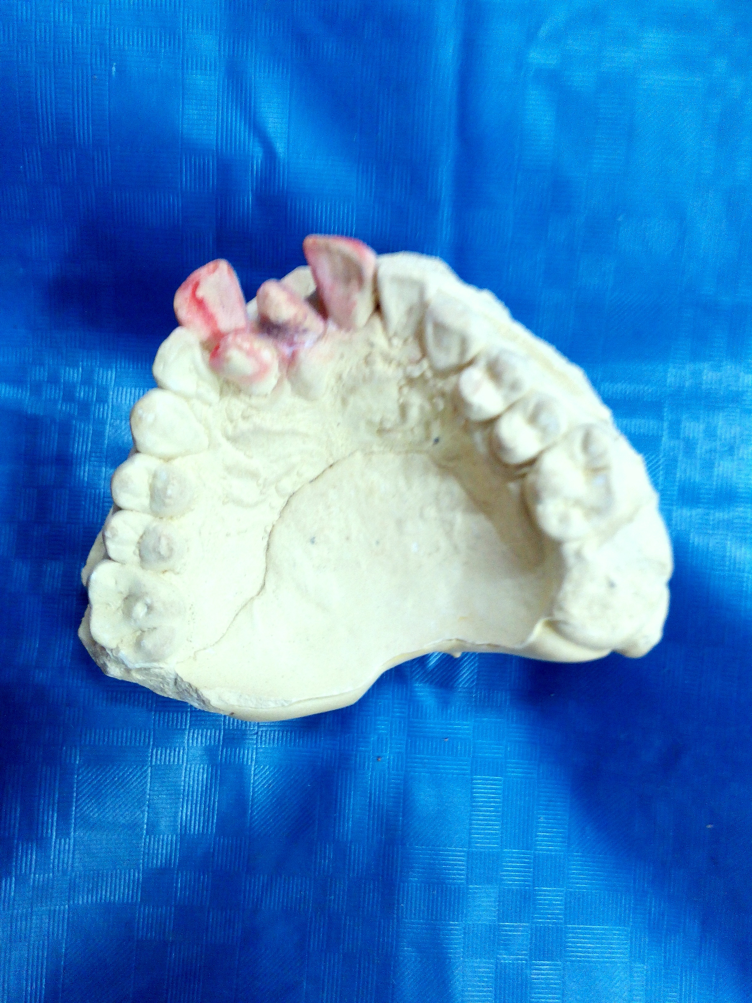
Hyperdontia is the condition of having supernumerary teeth, or teeth that appear in addition to the regular number of teeth (32 in the average adult). They can appear in any area of the dental arch and can affect any dental organ. The opposite of hyperdontia is hypodontia, where there is a congenital lack of teeth, which is a condition seen more commonly than hyperdontia.Pathology of the Hard Dental Tissues The scientific definition of hyperdontia is "any tooth or odontogenic structure that is formed from tooth germ in excess of usual number for any given region of the dental arch."R. S. Omer, R. P. Anthonappa, and N. M. King, "Determination of the optimum time for surgical removal of unerupted anterior supernumerary teeth," Pediatric Dentistry, vol. 32, no. 1, pp. 14–20, 2010. The additional teeth, which may be few or many, can occur on any place in the dental arch. Their arrangement may be symmetrical or non-symmetrical. Signs and symptoms The presence of a supernumerary ... [...More Info...] [...Related Items...] OR: [Wikipedia] [Google] [Baidu] |
Permanent Teeth
Permanent teeth or adult teeth are the second set of teeth formed in diphyodont mammals. In humans and old world simians, there are thirty-two permanent teeth, consisting of six maxillary and six mandibular molars, four maxillary and four mandibular premolars, two maxillary and two mandibular canines, four maxillary and four mandibular incisors. Timeline The first permanent tooth usually appears in the mouth at around 5-6 years of age, and the mouth will then be in a transition time with both primary (or deciduous dentition) teeth and permanent teeth during the mixed dentition period until the last primary tooth is lost or shed. The first of the permanent teeth to erupt are the permanent first molars, right behind the last 'milk' molars of the primary dentition. These first permanent molars are important for the correct development of a permanent dentition. Up to thirteen years of age, 28 of the 32 permanent teeth will appear. The full permanent dentition is completed much l ... [...More Info...] [...Related Items...] OR: [Wikipedia] [Google] [Baidu] |
Fontanelle
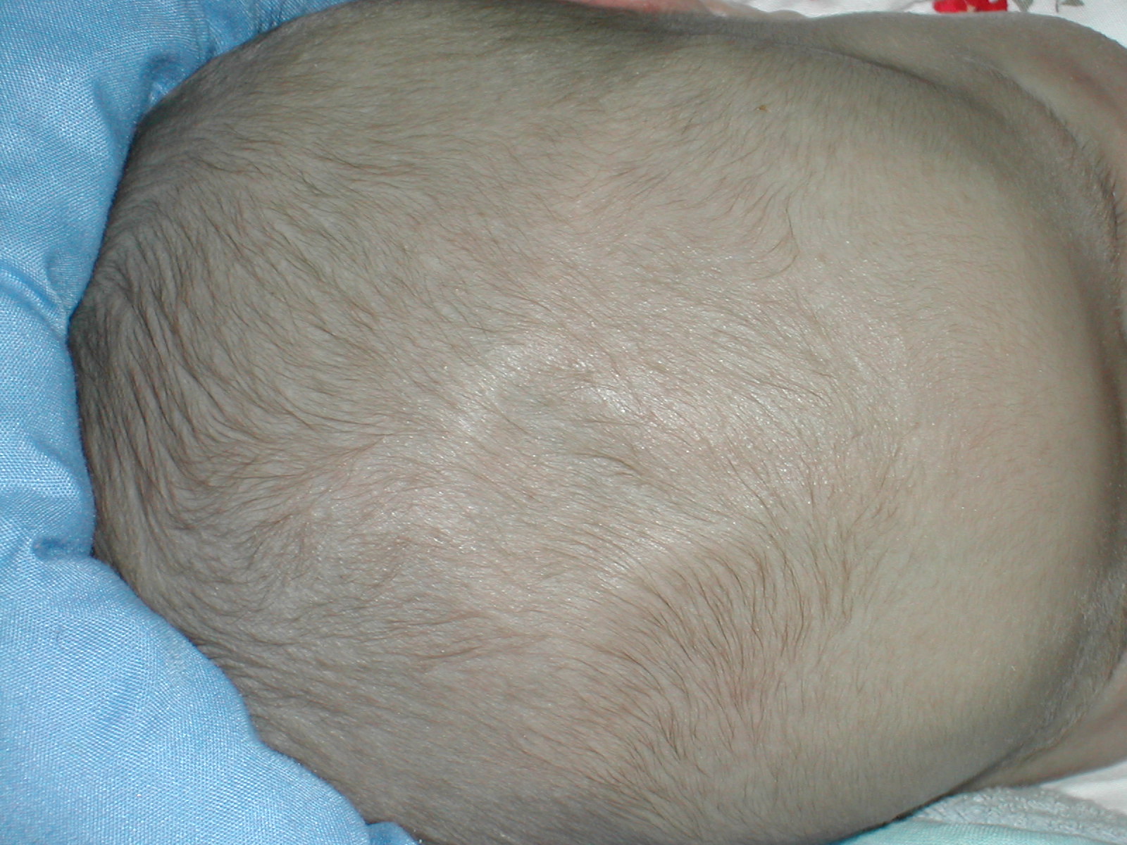
A fontanelle (or fontanel) (colloquially, soft spot) is an anatomical feature of the infant human skull comprising soft membranous gaps ( sutures) between the cranial bones that make up the calvaria of a fetus or an infant. Fontanelles allow for stretching and deformation of the neurocranium both during birth and later as the brain expands faster than the surrounding bone can grow. Premature complete ossification of the sutures is called craniosynostosis. After infancy, the anterior fontanelle is known as the bregma. Structure An infant's skull consists of five main bones: two frontal bones, two parietal bones, and one occipital bone. These are joined by fibrous sutures, which allow movement that facilitates childbirth and brain growth. * Posterior fontanelle is triangle-shaped. It lies at the junction between the sagittal suture and lambdoid suture. At birth, the skull features a small posterior fontanelle with an open area covered by a tough membrane, where the two pa ... [...More Info...] [...Related Items...] OR: [Wikipedia] [Google] [Baidu] |
Micrognathism
Micrognathism is a condition where the jaw is undersized. It is also sometimes called mandibular hypoplasia. It is common in infants, but is usually self-corrected during growth, due to the jaws' increasing in size. It may be a cause of abnormal tooth alignment and in severe cases can hamper feeding. It can also, both in adults and children, make intubation difficult, either during anesthesia or in emergency situations. Causes According to the NCBI, the following conditions feature micrognathism: * 11q partial monosomy syndrome * 3-methylglutaconic aciduria, type VIIB * 46,XY sex reversal 4 * 4p partial monosomy syndrome * Achard syndrome * Acrofacial dysostosis Cincinnati type * Acrofacial dysostosis Rodriguez type * Acrofacial dysostosis, Catania type * Acromegaloid facial appearance syndrome * Adams-Oliver syndrome 2 * Agnathia- otocephaly complex * ALG1-congenital disorder of glycosylation * Alveolar capillary dysplasia with pulmonary venous misalignment * Ami ... [...More Info...] [...Related Items...] OR: [Wikipedia] [Google] [Baidu] |
Prognathism
Prognathism is a positional relationship of the mandible or maxilla to the skeletal base where either of the jaws protrudes beyond a predetermined imaginary line in the coronal plane of the skull. In the case of ''mandibular'' prognathism (never maxillary prognathism), this is often also referred to as Habsburg chin, Habsburg's chin, Habsburg jaw or Habsburg's jaw especially when referenced with the context of its prevalence amongst historical members of the House of Habsburg. Mandibular prognathism is typically pathological, whereas maxillary prognathism is often the result of normal human population variation. In general dentistry, oral and maxillofacial surgery, and orthodontics, this is assessed clinically or radiographically ( cephalometrics). The word ''prognathism'' derives from the Greek πρό (''pro'', meaning 'forward') and γνάθος (''gnáthos'', 'jaw'). One or more types of prognathism can result in the common condition of malocclusion, in which an individua ... [...More Info...] [...Related Items...] OR: [Wikipedia] [Google] [Baidu] |
Hypermobility (joints)
Hypermobility, also known as double-jointedness, describes joints that stretch farther than normal. For example, some hypermobile people can bend their thumbs backwards to their Wrist, wrists, bend their knee joints backwards, put their leg behind the head, or perform other contortionist "tricks". It can affect one or more joints throughout the body. Hypermobile joints are common and occur in about 10 to 25% of the population. In a minority of people, pain and other symptoms are present. This may be a sign of hypermobility spectrum disorder (HSD). Hypermobile joints are a feature of genetic Connective tissue disease#Heritable connective tissue disorders, connective tissue disorders such as hypermobility spectrum disorder or Ehlers–Danlos syndrome (EDS). Until new diagnostic criteria were introduced, hypermobility syndrome was sometimes considered identical to hypermobile Ehlers–Danlos syndrome (hEDS), formerly called EDS Type 3. As no genetic test can distinguish the two condi ... [...More Info...] [...Related Items...] OR: [Wikipedia] [Google] [Baidu] |
Vestige
Vestigiality is the retention, during the process of evolution, of genetically determined structures or attributes that have lost some or all of the ancestral function in a given species. Assessment of the vestigiality must generally rely on comparison with homologous features in related species. The emergence of vestigiality occurs by normal evolutionary processes, typically by loss of function of a feature that is no longer subject to positive selection pressures when it loses its value in a changing environment. The feature may be selected against more urgently when its function becomes definitively harmful, but if the lack of the feature provides no advantage, and its presence provides no disadvantage, the feature may not be phased out by natural selection and persist across species. Examples of vestigial structures (also called degenerate, atrophied, or rudimentary organs) are the loss of functional wings in island-dwelling birds; the human vomeronasal organ; and the ... [...More Info...] [...Related Items...] OR: [Wikipedia] [Google] [Baidu] |
Anatomical Terms Of Location
Standard anatomical terms of location are used to describe unambiguously the anatomy of humans and other animals. The terms, typically derived from Latin or Greek roots, describe something in its standard anatomical position. This position provides a definition of what is at the front ("anterior"), behind ("posterior") and so on. As part of defining and describing terms, the body is described through the use of anatomical planes and axes. The meaning of terms that are used can change depending on whether a vertebrate is a biped or a quadruped, due to the difference in the neuraxis, or if an invertebrate is a non-bilaterian. A non-bilaterian has no anterior or posterior surface for example but can still have a descriptor used such as proximal or distal in relation to a body part that is nearest to, or furthest from its middle. International organisations have determined vocabularies that are often used as standards for subdisciplines of anatomy. For example, '' Termi ... [...More Info...] [...Related Items...] OR: [Wikipedia] [Google] [Baidu] |
Clavicle
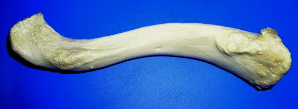
The clavicle, collarbone, or keybone is a slender, S-shaped long bone approximately long that serves as a strut between the scapula, shoulder blade and the sternum (breastbone). There are two clavicles, one on each side of the body. The clavicle is the only long bone in the body that lies horizontally. Together with the shoulder blade, it makes up the shoulder girdle. It is a palpable bone and, in people who have less fat in this region, the location of the bone is clearly visible. It receives its name from Latin ''clavicula'' 'little key' because the bone rotates along its axis like a key when the shoulder is Abduction (kinesiology), abducted. The clavicle is the most commonly fractured bone. It can easily be fractured by impacts to the shoulder from the force of falling on outstretched arms or by a direct hit. Structure The collarbone is a thin doubly curved long bone that connects the human arm, arm to the torso, trunk of the body. Located directly above the first rib, it ac ... [...More Info...] [...Related Items...] OR: [Wikipedia] [Google] [Baidu] |


