|
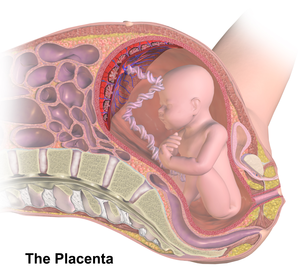
Chorionic Artery
The chorion is the outermost fetal membrane around the embryo in mammals, birds and reptiles (amniotes). It is also present around the embryo of other animals, like insects and molluscs. Structure In humans and other therian mammals, the chorion is one of the fetal membranes that exist during pregnancy between the developing fetus and mother. The chorion and the amnion together form the amniotic sac. In humans it is formed by extraembryonic mesoderm and the two layers of trophoblast that surround the embryo and other membranes; the chorionic villi emerge from the chorion, invade the endometrium, and allow the transfer of nutrients from maternal blood to fetal blood. Layers The chorion consists of two layers: an outer formed by the trophoblast, and an inner formed by the extra-embryonic mesoderm. The trophoblast is made up of an internal layer of cubical or prismatic cells, the cytotrophoblast or layer of Langhans, and an external multinucleated layer, the syncytiotrophoblast ... [...More Info...] [...Related Items...] OR: [Wikipedia] [Google] [Baidu] |
Fetal Membrane
The fetal membranes are the four extraembryonic membranes, associated with the developing embryo, and fetus in humans and other mammals. They are the amnion, chorion, allantois, and yolk sac. The amnion and the chorion are the chorioamniotic membranes that make up the amniotic sac which surrounds and protects the embryo. The fetal membranes are four of six accessory organs developed by the conceptus that are not part of the embryo itself, the other two are the placenta, and the umbilical cord. Structure The fetal membranes surround the developing embryo and form the fetal-maternal interface. The fetal membranes are derived from the trophoblast layer (outer layer of cells) of the implanting blastocyst. The trophoblast layer differentiates into amnion and the chorion, which then comprise the fetal membranes. The amnion is the innermost layer and, therefore, contacts the amniotic fluid, the fetus and the umbilical cord. The internal pressure of the amniotic fluid causes the am ... [...More Info...] [...Related Items...] OR: [Wikipedia] [Google] [Baidu] |
Cytotrophoblast
"Cytotrophoblast" is the name given to both the inner layer of the trophoblast (also called layer of Langhans) or the cells that live there. It is interior to the syncytiotrophoblast and external to the wall of the blastocyst in a developing embryo. The cytotrophoblast is considered to be the trophoblastic stem cell because the layer surrounding the blastocyst remains while daughter cells differentiate and proliferate to function in multiple roles. There are two lineages that cytotrophoblastic cells may differentiate through: fusion and invasive. The fusion lineage yields syncytiotrophoblast and the invasive lineage yields interstitial cytotrophoblast cells. Cytotrophoblastic cells play an important role in the implantation of an embryo in the uterus. Fusion lineage The formation of all syncytiotrophoblast is from the fusion of two or more cytotrophoblasts via this fusion pathway. This pathway is important because the syncytiotrophoblast plays an important role in fetal-materna ... [...More Info...] [...Related Items...] OR: [Wikipedia] [Google] [Baidu] |
Decidua Basalis
The decidua is the modified mucosal lining of the uterus (that is, modified endometrium) that forms every month, in preparation for pregnancy. It is shed off each month when there is no fertilized egg to support. The decidua is under the influence of progesterone. Endometrial cells become highly characteristic. The decidua forms the maternal part of the placenta and remains for the duration of the pregnancy. After birth the decidua is shed together with the placenta. Structure The part of the decidua that interacts with the trophoblast is the ''decidua basalis'' (also called ''decidua placentalis''), while the ''decidua capsularis'' grows over the embryo on the luminal side, enclosing it into the endometrium. The remainder of the decidua is termed the ''decidua parietalis'' or ''decidua vera'', and it will fuse with the decidua capsularis by the fourth month of gestation. Three morphologically distinct layers of the decidua basalis can then be described: * Compact outer layer ... [...More Info...] [...Related Items...] OR: [Wikipedia] [Google] [Baidu] |
Placenta With Fetal Membranes
The placenta (: placentas or placentae) is a temporary embryonic and later fetal organ that begins developing from the blastocyst shortly after implantation. It plays critical roles in facilitating nutrient, gas, and waste exchange between the physically separate maternal and fetal circulations, and is an important endocrine organ, producing hormones that regulate both maternal and fetal physiology during pregnancy. The placenta connects to the fetus via the umbilical cord, and on the opposite aspect to the maternal uterus in a species-dependent manner. In humans, a thin layer of maternal decidual (endometrial) tissue comes away with the placenta when it is expelled from the uterus following birth (sometimes incorrectly referred to as the 'maternal part' of the placenta). Placentas are a defining characteristic of placental mammals, but are also found in marsupials and some non-mammals with varying levels of development. Mammalian placentas probably first evolved about 150 mil ... [...More Info...] [...Related Items...] OR: [Wikipedia] [Google] [Baidu] |
Umbilical Vein
The umbilical vein is a vein present during fetal development that carries oxygenated blood from the placenta into the growing fetus. The umbilical vein provides convenient access to the central circulation of a neonate for restoration of blood volume and for administration of glucose and drugs. The blood pressure inside the umbilical vein is approximately 20 mmHg.Wang, Y. Vascular biology of the placenta. in Colloquium Series on Integrated Systems Physiology: from Molecule to Function. 2010. Morgan & Claypool Life Sciences. Fetal circulation The unpaired umbilical vein carries oxygen and nutrient rich blood derived from fetal-maternal blood exchange at the chorionic villi. More than two-thirds of fetal hepatic circulation is via the main portal vein, while the remainder is shunted from the left portal vein via the ductus venosus to the inferior vena cava, eventually being delivered to the fetal right atrium. Closure Closure of the umbilical vein usually occurs after the umbilic ... [...More Info...] [...Related Items...] OR: [Wikipedia] [Google] [Baidu] |
Cotyledon Arteries
The placenta of humans, and certain other mammals contains structures known as cotyledons, which transmit fetal blood and allow exchange of oxygen and nutrients with the maternal blood. Ruminants The Artiodactyla have a cotyledonary placenta. In this form of placenta, the chorionic villi form a number of separate circular structures (''cotyledons'') which are distributed over the surface of the chorionic sac. Sheep, goats and cattle have between 72 and 125 cotyledons whereas deer have 4-6 larger cotyledons. Human The form of the human placenta is generally classified as a discoid placenta. Within this, the ''cotyledons'' are the approximately 15-25 separations of the decidua basalis of the placenta, separated by placental septa SEPTA, the Southeastern Pennsylvania Transportation Authority, is a regional public transportation authority that operates bus, rapid transit, commuter rail, light rail, and electric trolleybus services for nearly four million people througho .... ... [...More Info...] [...Related Items...] OR: [Wikipedia] [Google] [Baidu] |
Chorionic Arteries
Chorionic (plate) vessels, also fetal surface vessels are blood vessels, including both arteries and veins, that carry blood through the chorion in the fetoplacental circulation. Chorionic arteries branch off the umbilical artery, and supply the capillaries of the chorionic villi. Increased vasocontractility of chorionic arteries may contribute to preeclampsia Pre-eclampsia is a multi-system disorder specific to pregnancy, characterized by the new onset of high blood pressure and often a significant amount of protein in the urine or by the new onset of high blood pressure along with significant end- .... References Embryology of cardiovascular system {{circulatory-stub ... [...More Info...] [...Related Items...] OR: [Wikipedia] [Google] [Baidu] |
Umbilical Arteries
The umbilical artery is a paired artery (with one for each half of the body) that is found in the abdominal and pelvic regions. In the fetus, it extends into the umbilical cord. Structure Development The umbilical arteries supply systemic arterial blood from the fetus to the placenta. Although this blood is sometimes referred to as deoxygenated blood it is not, and has the same oxygen saturation and nutrients as blood distributed to the other fetal tissues. There are usually two umbilical arteries present together with one umbilical vein in the umbilical cord. The umbilical arteries surround the urinary bladder and then carry all the deoxygenated blood out of the fetus through the umbilical cord. Inside the placenta, the umbilical arteries connect with each other at a distance of approximately 5 mm from the cord insertion in what is called the ''Hyrtl anastomosis''. Subsequently, they branch into chorionic arteries or ''intraplacental fetal arteries''. The umbilical art ... [...More Info...] [...Related Items...] OR: [Wikipedia] [Google] [Baidu] |
Vascularized
Angiogenesis is the physiological process through which new blood vessels form from pre-existing vessels, formed in the earlier stage of vasculogenesis. Angiogenesis continues the growth of the vasculature mainly by processes of sprouting and splitting, but processes such as coalescent angiogenesis, vessel elongation and vessel cooption also play a role. Vasculogenesis is the embryonic formation of endothelial cells from mesoderm cell precursors, and from neovascularization, although discussions are not always precise (especially in older texts). The first vessels in the developing embryo form through vasculogenesis, after which angiogenesis is responsible for most, if not all, blood vessel growth during development and in disease. Angiogenesis is a normal and vital process in growth and development, as well as in wound healing and in the formation of granulation tissue. However, it is also a fundamental step in the transition of tumors from a benign state to a malig ... [...More Info...] [...Related Items...] OR: [Wikipedia] [Google] [Baidu] |
Uterine Decidua
The decidua is the modified mucosal lining of the uterus (that is, modified endometrium) that forms every month, in preparation for pregnancy. It is shed off each month when there is no fertilized egg to support. The decidua is under the influence of progesterone. Endometrial cells become highly characteristic. The decidua forms the maternal part of the placenta and remains for the duration of the pregnancy. After birth the decidua is shed together with the placenta. Structure The part of the decidua that interacts with the trophoblast is the ''decidua basalis'' (also called ''decidua placentalis''), while the ''decidua capsularis'' grows over the embryo on the luminal side, enclosing it into the endometrium. The remainder of the decidua is termed the ''decidua parietalis'' or ''decidua vera'', and it will fuse with the decidua capsularis by the fourth month of gestation. Three morphologically distinct layers of the decidua basalis can then be described: * Compact outer layer ... [...More Info...] [...Related Items...] OR: [Wikipedia] [Google] [Baidu] |


