|
Centrosome
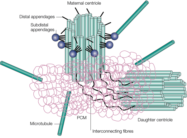
In cell biology, the centrosome (Latin centrum 'center' + Greek sōma 'body') (archaically cytocentre) is an organelle that serves as the main microtubule organizing center (MTOC) of the animal cell, as well as a regulator of cell-cycle progression. The centrosome provides structure for the cell. It is thought to have evolved only in the metazoan lineage of eukaryotic cells. Fungi and plants lack centrosomes and therefore use other structures to organize their microtubules. Although the centrosome has a key role in efficient mitosis in animal cells, it is not essential in certain fly and flatworm species. Centrosomes are composed of two centrioles arranged at right angles to each other, and surrounded by a dense, highly structured mass of protein termed the pericentriolar material (PCM). The PCM contains proteins responsible for microtubule nucleation and anchoring — including γ-tubulin, pericentrin and ninein. In general, each centriole of the centrosome is based on ... [...More Info...] [...Related Items...] OR: [Wikipedia] [Google] [Baidu] |
Microtubule
Microtubules are polymers of tubulin that form part of the cytoskeleton and provide structure and shape to eukaryotic cells. Microtubules can be as long as 50 micrometres, as wide as 23 to 27 nanometer, nm and have an inner diameter between 11 and 15 nm. They are formed by the polymerization of a Protein dimer, dimer of two globular proteins, Tubulin#Eukaryotic, alpha and beta tubulin into #Structure, protofilaments that can then associate laterally to form a hollow tube, the microtubule. The most common form of a microtubule consists of 13 protofilaments in the tubular arrangement. Microtubules play an important role in a number of cellular processes. They are involved in maintaining the structure of the cell and, together with microfilaments and intermediate filaments, they form the cytoskeleton. They also make up the internal structure of cilia and flagella. They provide platforms for intracellular transport and are involved in a variety of cellular processes, in ... [...More Info...] [...Related Items...] OR: [Wikipedia] [Google] [Baidu] |
Pericentrin
Pericentrin (kendrin), also known as PCNT and pericentrin-B (PCNTB), is a protein which in humans is encoded by the ''PCNT'' gene on chromosome 21. This protein localizes to the centrosome and recruits proteins to the pericentriolar matrix (PCM) to ensure proper centrosome and mitotic spindle formation, and thus, uninterrupted cell cycle progression. This gene is implicated in many diseases and disorders, including congenital disorders such as microcephalic osteodysplastic primordial dwarfism type II (MOPDII) and Seckel syndrome. Structure PCNT is a 360 kDa protein which contains a series of coiled coil domains and a highly conserved PCM targeting motif called the PACT domain near its C-terminus. The PACT domain is responsible for targeting the protein to the centrosomes and attaching it to the centriole walls during interphase. In addition, PCNT possesses five nuclear export sequences which all contribute to its nuclear export into the cytoplasm, as well as one nucl ... [...More Info...] [...Related Items...] OR: [Wikipedia] [Google] [Baidu] |
Microtubule Organizing Center
The microtubule-organizing center (MTOC) is a structure found in eukaryotic cells from which microtubules emerge. MTOCs have two main functions: the organization of eukaryotic flagella and cilia and the organization of the mitotic and meiotic spindle apparatus, which separate the chromosomes during cell division. The MTOC is a major site of microtubule nucleation and can be visualized in cells by immunohistochemical detection of γ-tubulin. The morphological characteristics of MTOCs vary between the different phyla and kingdoms. In animals, the two most important types of MTOCs are 1) the basal bodies associated with cilia and flagella and 2) the centrosome associated with spindle formation. Organization Microtubule-organizing centers function as the site where microtubule formation begins, as well as a location where free-ends of microtubules attract to. Within the cells, microtubule-organizing centers can take on many different forms. An array of microtubules can arrange the ... [...More Info...] [...Related Items...] OR: [Wikipedia] [Google] [Baidu] |
Mitosis
Mitosis () is a part of the cell cycle in eukaryote, eukaryotic cells in which replicated chromosomes are separated into two new Cell nucleus, nuclei. Cell division by mitosis is an equational division which gives rise to genetically identical cells in which the total number of chromosomes is maintained. Mitosis is preceded by the S phase of interphase (during which DNA replication occurs) and is followed by telophase and cytokinesis, which divide the cytoplasm, organelles, and cell membrane of one cell into two new cell (biology), cells containing roughly equal shares of these cellular components. The different stages of mitosis altogether define the mitotic phase (M phase) of a cell cycle—the cell division, division of the mother cell into two daughter cells genetically identical to each other. The process of mitosis is divided into stages corresponding to the completion of one set of activities and the start of the next. These stages are preprophase (specific to plant ce ... [...More Info...] [...Related Items...] OR: [Wikipedia] [Google] [Baidu] |
Prophase
Prophase () is the first stage of cell division in both mitosis and meiosis. Beginning after interphase, DNA has already been replicated when the cell enters prophase. The main occurrences in prophase are the condensation of the chromatin reticulum and the disappearance of the nucleolus. Staining and microscopy Microscopy can be used to visualize condensed chromosomes as they move through meiosis and mitosis. Various DNA stains are used to treat cells such that condensing chromosomes can be visualized as the move through prophase. The giemsa G-banding technique is commonly used to identify mammalian chromosomes, but utilizing the technology on plant cells was originally difficult due to the high degree of chromosome compaction in plant cells. G-banding was fully realized for plant chromosomes in 1990. During both meiotic and mitotic prophase, giemsa staining can be applied to cells to elicit G-banding in chromosomes. Silver staining, a more modern technology, in conj ... [...More Info...] [...Related Items...] OR: [Wikipedia] [Google] [Baidu] |
Centriole
In cell biology a centriole is a cylindrical organelle composed mainly of a protein called tubulin. Centrioles are found in most eukaryotic cells, but are not present in conifers ( Pinophyta), flowering plants ( angiosperms) and most fungi, and are only present in the male gametes of charophytes, bryophytes, seedless vascular plants, cycads, and ''Ginkgo''. A bound pair of centrioles, surrounded by a highly ordered mass of dense material, called the pericentriolar material (PCM), makes up a structure called a centrosome. Centrioles are typically made up of nine sets of short microtubule triplets, arranged in a cylinder. Deviations from this structure include crabs and ''Drosophila melanogaster'' embryos, with nine doublets, and '' Caenorhabditis elegans'' sperm cells and early embryos, with nine singlets. Additional proteins include centrin, cenexin and tektin. The main function of centrioles is to produce cilia during interphase and the aster and the spindle durin ... [...More Info...] [...Related Items...] OR: [Wikipedia] [Google] [Baidu] |
Mitotic Spindle
In cell biology, the spindle apparatus is the cytoskeletal structure of eukaryotic cells that forms during cell division to separate sister chromatids between daughter cells. It is referred to as the mitotic spindle during mitosis, a process that produces genetically identical daughter cells, or the meiotic spindle during meiosis, a process that produces gametes with half the number of chromosomes of the parent cell. Besides chromosomes, the spindle apparatus is composed of hundreds of proteins. Microtubules comprise the most abundant components of the machinery. Spindle structure Attachment of microtubules to chromosomes is mediated by kinetochores, which actively monitor spindle formation and prevent premature anaphase onset. Microtubule polymerization and depolymerization dynamic drive chromosome congression. Depolymerization of microtubules generates tension at kinetochores; bipolar attachment of sister kinetochores to microtubules emanating from opposite cell poles ... [...More Info...] [...Related Items...] OR: [Wikipedia] [Google] [Baidu] |
Microtubule Nucleation
In cell biology, microtubule nucleation is the event that initiates '' de novo'' formation of microtubules (MTs). These filaments of the cytoskeleton typically form through polymerization of α- and β-tubulin dimers, the basic building blocks of the microtubule, which initially interact to nucleate a seed from which the filament elongates. Microtubule nucleation occurs spontaneously ''in vitro'', with solutions of purified tubulin giving rise to full-length polymers. The tubulin dimers that make up the polymers have an intrinsic capacity to self-aggregate and assemble into cylindrical tubes, provided there is an adequate supply of GTP. The kinetics barriers of such a process, however, mean that the rate at which microtubules spontaneously nucleate is relatively low. Role of γ-tubulin and the γ-tubulin ring complex (γ-TuRC) ''In vivo'', cells get around this kinetic barrier by using various proteins to aid microtubule nucleation. The primary pathway by which microtubule nuclea ... [...More Info...] [...Related Items...] OR: [Wikipedia] [Google] [Baidu] |
Ninein
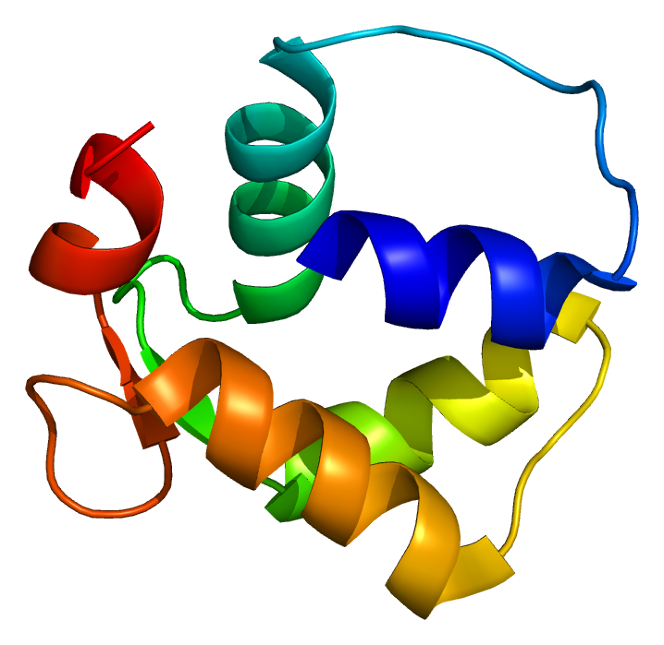
Ninein is a protein that in humans is encoded by the ''NIN'' gene. Function Ninein, together with its paralog Ninein-like protein is one of the proteins important for centrosomal function. Localization of this protein to the centrosome requires three leucine zippers in the central coiled-coil domain. Multiple alternatively spliced transcript variants that encode different isoforms have been reported. This protein is important for positioning and anchoring the microtubules minus-ends in epithelial cell Epithelium or epithelial tissue is a thin, continuous, protective layer of Cell (biology), cells with little extracellular matrix. An example is the epidermis, the outermost layer of the skin. Epithelial (Mesothelium, mesothelial) tissues line ...s. References Further reading * * * * * * * * * * * * * * * * * EF-hand-containing proteins {{gene-14-stub ... [...More Info...] [...Related Items...] OR: [Wikipedia] [Google] [Baidu] |
Pericentriolar Material
Pericentriolar material (PCM, sometimes also called pericent matrix) is a highly structured, dense mass of protein which makes up the part of the animal centrosome that surrounds the two centrioles. The PCM contains proteins responsible for microtubule nucleation and Microtubule anchoring, anchoring including γ-tubulin, pericentrin and ninein. Although the PCM appears amorphous by electron microscopy, super-resolution microscopy finds that it is highly organized. The PCM have 9-fold symmetry that mimics the symmetry of the centriole. Some PCM proteins are organized such that one end of the protein is found near the centriole and the other end is farther away from the centriole. The PCM size is dynamic during the cell cycle. After cell division, the PCM size is reduced in a process named centrosome cycle#Centrosome reduction, centrosome reduction.Atypical centrioles during sexual reproduction Tomer Avidor-Reiss*, Atul Khire, Emily L. Fishman and Kyoung H. Jo Curr Biol. 2015 Nov 16;25 ... [...More Info...] [...Related Items...] OR: [Wikipedia] [Google] [Baidu] |
Centrin
Centrins, also known as caltractins, are a family of calcium-binding phosphoproteins found in the centrosome of eukaryotes. Centrins are small calcium binding proteins that are ubiquitous centrosome components. There are about 350 “signature” proteins that are unique to eukaryotic cells but have no significant homology to proteins in archaea and bacteria. They are a type of protein that is essential and present in almost all eukaryotic cells and are found in the centrioles and pericentriolar lattice. Human centrin genes are CETN1, CETN2 and CETN3. Humans and mice have three centrin genes: Cetn-1, which is typically only expressed in male germ cells, and Cetn-2 and Cetn-3, which are typically only expressed in somatic cells. Centrin-2 is a recombinant GFP-centrin-2 and centriole protein that localizes to centrioles throughout the cell cycle, while centrin-3 seems to stick to the pericentriolar material that surrounds the centrioles. History Centrin was first isolated and ... [...More Info...] [...Related Items...] OR: [Wikipedia] [Google] [Baidu] |
Chromosome
A chromosome is a package of DNA containing part or all of the genetic material of an organism. In most chromosomes, the very long thin DNA fibers are coated with nucleosome-forming packaging proteins; in eukaryotic cells, the most important of these proteins are the histones. Aided by chaperone proteins, the histones bind to and condense the DNA molecule to maintain its integrity. These eukaryotic chromosomes display a complex three-dimensional structure that has a significant role in transcriptional regulation. Normally, chromosomes are visible under a light microscope only during the metaphase of cell division, where all chromosomes are aligned in the center of the cell in their condensed form. Before this stage occurs, each chromosome is duplicated ( S phase), and the two copies are joined by a centromere—resulting in either an X-shaped structure if the centromere is located equatorially, or a two-armed structure if the centromere is located distally; the jo ... [...More Info...] [...Related Items...] OR: [Wikipedia] [Google] [Baidu] |


