|
Canaliculus (bone)
Bone canaliculi are microscopic canals between the lacunae of ossified bone. The radiating processes of the osteocytes (called filopodia) project into these canals. These cytoplasmic processes are joined together by gap junctions. Osteocytes do not entirely fill up the canaliculi. The remaining space is known as the periosteocytic space, which is filled with periosteocytic fluid. This fluid contains substances too large to be transported through the gap junctions that connect the osteocytes. In cartilage, the lacunae and hence, the chondrocytes, are isolated from each other. Materials picked up by osteocytes adjacent to blood vessels are distributed throughout the bone matrix via the canaliculi. Diameter of canaliculi in human bone is approximately 200 to 900 nm. In bovine tibia diameter of canaliculi was found to vary from 155 to 844 nm (average 426 nm). In mice humeri it varies from 80 to 710 nm (average 259 nm), while diameter of osteocytic processes varies from 50 ... [...More Info...] [...Related Items...] OR: [Wikipedia] [Google] [Baidu] |
Lacuna (histology)
In histology, a lacuna is a small space, containing an osteocyte in bone, or chondrocyte in cartilage. Bone The lacuna are situated between the lamella of osteon, lamellae, and consist of a number of oblong spaces. In an ordinary microscopic section, viewed by transmitted light, they appear as fusiform opaque spots. Each lacuna is occupied during life by a branched cell, termed an osteocyte, bone-cell or bone-corpuscle. Lacunae are connected to one another by small canals called canaliculus (bone), canaliculi. A lacuna never contains more than one osteocyte. Sinuses are an example of lacuna. Cartilage The cartilage cells or chondrocytes are contained in cavities in the matrix, called cartilage lacunae; around these, the matrix is arranged in concentric lines as if it had been formed in successive portions around the cartilage cells. This constitutes the so-called capsule of the space. Each lacuna is generally occupied by a single cell, but during the division of the cells, it may ... [...More Info...] [...Related Items...] OR: [Wikipedia] [Google] [Baidu] |
Ossified
Ossification (also called osteogenesis or bone mineralization) in bone remodeling is the process of laying down new bone material by cells named osteoblasts. It is synonymous with bone tissue formation. There are two processes resulting in the formation of normal, healthy bone tissue: Intramembranous ossification is the direct laying down of bone into the primitive connective tissue ( mesenchyme), while endochondral ossification involves cartilage as a precursor. In fracture healing, endochondral osteogenesis is the most commonly occurring process, for example in fractures of long bones treated by plaster of Paris, whereas fractures treated by open reduction and internal fixation with metal plates, screws, pins, rods and nails may heal by intramembranous osteogenesis. Heterotopic ossification is a process resulting in the formation of bone tissue that is often atypical, at an extraskeletal location. Calcification is often confused with ossification. Calcification ... [...More Info...] [...Related Items...] OR: [Wikipedia] [Google] [Baidu] |
Bone
A bone is a rigid organ that constitutes part of the skeleton in most vertebrate animals. Bones protect the various other organs of the body, produce red and white blood cells, store minerals, provide structure and support for the body, and enable mobility. Bones come in a variety of shapes and sizes and have complex internal and external structures. They are lightweight yet strong and hard and serve multiple functions. Bone tissue (osseous tissue), which is also called bone in the uncountable sense of that word, is hard tissue, a type of specialised connective tissue. It has a honeycomb-like matrix internally, which helps to give the bone rigidity. Bone tissue is made up of different types of bone cells. Osteoblasts and osteocytes are involved in the formation and mineralisation of bone; osteoclasts are involved in the resorption of bone tissue. Modified (flattened) osteoblasts become the lining cells that form a protective layer on the bone surface. The mine ... [...More Info...] [...Related Items...] OR: [Wikipedia] [Google] [Baidu] |
Gap Junction
Gap junctions are membrane channels between adjacent cells that allow the direct exchange of cytoplasmic substances, such small molecules, substrates, and metabolites. Gap junctions were first described as ''close appositions'' alongside tight junctions, however, electron microscopy studies in 1967 led to gap junctions being named as such to be distinguished from tight junctions. They bridge a 2-4 nm gap between cell membranes. Gap junctions use protein complexes known as connexons, composed of connexin proteins to connect one cell to another. Gap junction proteins include the more than 26 types of connexin, as well as at least 12 non-connexin components that make up the gap junction complex or ''nexus,'' including the tight junction protein ZO-1—a protein that holds membrane content together and adds structural clarity to a cell, sodium channels, and aquaporin. More gap junction proteins have become known due to the development of next-generation sequencing. Connexins ... [...More Info...] [...Related Items...] OR: [Wikipedia] [Google] [Baidu] |
Osteocyte
An osteocyte, an oblate-shaped type of bone cell with dendritic processes, is the most commonly found cell in mature bone. It can live as long as the organism itself. The adult human body has about 42 billion of them. Osteocytes do not divide and have an average half life of 25 years. They are derived from osteoprogenitor cells, some of which differentiate into active osteoblasts (which may further differentiate to osteocytes). Osteoblasts/osteocytes develop in mesenchyme. In mature bones, osteocytes and their processes reside inside spaces called lacunae (Latin for a ''pit'') and canaliculi, respectively. Osteocytes are simply osteoblasts trapped in the matrix that they secrete. They are networked to each other via long cytoplasmic extensions that occupy tiny canals called canaliculi, which are used for exchange of nutrients and waste through gap junctions. Although osteocytes have reduced synthetic activity and (like osteoblasts) are not capable of mitotic division, they ... [...More Info...] [...Related Items...] OR: [Wikipedia] [Google] [Baidu] |
Cartilage
Cartilage is a resilient and smooth type of connective tissue. Semi-transparent and non-porous, it is usually covered by a tough and fibrous membrane called perichondrium. In tetrapods, it covers and protects the ends of long bones at the joints as articular cartilage, and is a structural component of many body parts including the rib cage, the neck and the bronchial tubes, and the intervertebral discs. In other taxa, such as chondrichthyans and cyclostomes, it constitutes a much greater proportion of the skeleton. It is not as hard and rigid as bone, but it is much stiffer and much less flexible than muscle. The matrix of cartilage is made up of glycosaminoglycans, proteoglycans, collagen fibers and, sometimes, elastin. It usually grows quicker than bone. Because of its rigidity, cartilage often serves the purpose of holding tubes open in the body. Examples include the rings of the trachea, such as the cricoid cartilage and carina. Cartilage is composed of specialized c ... [...More Info...] [...Related Items...] OR: [Wikipedia] [Google] [Baidu] |
Chondrocytes
Chondrocytes (, ) are the only cells found in healthy cartilage. They produce and maintain the cartilaginous matrix, which consists mainly of collagen and proteoglycans. Although the word '' chondroblast'' is commonly used to describe an immature chondrocyte, the term is imprecise, since the progenitor of chondrocytes (which are mesenchymal stem cells) can differentiate into various cell types, including osteoblasts. Development From least- to terminally-differentiated, the chondrocytic lineage is: # Colony-forming unit-fibroblast # Mesenchymal stem cell / marrow stromal cell # Chondrocyte # Hypertrophic chondrocyte Mesenchymal (mesoderm origin) stem cells are undifferentiated, meaning they can differentiate into a variety of generative cells commonly known as osteochondrogenic (or osteogenic, chondrogenic, osteoprogenitor, etc.) cells. When referring to bone, or in this case cartilage, the originally undifferentiated mesenchymal stem cells lose their pluripotency, proliferat ... [...More Info...] [...Related Items...] OR: [Wikipedia] [Google] [Baidu] |
Tibia
The tibia (; : tibiae or tibias), also known as the shinbone or shankbone, is the larger, stronger, and anterior (frontal) of the two Leg bones, bones in the leg below the knee in vertebrates (the other being the fibula, behind and to the outside of the tibia); it connects the knee with the ankle bones, ankle. The tibia is found on the anatomical terms of location#Medial, medial side of the leg next to the fibula and closer to the median plane. The tibia is connected to the fibula by the interosseous membrane of leg, forming a type of fibrous joint called a syndesmosis with very little movement. The tibia is named for the flute ''aulos, tibia''. It is the second largest bone in the human body, after the femur. The leg bones are the strongest long bones as they support the rest of the body. Structure In human anatomy, the tibia is the second largest bone next to the femur. As in other vertebrates the tibia is one of two bones in the lower leg, the other being the fibula, and is a ... [...More Info...] [...Related Items...] OR: [Wikipedia] [Google] [Baidu] |
Humerus
The humerus (; : humeri) is a long bone in the arm that runs from the shoulder to the elbow. It connects the scapula and the two bones of the lower arm, the radius (bone), radius and ulna, and consists of three sections. The humeral upper extremity of humerus, upper extremity consists of a rounded head, a narrow neck, and two short processes (tubercles, sometimes called tuberosities). The body of humerus, body is cylindrical in its upper portion, and more prism (geometry), prismatic below. The lower extremity of humerus, lower extremity consists of 2 epicondyles, 2 processes (trochlea of the humerus, trochlea and capitulum of the humerus, capitulum), and 3 fossae (radial fossa, coronoid fossa, and olecranon fossa). As well as its true anatomical neck, the constriction below the greater and lesser tubercles of the humerus is referred to as its Surgical neck of the humerus, surgical neck due to its tendency to fracture, thus often becoming the focus of surgeons. Etymology The word ... [...More Info...] [...Related Items...] OR: [Wikipedia] [Google] [Baidu] |
Tooth
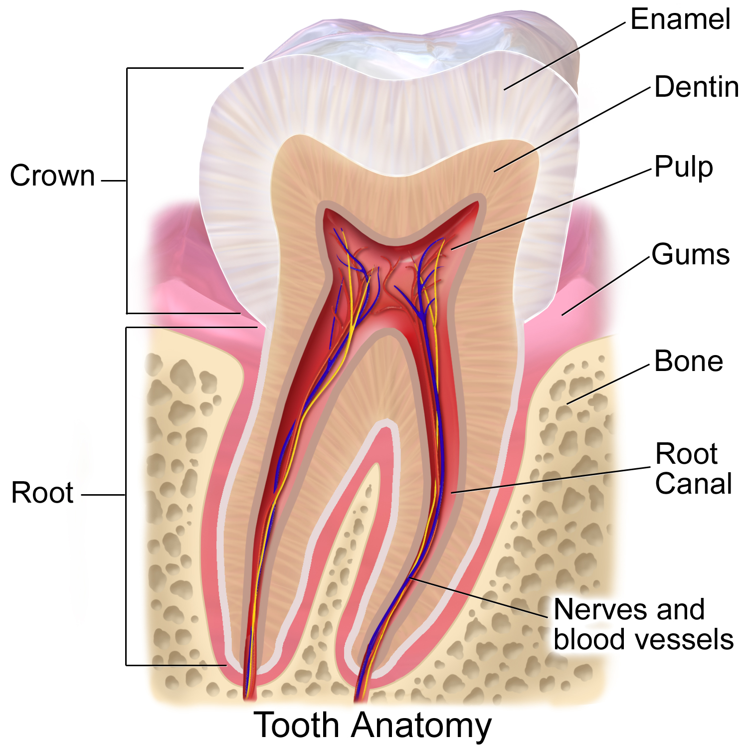
A tooth (: teeth) is a hard, calcified structure found in the jaws (or mouths) of many vertebrates and used to break down food. Some animals, particularly carnivores and omnivores, also use teeth to help with capturing or wounding prey, tearing food, for defensive purposes, to intimidate other animals often including their own, or to carry prey or their young. The roots of teeth are covered by gums. Teeth are not made of bone, but rather of multiple tissues of varying density and hardness that originate from the outermost embryonic germ layer, the ectoderm. The general structure of teeth is similar across the vertebrates, although there is considerable variation in their form and position. The teeth of mammals have deep roots, and this pattern is also found in some fish, and in crocodilians. In most teleost fish, however, the teeth are attached to the outer surface of the bone, while in lizards they are attached to the inner surface of the jaw by one side. In cartila ... [...More Info...] [...Related Items...] OR: [Wikipedia] [Google] [Baidu] |
Tooth Enamel
Tooth enamel is one of the four major Tissue (biology), tissues that make up the tooth in humans and many animals, including some species of fish. It makes up the normally visible part of the tooth, covering the Crown (tooth), crown. The other major tissues are dentin, cementum, and Pulp (tooth), dental pulp. It is a very hard, white to off-white, highly mineralised substance that acts as a barrier to protect the tooth but can become susceptible to degradation, especially by acids from food and drink. In rare circumstances enamel fails to form, leaving the underlying dentin exposed on the surface. Features Enamel is the hardest substance in the human body and contains the highest percentage of minerals (at 96%),Ross ''et al.'', p. 485 with water and organic material composing the rest.Ten Cate's Oral Histology, Nancy, Elsevier, pp. 70–94 The primary mineral is hydroxyapatite, which is a crystalline calcium phosphate. Enamel is formed on the tooth while the tooth develops wit ... [...More Info...] [...Related Items...] OR: [Wikipedia] [Google] [Baidu] |


