|
CCR6
Chemokine receptor 6 also known as CCR6 is a CC chemokine receptor protein which in humans is encoded by the ''CCR6'' gene. CCR6 has also recently been designated CD196 ( cluster of differentiation 196). The gene is located on the long arm of Chromosome 6 (6q27) on the Watson (plus) strand. It is 139,737 bases long and encodes a protein of 374 amino acids (molecular weight 42,494 Da). Function This protein belongs to family A of G protein-coupled receptor superfamily. The gene is expressed in lymphatic and non-lymphatic tissue as spleen, lymph nodes, pancreas, colon, appendix, small intestine. CCR6 is expressed on B-cells, immature dendritic cells (DC), T-cells ( Th1, Th2, Th17, Treg), natural killer T cells ( NKT cells) and neutrophils. The ligand of this receptor is CCL20 or in the other name - macrophage inflammatory protein 3 alpha ( MIP-3 alpha). This chemokine receptor is special because it binds only one chemokine ligand CCL20 in compare to other chemokine recep ... [...More Info...] [...Related Items...] OR: [Wikipedia] [Google] [Baidu] |
CC Chemokine Receptors
CC chemokine receptors (or beta chemokine receptors) are integral membrane proteins that specifically bind and respond to cytokines of the CC chemokine family. They represent one subfamily of chemokine receptors, a large family of G protein-linked receptors that are known as seven transmembrane (7-TM) proteins since they span the cell membrane seven times. To date, ten true members of the CC chemokine receptor subfamily have been described. These are named CCR1 to CCR10 according to the IUIS/WHO Subcommittee on Chemokine Nomenclature. Mechanism The CC chemokine receptors all work by activating the G protein Gi. Types Overview table CCR1 CCR1 was the first CC chemokine receptor identified and binds multiple inflammatory/inducible (see inducible gene) CC chemokines (including CCL4, CCL5, CCL6, CCL14, CCL15, CCL16 and CCL23). In humans, this receptor can be found on peripheral blood lymphocytes and monocytes. There is some suggestion that this c ... [...More Info...] [...Related Items...] OR: [Wikipedia] [Google] [Baidu] |
MIP-3 Alpha
Chemokine (C-C motif) ligand 20 (CCL20) or liver activation regulated chemokine (LARC) or Macrophage Inflammatory Protein-3 (MIP3A) is a small cytokine belonging to the CC chemokine family. It is strongly chemotactic for lymphocytes and weakly attracts neutrophils. CCL20 is implicated in the formation and function of mucosal lymphoid tissues via chemoattraction of lymphocytes and dendritic cells towards the epithelial cells surrounding these tissues. CCL20 elicits its effects on its target cells by binding and activating the chemokine receptor CCR6. Gene expression of CCL20 can be induced by microbial factors such as lipopolysaccharide (LPS), and inflammatory cytokines such as tumor necrosis factor and interferon-γ, and down-regulated by IL-10. CCL20 is expressed in several tissues with highest expression observed in peripheral blood lymphocytes, lymph nodes, liver, appendix, and fetal lung and lower levels in thymus, testis, prostate and gut. The gene for CCL20 (''s ... [...More Info...] [...Related Items...] OR: [Wikipedia] [Google] [Baidu] |
CCL20
Chemokine (C-C motif) ligand 20 (CCL20) or liver activation regulated chemokine (LARC) or Macrophage Inflammatory Protein-3 (MIP3A) is a small cytokine belonging to the CC chemokine family. It is strongly chemotactic for lymphocytes and weakly attracts neutrophils. CCL20 is implicated in the formation and function of mucosal lymphoid tissues via chemoattraction of lymphocytes and dendritic cells towards the epithelial cells surrounding these tissues. CCL20 elicits its effects on its target cells by binding and activating the chemokine receptor CCR6. Gene expression of CCL20 can be induced by microbial factors such as lipopolysaccharide (LPS), and inflammatory cytokines such as tumor necrosis factor and interferon-γ, and down-regulated by IL-10. CCL20 is expressed in several tissues with highest expression observed in peripheral blood lymphocytes, lymph nodes, liver, appendix, and fetal lung and lower levels in thymus, testis, prostate and gut. The gene for CCL20 ... [...More Info...] [...Related Items...] OR: [Wikipedia] [Google] [Baidu] |
T Helper 17 Cell
T helper 17 cells (Th17) are a subset of pro-inflammatory T helper cells defined by their production of interleukin 17 (IL-17). They are related to T regulatory cells and the signals that cause Th17s to differentiate actually inhibit Treg differentiation. However, Th17s are developmentally distinct from Th1 and Th2 lineages. Th17 cells play an important role in maintaining mucosal barriers and contributing to pathogen clearance at mucosal surfaces; such protective and non-pathogenic Th17 cells have been termed as Treg17 cells. They have also been implicated in autoimmune and inflammatory disorders. The loss of Th17 cell populations at mucosal surfaces has been linked to chronic inflammation and microbial translocation. These regulatory Th17 cells can be generated by TGF-beta plus IL-6 in vitro. Differentiation Like conventional regulatory T cells (Treg), induction of regulatory Treg17 cells could play an important role in modulating and preventing certain autoimmune diseases. ... [...More Info...] [...Related Items...] OR: [Wikipedia] [Google] [Baidu] |
Neutrophil
Neutrophils (also known as neutrocytes or heterophils) are the most abundant type of granulocytes and make up 40% to 70% of all white blood cells in humans. They form an essential part of the innate immune system, with their functions varying in different animals. They are formed from stem cells in the bone marrow and differentiated into subpopulations of neutrophil-killers and neutrophil-cagers. They are short-lived and highly mobile, as they can enter parts of tissue where other cells/molecules cannot. Neutrophils may be subdivided into segmented neutrophils and banded neutrophils (or bands). They form part of the polymorphonuclear cells family (PMNs) together with basophils and eosinophils. The name ''neutrophil'' derives from staining characteristics on hematoxylin and eosin ( H&E) histological or cytological preparations. Whereas basophilic white blood cells stain dark blue and eosinophilic white blood cells stain bright red, neutrophils stain a neutral pink. Normally, n ... [...More Info...] [...Related Items...] OR: [Wikipedia] [Google] [Baidu] |
MicroRNA
MicroRNA (miRNA) are small, single-stranded, non-coding RNA molecules containing 21 to 23 nucleotides. Found in plants, animals and some viruses, miRNAs are involved in RNA silencing and post-transcriptional regulation of gene expression. miRNAs base-pair to complementary sequences in mRNA molecules, then gene silence said mRNA molecules by one or more of the following processes: (1) cleavage of mRNA strand into two pieces, (2) destabilization of mRNA by shortening its poly(A) tail, or (3) translation of mRNA into proteins. This last method of gene silencing is the least efficient of the three, and requires the aid of ribosomes. miRNAs resemble the small interfering RNAs (siRNAs) of the RNA interference (RNAi) pathway, except miRNAs derive from regions of RNA transcripts that fold back on themselves to form short hairpins, whereas siRNAs derive from longer regions of double-stranded RNA. The human genome may encode over 1900 miRNAs, although more recent analysis s ... [...More Info...] [...Related Items...] OR: [Wikipedia] [Google] [Baidu] |
Langerhans Cell
A Langerhans cell (LC) is a tissue-resident macrophage of the skin. These cells contain organelles called Birbeck granules. They are present in all layers of the epidermis and are most prominent in the stratum spinosum. They also occur in the papillary dermis, particularly around blood vessels, as well as in the mucosa of the mouth, foreskin, and vaginal epithelium. They can be found in other tissues, such as lymph nodes, particularly in association with the condition Langerhans cell histiocytosis (LCH). Function In skin infection An infection is the invasion of tissues by pathogens, their multiplication, and the reaction of host tissues to the infectious agent and the toxins they produce. An infectious disease, also known as a transmissible disease or communicable d ...s, the local Langerhans cells take up and process microbe, microbial antigens to become fully functional antigen-presenting cells. Generally, tissue-resident macrophages are involved in immune ... [...More Info...] [...Related Items...] OR: [Wikipedia] [Google] [Baidu] |
Interferon Gamma
Interferon gamma (IFN-γ) is a dimerized soluble cytokine that is the only member of the type II class of interferons. The existence of this interferon, which early in its history was known as immune interferon, was described by E. F. Wheelock as a product of human leukocytes stimulated with phytohemagglutinin, and by others as a product of antigen-stimulated lymphocytes. It was also shown to be produced in human lymphocytes. or tuberculin-sensitized mouse peritoneal lymphocytes challenged with Mantoux test (PPD); the resulting supernatants were shown to inhibit growth of vesicular stomatitis virus. Those reports also contained the basic observation underlying the now widely employed IFN-γ release assay used to test for tuberculosis. In humans, the IFN-γ protein is encoded by the ''IFNG'' gene. Through cell signaling, IFN-γ plays a role in regulating the immune response of its target cell. A key signaling pathway that is activated by type II IFN is the JAK-STAT ... [...More Info...] [...Related Items...] OR: [Wikipedia] [Google] [Baidu] |
Interleukin 4
The interleukin 4 (IL4, IL-4) is a cytokine that induces differentiation of naive helper T cells ( Th0 cells) to Th2 cells. Upon activation by IL-4, Th2 cells subsequently produce additional IL-4 in a positive feedback loop. IL-4 is produced primarily by mast cells, Th2 cells, eosinophils and basophils. It is closely related and has functions similar to IL-13. Function Interleukin 4 has many biological roles, including the stimulation of activated B cell and T cell proliferation, and the differentiation of B cells into plasma cells. It is a key regulator in humoral and adaptive immunity. IL-4 induces B cell class switching to IgE, and up-regulates MHC class II production. IL-4 decreases the production of Th1 cells, macrophages, IFNγ, and dendritic cells IL-12. Overproduction of IL-4 is associated with allergies. * Inflammation and wound repair Tissue macrophages play an important role in chronic inflammation and wound ... [...More Info...] [...Related Items...] OR: [Wikipedia] [Google] [Baidu] |
B-cell
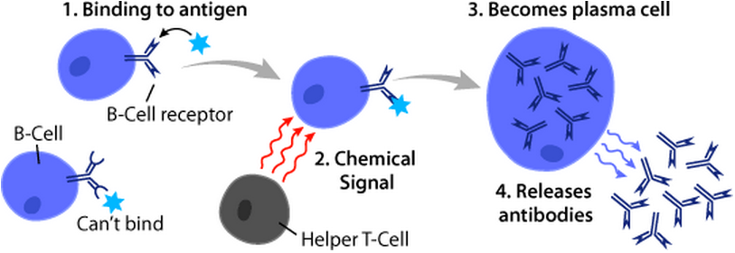
B cells, also known as B lymphocytes, are a type of white blood cell of the lymphocyte subtype. They function in the humoral immunity component of the adaptive immune system. B cells produce antibody molecules which may be either secreted or inserted into the plasma membrane where they serve as a part of B-cell receptors. When a naïve or memory B cell is activated by an antigen, it proliferates and differentiates into an antibody-secreting effector cell, known as a plasmablast or plasma cell. Additionally, B cells present antigens (they are also classified as professional antigen-presenting cells (APCs)) and secrete cytokines. In mammals, B cells mature in the bone marrow, which is at the core of most bones. In birds, B cells mature in the bursa of Fabricius, a lymphoid organ where they were first discovered by Chang and Glick, which is why the 'B' stands for bursa and not bone marrow as commonly believed. B cells, unlike the other two classes of lymphocytes, T ce ... [...More Info...] [...Related Items...] OR: [Wikipedia] [Google] [Baidu] |
Regulatory T Cell
The regulatory T cells (Tregs or Treg cells), formerly known as suppressor T cells, are a subpopulation of T cells that modulate the immune system, maintain tolerance to self-antigens, and prevent autoimmune disease. Treg cells are immunosuppressive and generally suppress or downregulate induction and proliferation of effector T cells. Treg cells express the biomarkers CD4, FOXP3, and CD25 and are thought to be derived from the same lineage as naïve CD4+ cells. Because effector T cells also express CD4 and CD25, Treg cells are very difficult to effectively discern from effector CD4+, making them difficult to study. Research has found that the cytokine transforming growth factor beta (TGF-β) is essential for Treg cells to differentiate from naïve CD4+ cells and is important in maintaining Treg cell homeostasis. Mouse models have suggested that modulation of Treg cells can treat autoimmune disease and cancer and can facilitat ... [...More Info...] [...Related Items...] OR: [Wikipedia] [Google] [Baidu] |
Natural Killer T Cell
Natural killer T (NKT) cells are a heterogeneous group of T cells that share properties of both T cells and natural killer cells. Many of these cells recognize the non-polymorphic CD1d molecule, an antigen-presenting molecule that binds self and foreign lipids and glycolipids. They constitute only approximately 1% of all peripheral blood T cells. Natural killer T cells should neither be confused with natural killer cells nor killer T cells (cytotoxic T cells). Nomenclature The term "NK T cells" was first used in mice to define a subset of T cells that expressed the natural killer (NK) cell-associated marker NK1.1 (CD161). It is now generally accepted that the term "NKT cells" refers to CD1d-restricted T cells, present in mice and humans, some of which coexpress a heavily biased, semi-invariant T-cell receptor and NK cell markers. Molecular characterization NKT cells are a subset of T cells that coexpress an αβ T-cell receptor, but also express a variety of molecular ma ... [...More Info...] [...Related Items...] OR: [Wikipedia] [Google] [Baidu] |


