|
Anatomical Variations
An anatomical variation, anatomical variant, or anatomical variability is a presentation of body structure with morphological features different from those that are typically described in the majority of individuals. Anatomical variations are categorized into three types including morphometric (size or shape), consistency (present or absent), and spatial (proximal/distal or right/left). Variations are seen as normal in the sense that they are found consistently among different individuals, are mostly without symptoms, and are termed anatomical variations rather than abnormalities. Anatomical variations are mainly caused by genetics and may vary considerably between different populations. The rate of variation considerably differs between single organs, particularly in muscles. Knowledge of anatomical variations is important in order to distinguish them from pathological conditions. A very early paper published in 1898, presented anatomic variations to have a wide range and signi ... [...More Info...] [...Related Items...] OR: [Wikipedia] [Google] [Baidu] |
Morphology (biology)
Morphology (from Ancient Greek μορφή (morphḗ) "form", and λόγος (lógos) "word, study, research") is the study of the form and structure of organisms and their specific structural features. This includes aspects of the outward appearance (shape, structure, color, pattern, size), as well as the form and structure of internal parts like bones and organs, i.e., anatomy. This is in contrast to physiology, which deals primarily with function. Morphology is a branch of life science dealing with the study of the overall structure of an organism or taxon and its component parts. History The etymology of the word "morphology" is from the Ancient Greek (), meaning "form", and (), meaning "word, study, research". While the concept of form in biology, opposed to function, dates back to Aristotle (see Aristotle's biology), the field of morphology was developed by Johann Wolfgang von Goethe (1790) and independently by the German anatomist and physiologist Karl Fried ... [...More Info...] [...Related Items...] OR: [Wikipedia] [Google] [Baidu] |
Plantaris Muscle
The plantaris is one of the superficial muscles of the superficial posterior compartment of the leg, one of the fascial compartments of the leg. It is composed of a thin muscle belly and a long thin tendon. While not as thick as the achilles tendon, the plantaris tendon (which tends to be between in length) is the longest tendon in the human body. Not including the tendon, the plantaris muscle is approximately long and is absent in 8-12% of the population. It is one of the plantar flexors in the posterior compartment of the leg, along with the gastrocnemius and soleus muscles. The plantaris is considered to have become an unimportant muscle when human ancestors switched from climbing trees to bipedalism and in anatomically modern humans it mainly acts with the gastrocnemius. Structure The plantaris muscle arises from the inferior part of the lateral supracondylar ridge of the femur at a position slightly superior to the origin of the lateral head of gastrocnemius. It pas ... [...More Info...] [...Related Items...] OR: [Wikipedia] [Google] [Baidu] |
Visible Difference
A visible difference refers to a physical characteristic, such as a scar, mark, disfigurement, or condition on the face or body that distinguishes an individual from what is conventionally considered to be the societal norm. These visible differences can arise due to a multitude of factors including but not limited to congenital conditions, accidents, diseases, or surgical procedures. Such differences often carry significant psychological, social, and sometimes physical implications for the affected individual. Terminology While the terms visible difference and disfigurement are sometimes used interchangeably, they are not synonymous and possess distinct connotations both in medical and social contexts. The term visible difference is a more neutral, encompassing descriptor for any physical characteristic that sets an individual apart from what is generally considered the societal norm. This could be due to congenital conditions, acquired conditions, diseases, or even personal ch ... [...More Info...] [...Related Items...] OR: [Wikipedia] [Google] [Baidu] |
Supernumerary Body Part
Supernumerary body parts are most commonly a congenital disorder involving the growth of an additional part of the body and a deviation from the body plan. Body parts may be easily visible or hidden away, such as internal organs. Many additional body parts form by the same process as conjoined twins: the zygote begins to split but fails to completely separate. This condition may also be a symptom of repeated occurrences of continuous inbreeding in a genetic line. Specific types of occurrence Specific types of additional body parts include: * Accessory breast – one or more additional breastsOnline Mendelian Inheritance in Man. Nipples, Supernumerary. Accessed 10 July 2006. *Accessory spleen – one or more additional spleens *Cervical rib – an additional rib *Diphallia – having two penes/penises * Hyperdontia – additional teeth *Pelvic digit – a bony growth in the soft tissue of the pelvic region *Polycephaly – an extra head *Polydactyly – additional fingers or to ... [...More Info...] [...Related Items...] OR: [Wikipedia] [Google] [Baidu] |
Lung
The lungs are the primary Organ (biology), organs of the respiratory system in many animals, including humans. In mammals and most other tetrapods, two lungs are located near the Vertebral column, backbone on either side of the heart. Their function in the respiratory system is to extract oxygen from the atmosphere and transfer it into the bloodstream, and to release carbon dioxide from the bloodstream into the atmosphere, in a process of gas exchange. Respiration is driven by different muscular systems in different species. Mammals, reptiles and birds use their musculoskeletal systems to support and foster breathing. In early tetrapods, air was driven into the lungs by the pharyngeal muscles via buccal pumping, a mechanism still seen in amphibians. In humans, the primary muscle that drives breathing is the Thoracic diaphragm, diaphragm. The lungs also provide airflow that makes Animal communication#Auditory, vocalisation including speech possible. Humans have two lungs, a ri ... [...More Info...] [...Related Items...] OR: [Wikipedia] [Google] [Baidu] |
Knee Joint
In humans and other primates, the knee joins the thigh with the leg and consists of two joints: one between the femur and tibia (tibiofemoral joint), and one between the femur and patella (patellofemoral joint). It is the largest joint in the human body. The knee is a modified hinge joint, which permits flexion and extension as well as slight internal and external rotation. The knee is vulnerable to injury and to the development of osteoarthritis. It is often termed a ''compound joint'' having tibiofemoral and patellofemoral components. (The fibular collateral ligament is often considered with tibiofemoral components.) Structure The knee is a modified hinge joint, a type of synovial joint, which is composed of three functional compartments: the patellofemoral articulation, consisting of the patella, or "kneecap", and the patellar groove on the front of the femur through which it slides; and the medial and lateral tibiofemoral articulations linking the femur, or thigh b ... [...More Info...] [...Related Items...] OR: [Wikipedia] [Google] [Baidu] |
Lateral Meniscus
The lateral meniscus (external semilunar fibrocartilage) is a fibrocartilaginous band that spans the lateral side of the interior of the knee joint. It is one of two menisci of the knee, the other being the medial meniscus. It is nearly circular and covers a larger portion of the articular surface than the medial. It can occasionally be injured or torn by twisting the knee or applying direct force, as seen in contact sports. Structure The lateral meniscus is grooved laterally for the tendon of the popliteus, which separates it from the fibular collateral ligament. Its anterior end is attached in front of the intercondyloid eminence of the tibia, lateral to, and behind, the anterior cruciate ligament, with which it blends; the posterior end is attached behind the intercondyloid eminence of the tibia and in front of the posterior end of the medial meniscus. The anterior attachment of the lateral meniscus is twisted on itself so that its free margin looks backward and upward, its ... [...More Info...] [...Related Items...] OR: [Wikipedia] [Google] [Baidu] |
Discoid Meniscus
Discoid meniscus is a rare human anatomic variant that usually affects the lateral meniscus of the knee. Usually a person with this anomaly has no complaints; however, it may present as pain, swelling, or a snapping sound heard from the affected knee. Strong suggestive findings on magnetic resonance imaging includes a thickened meniscal body seen on more than two contiguous sagittal slices. Description The Watanabe classification of discoid lateral meniscus is: (A) Incomplete, (B) Complete, and (C) Wrisberg-ligament variant Normally, the meniscus (anatomy), meniscus is a thin crescent-shaped piece of cartilage that lies between the weight bearing joint surfaces of the femur and the tibia. It is attached to the lining of the knee joint along its periphery and serves to absorb about a third of the impact load that the joint cartilage surface sees and also provides some degree of stabilization for the knee. There are two menisci in the knee joint, with one on the outside (away fro ... [...More Info...] [...Related Items...] OR: [Wikipedia] [Google] [Baidu] |
Lumbar Vertebra
The lumbar vertebrae are located between the thoracic vertebrae and pelvis. They form the lower part of the back in humans, and the tail end of the back in quadrupeds. In humans, there are five lumbar vertebrae. The term is used to describe the anatomy of humans and quadrupeds, such as horses, pigs, or cattle. These bones are found in particular cuts of meat, including tenderloin or sirloin steak. Human anatomy In human anatomy, the five vertebrae are between the rib cage and the pelvis. They are the largest segments of the vertebral column and are characterized by the absence of the foramen transversarium within the transverse process (since it is only found in the cervical region) and by the absence of facets on the sides of the body (as found only in the thoracic region). They are designated L1 to L5, starting at the top. The lumbar vertebrae help support the weight of the body, and permit movement. General characteristics The adjacent figure depicts the general charac ... [...More Info...] [...Related Items...] OR: [Wikipedia] [Google] [Baidu] |
Mammogram
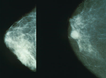
Mammography (also called mastography; DICOM modality: MG) is the process of using low-energy X-rays (usually around 30 kVp) to examine the human breast for diagnosis and screening. The goal of mammography is the early detection of breast cancer, typically through detection of characteristic masses, microcalcifications, asymmetries, and distortions. As with all X-rays, mammograms use doses of ionizing radiation to create images. These images are then analyzed for abnormal findings. It is usual to employ lower-energy X-rays, typically Mo (K-shell X-ray energies of 17.5 and 19.6 keV) and Rh (20.2 and 22.7 keV) than those used for radiography of bones. Mammography may be 2D or 3D ( tomosynthesis), depending on the available equipment or purpose of the examination. Ultrasound, ductography, positron emission mammography (PEM), and magnetic resonance imaging (MRI) are adjuncts to mammography. Ultrasound is typically used for further evaluation of masses found on mammography or palpa ... [...More Info...] [...Related Items...] OR: [Wikipedia] [Google] [Baidu] |
Sternalis Muscle
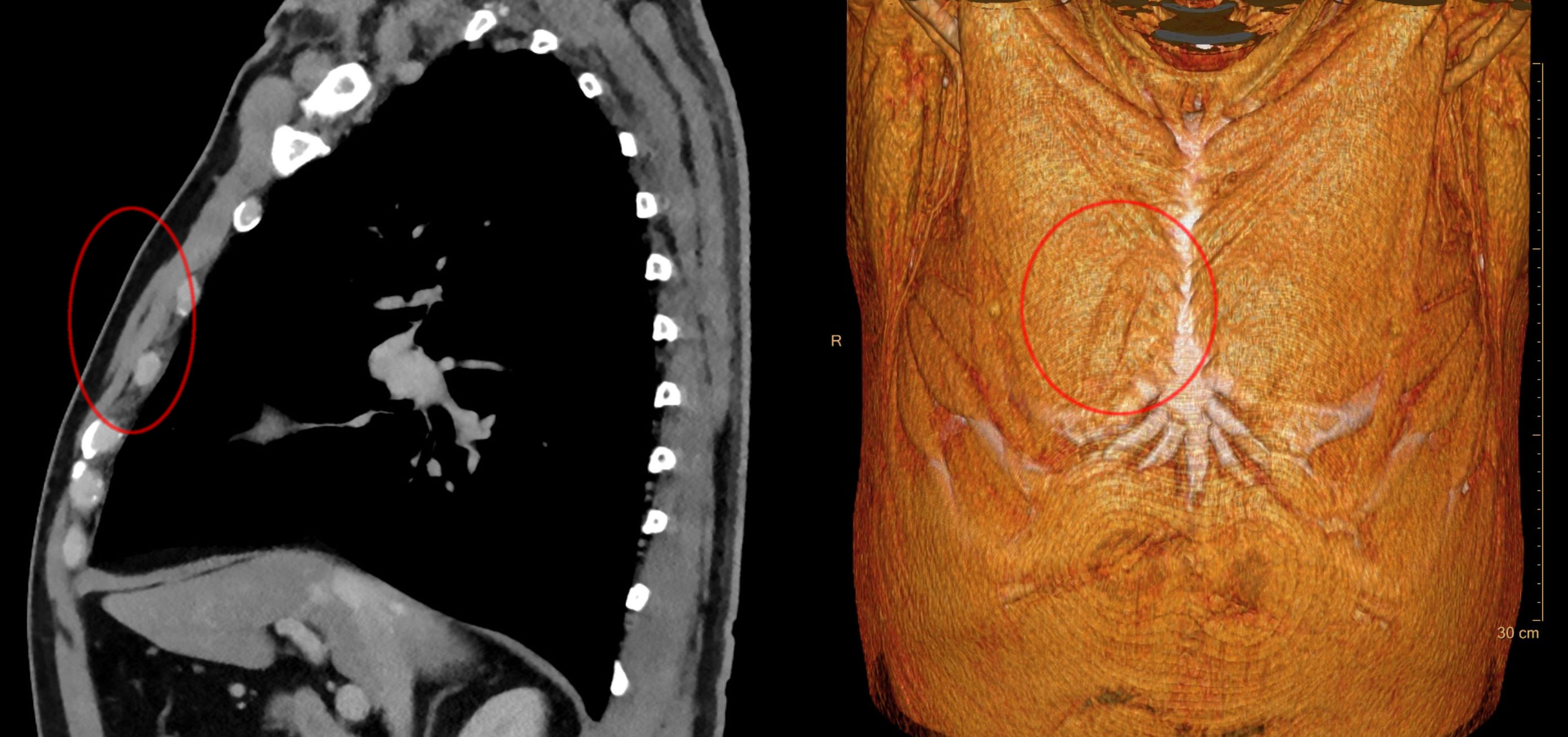
The rectus sternalis muscle is an anatomical variation that lies in front of the sternal end of the pectoralis major parallel to the margin of the sternum. The sternalis muscle may be a variation of the pectoralis major or of the rectus abdominis. Structure The sternalis is a muscle that runs along the anterior aspect of the body of the sternum. It lies superficially and parallel to the sternum. Its origin and insertion are variable. The sternalis muscle often originates from the upper part of the sternum and can display varying insertions such as the pectoral fascia, lower ribs, costal cartilages, rectus sheath, aponeurosis of the abdominal external oblique muscle. It may be present unilaterally or bilaterally. There is still a great deal of disagreement about its innervation and its embryonic origin. In a review, it was reported that the muscle was innervated by the external or internal thoracic nerves in 55% of the cases, by the intercostal nerves in 43% of the cases, whi ... [...More Info...] [...Related Items...] OR: [Wikipedia] [Google] [Baidu] |


