|
Biliverdin
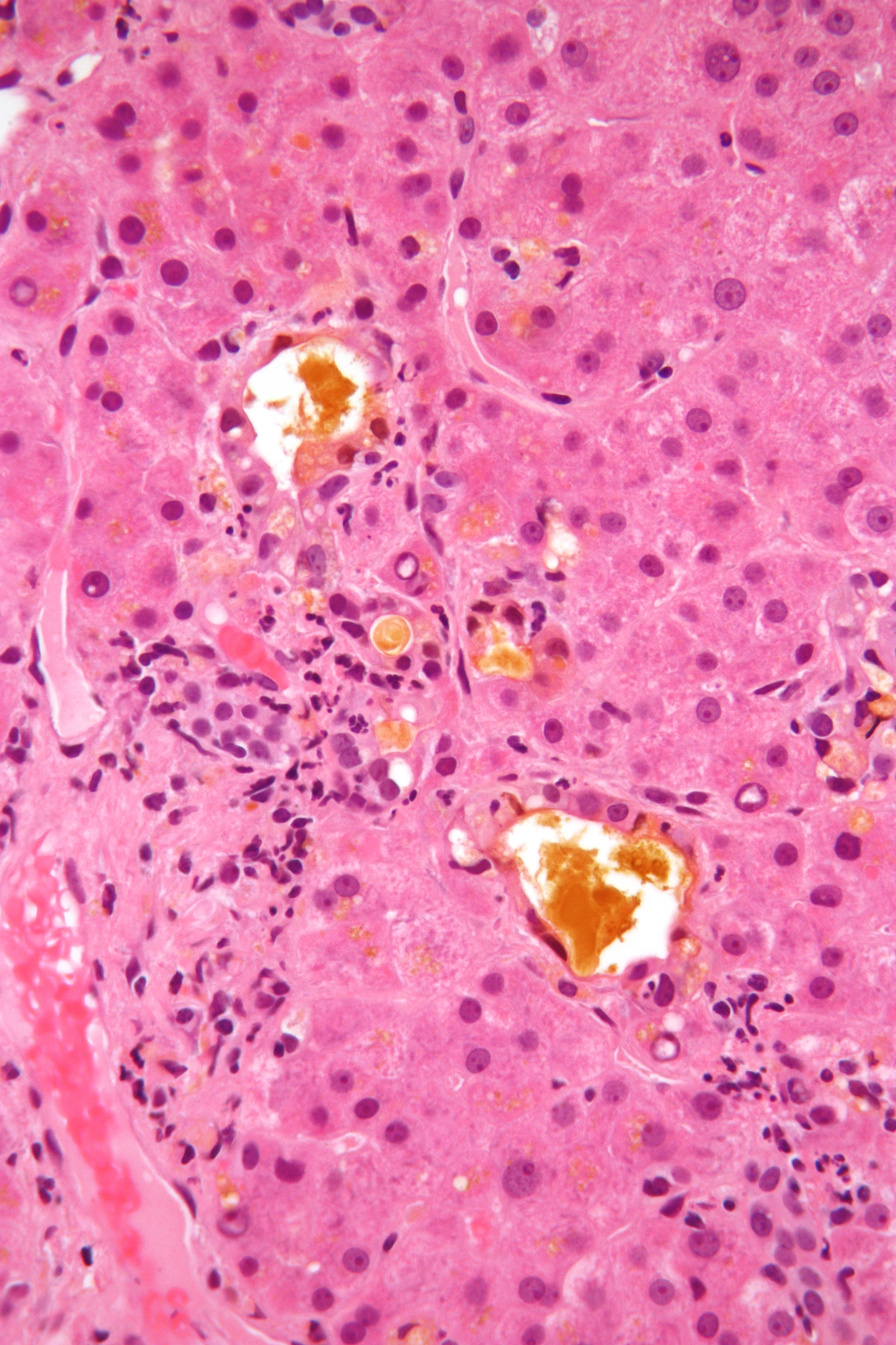
Biliverdin (latin for green bile) is a green tetrapyrrolic bile pigment, and is a product of heme catabolism.Boron W, Boulpaep E. Medical Physiology: a cellular and molecular approach, 2005. 984-986. Elsevier Saunders, United States. It is the pigment responsible for a greenish color sometimes seen in bruises. Metabolism Biliverdin results from the breakdown of the heme moiety of hemoglobin in erythrocytes. Macrophages break down senescent erythrocytes and break the heme down into biliverdin along with hemosiderin, in which biliverdin normally rapidly reduces to free bilirubin. Biliverdin is seen briefly in some bruises as a green color. In bruises, its breakdown into bilirubin leads to a yellowish color. Role in disease Biliverdin has been found in excess in the blood of humans suffering from hepatic diseases. Jaundice is caused by the accumulation of biliverdin or bilirubin (or both) in the circulatory system and tissues. Jaundiced skin and sclera (whites of th ... [...More Info...] [...Related Items...] OR: [Wikipedia] [Google] [Baidu] |
Biliverdin Reductase
Biliverdin reductase (BVR) is an enzyme () found in all tissues under normal conditions, but especially in reticulo-macrophages of the liver and spleen. BVR facilitates the conversion of biliverdin to bilirubin via the reduction of a double-bond between the second and third pyrrole ring into a single-bond. There are two isozymes, in humans, each encoded by its own gene, biliverdin reductase A (BLVRA) and biliverdin reductase B (BLVRB). Mechanism of catalysis BVR acts on biliverdin by reducing its double-bond between the pyrrole rings into a single-bond. It accomplishes this using NADPH + H+ as an electron donor, forming bilirubin and NADP+ as products. BVR catalyzes this reaction through an overlapping binding site including Lys18, Lys22, Lys179, Arg183, and Arg185 as key residues. This binding site attaches to biliverdin, and causes its dissociation from heme oxygenase (HO) (which catalyzes reaction of ferric heme --> biliverdin), causing the subsequent reduction to bil ... [...More Info...] [...Related Items...] OR: [Wikipedia] [Google] [Baidu] |
Bilirubin
Bilirubin (BR) (Latin for "red bile") is a red-orange compound that occurs in the normal catabolic pathway that breaks down heme in vertebrates. This catabolism is a necessary process in the body's clearance of waste products that arise from the destruction of aged or abnormal red blood cells. In the first step of bilirubin synthesis, the heme molecule is stripped from the hemoglobin molecule. Heme then passes through various processes of porphyrin catabolism, which varies according to the region of the body in which the breakdown occurs. For example, the molecules excreted in the urine differ from those in the feces. The production of biliverdin from heme is the first major step in the catabolic pathway, after which the enzyme biliverdin reductase performs the second step, producing bilirubin from biliverdin.Boron W, Boulpaep E. Medical Physiology: a cellular and molecular approach, 2005. 984–986. Elsevier Saunders, United States. Ultimately, bilirubin is broken down within ... [...More Info...] [...Related Items...] OR: [Wikipedia] [Google] [Baidu] |
Heme Breakdown
Heme, or haem (pronounced / hi:m/ ), is a precursor to hemoglobin, which is necessary to bind oxygen in the bloodstream. Heme is biosynthesized in both the bone marrow and the liver. In biochemical terms, heme is a coordination complex "consisting of an iron ion coordinated to a porphyrin acting as a tetradentate ligand, and to one or two axial ligands." The definition is loose, and many depictions omit the axial ligands. Among the metalloporphyrins deployed by metalloproteins as prosthetic groups, heme is one of the most widely used and defines a family of proteins known as hemoproteins. Hemes are most commonly recognized as components of hemoglobin, the red pigment in blood, but are also found in a number of other biologically important hemoproteins such as myoglobin, cytochromes, catalases, heme peroxidase, and endothelial nitric oxide synthase. The word ''haem'' is derived from Greek ''haima'' meaning "blood". Function Hemoproteins have diverse biological functi ... [...More Info...] [...Related Items...] OR: [Wikipedia] [Google] [Baidu] |
Prasinohaema
''Prasinohaema'' (Greek: "green blood") is a genus of skinks characterized by having green blood. This condition is caused by an excess buildup of the bile pigment biliverdin. ''Prasinohaema'' species have plasma biliverdin concentrations approximately 1.5-30 times greater than fish species with green blood plasma and 40 times greater than humans with green jaundice. The benefit provided by the high pigment concentration is unknown, but one possibility is that it protects against malaria. Geographic range Species in the genus ''Prasinohaema'' are endemic to New Guinea and the Solomon Islands. Species Species in the genus include:. www.reptile-database.org. *'' Prasinohaema flavipes'' – common green tree skink *'' Prasinohaema parkeri'' – Parker's green tree skink *'' Prasinohaema prehensicauda'' – prehensile green tree skink *'' Prasinohaema semoni'' – Semon's green tree skink *'' Prasinohaema virens'' - green-blooded skink, green tree skink ''Nota bene'': A bin ... [...More Info...] [...Related Items...] OR: [Wikipedia] [Google] [Baidu] |
Jaundice
Jaundice, also known as icterus, is a yellowish or greenish pigmentation of the skin and sclera due to high bilirubin levels. Jaundice in adults is typically a sign indicating the presence of underlying diseases involving abnormal heme metabolism, liver dysfunction, or biliary-tract obstruction. The prevalence of jaundice in adults is rare, while jaundice in babies is common, with an estimated 80% affected during their first week of life. The most commonly associated symptoms of jaundice are itchiness, pale feces, and dark urine. Normal levels of bilirubin in blood are below 1.0 mg/ dl (17 μmol/ L), while levels over 2–3 mg/dl (34–51 μmol/L) typically result in jaundice. High blood bilirubin is divided into two types – unconjugated and conjugated bilirubin. Causes of jaundice vary from relatively benign to potentially fatal. High unconjugated bilirubin may be due to excess red blood cell breakdown, large bruises, genetic conditions su ... [...More Info...] [...Related Items...] OR: [Wikipedia] [Google] [Baidu] |
Bruise
A bruise, also known as a contusion, is a type of hematoma of tissue, the most common cause being capillaries damaged by trauma, causing localized bleeding that extravasates into the surrounding interstitial tissues. Most bruises occur close enough to the epidermis such that the bleeding causes a visible discoloration. The bruise then remains visible until the blood is either absorbed by tissues or cleared by immune system action. Bruises which do not blanch under pressure can involve capillaries at the level of skin, subcutaneous tissue, muscle, or bone. Bruises are not to be confused with other similar-looking lesions. (Such lesions include petechia (less than , resulting from numerous and diverse etiologies such as adverse reactions from medications such as warfarin, straining, asphyxiation, platelet disorders and diseases such as ''cytomegalovirus''), purpura (, classified as palpable purpura or non-palpable purpura and indicates various pathologic conditions such as ... [...More Info...] [...Related Items...] OR: [Wikipedia] [Google] [Baidu] |
Erythrocytes
Red blood cells (RBCs), also referred to as red cells, red blood corpuscles (in humans or other animals not having nucleus in red blood cells), haematids, erythroid cells or erythrocytes (from Greek ''erythros'' for "red" and ''kytos'' for "hollow vessel", with ''-cyte'' translated as "cell" in modern usage), are the most common type of blood cell and the vertebrate's principal means of delivering oxygen (O2) to the body tissues—via blood flow through the circulatory system. RBCs take up oxygen in the lungs, or in fish the gills, and release it into tissues while squeezing through the body's capillaries. The cytoplasm of a red blood cell is rich in hemoglobin, an iron-containing biomolecule that can bind oxygen and is responsible for the red color of the cells and the blood. Each human red blood cell contains approximately 270 million hemoglobin molecules. The cell membrane is composed of proteins and lipids, and this structure provides properties essential for physio ... [...More Info...] [...Related Items...] OR: [Wikipedia] [Google] [Baidu] |
Hemosiderin
Hemosiderin image of a kidney viewed under a microscope. The brown areas represent hemosiderin Hemosiderin or haemosiderin is an iron-storage complex that is composed of partially digested ferritin and lysosomes. The breakdown of heme gives rise to biliverdin and iron. The body then traps the released iron and stores it as hemosiderin in tissues. Hemosiderin is also generated from the abnormal metabolic pathway of ferritin. It is only found within cells (as opposed to circulating in blood) and appears to be a complex of ferritin, denatured ferritin and other material. The iron within deposits of hemosiderin is very poorly available to supply iron when needed. Hemosiderin can be identified histologically with ''Perls' Prussian blue stain''; iron in hemosiderin turns blue to black when exposed to potassium ferrocyanide. In normal animals, hemosiderin deposits are small and commonly inapparent without special stains. Excessive accumulation of hemosiderin is usually detected within ... [...More Info...] [...Related Items...] OR: [Wikipedia] [Google] [Baidu] |
Tetrapyrrole
Tetrapyrroles are a class of chemical compounds that contain four pyrrole or pyrrole-like rings. The pyrrole/pyrrole derivatives are linked by ( =- or -- units), in either a linear or a cyclic fashion. Pyrroles are a five-atom ring with four carbon atoms and one nitrogen atom. Tetrapyrroles are common cofactors in biochemistry and their biosynthesis and degradation feature prominently in the chemistry of life. Some tetrapyrroles form the active core of compounds with crucial biochemical roles in living systems, such as hemoglobin and chlorophyll. In these two molecules, in particular, the pyrrole macrocycle ring frames a metal atom, that forms a coordination compound with the pyrroles and plays a central role in the biochemical function of those molecules. Structure Linear tetrapyrroles (called bilanes) include: *Heme breakdown products (e.g., bilirubin, biliverdin) * Phycobilins (found in cyanobacteria) * Luciferins as found in dinoflagellates and euphausiid shrimps ... [...More Info...] [...Related Items...] OR: [Wikipedia] [Google] [Baidu] |
Bile
Bile (from Latin ''bilis''), or gall, is a dark-green-to-yellowish-brown fluid produced by the liver of most vertebrates that aids the digestion of lipids in the small intestine. In humans, bile is produced continuously by the liver (liver bile) and stored and concentrated in the gallbladder. After eating, this stored bile is discharged into the duodenum. The composition of hepatic bile is (97–98)% water, 0.7% bile salts, 0.2% bilirubin, 0.51% fats (cholesterol, fatty acids, and lecithin), and 200 meq/L inorganic salts. The two main pigments of bile are bilirubin, which is yellow, and its oxidised form biliverdin, which is green. When mixed, they are responsible for the brown color of feces. About 400 to 800 millilitres of bile is produced per day in adult human beings. Function Bile or gall acts to some extent as a surfactant, helping to emulsify the lipids in food. Bile salt anions are hydrophilic on one side and hydrophobic on the other side; consequently, they ... [...More Info...] [...Related Items...] OR: [Wikipedia] [Google] [Baidu] |
Skinks
Skinks are lizards belonging to the family Scincidae, a family in the infraorder Scincomorpha. With more than 1,500 described species across 100 different taxonomic genera, the family Scincidae is one of the most diverse families of lizards. Skinks are characterized by their smaller legs in comparison to typical lizards and are found in different habitats except arctic and subarctic regions. Description Skinks look like lizards of the family Lacertidae (sometimes called ''true lizards''), but most species of skinks have no pronounced neck and relatively small legs. Several genera (e.g., '' Typhlosaurus'') have no limbs at all. This is not true for all skinks, however, as some species such as the red-eyed crocodile skink have a head that is very distinguished from the body. These lizards also have legs that are relatively small proportional to their body size. Skinks' skulls are covered by substantial bony scales, usually matching up in shape and size, while overlapping. Other ge ... [...More Info...] [...Related Items...] OR: [Wikipedia] [Google] [Baidu] |


