|
Beck's Triad (cardiology)
Beck's triad is a collection of three medical signs associated with acute cardiac tamponade, a medical emergency when excessive fluid accumulates in the pericardial sac around the heart and impairs its ability to pump blood. The signs are low arterial blood pressure, distended neck veins, and distant, muffled heart sounds. Narrowed pulse pressure might also be observed. The concept was developed in 1935 by Claude Beck, a resident and later Professor of Cardiovascular Surgery at Case Western Reserve University. Components The components are: # Hypotension with a narrowed pulse pressure # Jugular venous distention (JVD) # Muffled heart sounds Physiology The rising central venous pressure is evidenced by distended jugular veins while in a non-supine position. It is caused by reduced diastolic filling of the right ventricle, due to pressure from the adjacent expanding pericardial sac. This results in a backup of fluid into the veins draining into the heart, most notably, the ... [...More Info...] [...Related Items...] OR: [Wikipedia] [Google] [Baidu] |
Hemopericardium
Hemopericardium refers to blood in the pericardial sac of the heart. It is clinically similar to a pericardial effusion, and, depending on the volume and rapidity with which it develops, may cause cardiac tamponade. The condition can be caused by full-thickness necrosis (death) of the myocardium (heart muscle) after myocardial infarction, chest trauma, and by over-prescription of anticoagulants. Other causes include ruptured aneurysm of sinus of Valsalva and other aneurysms of the aortic arch.Gray's Anatomy, 1902 ed. Hemopericardium can be diagnosed with a chest X-ray or a chest ultrasound, and is most commonly treated with pericardiocentesis. While hemopericardium itself is not deadly, it can lead to cardiac tamponade, a condition that is fatal if left untreated. Symptoms and signs Symptoms of hemopericardium often include difficulty breathing, abnormally rapid breathing, and fatigue, each of which can be a sign of a serious medical condition not limited to hemopericardium. ... [...More Info...] [...Related Items...] OR: [Wikipedia] [Google] [Baidu] |
Central Venous Pressure
Central venous pressure (CVP) is the blood pressure in the venae cavae, near the right atrium of the heart. CVP reflects the amount of blood returning to the heart and the ability of the heart to pump the blood back into the arterial system. CVP is often a good approximation of right atrial pressure (RAP), although the two terms are not identical, as a pressure differential can sometimes exist between the venae cavae and the right atrium. CVP and RAP can differ when arterial tone is altered. This can be graphically depicted as changes in the slope of the venous return plotted against right atrial pressure (where central venous pressure increases, but right atrial pressure stays the same; VR = CVP − RAP). CVP has been, and often still is, used as a surrogate for preload, and changes in CVP in response to infusions of intravenous fluid have been used to predict volume-responsiveness (i.e. whether more fluid will improve cardiac output). However, there is increasing evidence t ... [...More Info...] [...Related Items...] OR: [Wikipedia] [Google] [Baidu] |
Medical Emergencies
A medical emergency is an acute injury or illness that poses an immediate risk to a person's life or long-term health, sometimes referred to as a situation risking "life or limb". These emergencies may require assistance from another, qualified person, as some of these emergencies, such as cardiovascular (heart), respiratory, and gastrointestinal cannot be dealt with by the victim themselves.AAOS 10th Edition Orange Book Dependent on the severity of the emergency, and the quality of any treatment given, it may require the involvement of multiple levels of care, from first aiders through emergency medical technicians, paramedics, emergency physicians and anesthesiologists. Any response to an emergency medical situation will depend strongly on the situation, the patient involved, and availability of resources to help them. It will also vary depending on whether the emergency occurs whilst in hospital under medical care, or outside medical care (for instance, in the street or alone ... [...More Info...] [...Related Items...] OR: [Wikipedia] [Google] [Baidu] |
Symptoms And Signs: Cardiac
Signs and symptoms are diagnostic indications of an illness, injury, or condition. Signs are objective and externally observable; symptoms are a person's reported subjective experiences. A sign for example may be a higher or lower temperature than normal, raised or lowered blood pressure or an abnormality showing on a medical scan. A symptom is something out of the ordinary that is experienced by an individual such as feeling feverish, a headache or other pains in the body, which occur as the body's immune system fights off an infection. Signs and symptoms Signs A medical sign is an objective observable indication of a disease, injury, or medical condition that may be detected during a physical examination. These signs may be visible, such as a rash or bruise, or otherwise detectable such as by using a stethoscope or taking blood pressure. Medical signs, along with symptoms, help in forming a diagnosis. Some examples of signs are nail clubbing of either the fingernails or toe ... [...More Info...] [...Related Items...] OR: [Wikipedia] [Google] [Baidu] |
Pathognomonic
Pathognomonic (synonym ''pathognomic'') is a term, often used in medicine, that means "characteristic for a particular disease". A pathognomonic sign is a particular sign whose presence means that a particular disease is present beyond any doubt. The absence of a pathognomonic sign does not rule out the disease. Labelling a sign or symptom "pathognomonic" represents a marked intensification of a "diagnostic" sign or symptom. The word is an adjective of Greek origin derived from πάθος ''pathos'' 'disease' and γνώμων ''gnomon'' 'indicator' (from γιγνώσκω ''gignosko'' 'I know, I recognize'). Practical use While some findings may be classic, typical or highly suggestive in a certain condition, they may not occur ''uniquely'' in this condition and therefore may not directly imply a specific diagnosis. A pathognomonic sign or symptom has very high positive predictive value and high specificity but does not need to have high sensitivity: for example it can som ... [...More Info...] [...Related Items...] OR: [Wikipedia] [Google] [Baidu] |
Hypovolemia
Hypovolemia, also known as volume depletion or volume contraction, is a state of abnormally low extracellular fluid in the body. This may be due to either a loss of both salt and water or a decrease in blood volume. Hypovolemia refers to the loss of extracellular fluid and should not be confused with dehydration. Hypovolemia is caused by a variety of events, but these can be simplified into two categories: those that are associated with Renal physiology, kidney function and those that are not. The signs and symptoms of hypovolemia worsen as the amount of fluid lost increases. Immediately or shortly after mild fluid loss (from blood donation, diarrhea, vomiting, bleeding from trauma, etc.), one may experience headache, fatigue, weakness, dizziness, or thirst. Untreated hypovolemia or excessive and rapid losses of volume may lead to hypovolemic shock. Signs and symptoms of hypovolemic shock include Tachycardia, increased heart rate, Hypotension, low blood pressure, Pallor, pale or c ... [...More Info...] [...Related Items...] OR: [Wikipedia] [Google] [Baidu] |
Jugular Vein
The jugular veins () are veins that take blood from the head back to the heart via the superior vena cava. The internal jugular vein descends next to the internal carotid artery and continues posteriorly to the sternocleidomastoid muscle. Structure and function There are two sets of jugular veins: external and internal. The left and right external jugular veins drain into the subclavian veins. The internal jugular veins join with the subclavian veins more medially to form the brachiocephalic veins. Finally, the left and right brachiocephalic veins join to form the superior vena cava, which delivers deoxygenated blood to the right atrium of the heart. The jugular vein has tributaries consisting of petrosal sinus, facial, lingual, pharylingual, the thyroid, and sometimes the occipital vein. Internal The internal jugular vein is formed by the anastomosis of blood from the sigmoid sinus of the dura mater and the ... [...More Info...] [...Related Items...] OR: [Wikipedia] [Google] [Baidu] |
Pericardial Sac
The pericardium (: pericardia), also called pericardial sac, is a double-walled sac containing the heart and the roots of the great vessels. It has two layers, an outer layer made of strong inelastic connective tissue (fibrous pericardium), and an inner layer made of serous membrane (serous pericardium). It encloses the pericardial cavity, which contains pericardial fluid, and defines the middle mediastinum. It separates the heart from interference of other structures, protects it against infection and blunt trauma, and lubricates the heart's movements. The English name originates from the Ancient Greek prefix ''peri-'' (περί) 'around' and the suffix ''-cardion'' (κάρδιον) 'heart'. Anatomy The pericardium is a tough fibroelastic sac which covers the heart from all sides except at the cardiac root (where the great vessels join the heart) and the bottom (where only the serous pericardium exists to cover the upper surface of the central tendon of diaphragm). The ... [...More Info...] [...Related Items...] OR: [Wikipedia] [Google] [Baidu] |
Right Ventricle
A ventricle is one of two large chambers located toward the bottom of the heart that collect and expel blood towards the peripheral beds within the body and lungs. The blood pumped by a ventricle is supplied by an atrium (heart), atrium, an adjacent chamber in the upper heart that is smaller than a ventricle. Interventricular means between the ventricles (for example the interventricular septum), while intraventricular means within one ventricle (for example an intraventricular block). In a four-chambered heart, such as that in humans, there are two ventricles that operate in a double circulatory system: the right ventricle pumps blood into the pulmonary circulation to the lungs, and the left ventricle pumps blood into the systemic circulation through the aorta. Structure Ventricles have thicker walls than atria and generate higher blood pressures. The physiological load on the ventricles requiring pumping of blood throughout the body and lungs is much greater than the pressure g ... [...More Info...] [...Related Items...] OR: [Wikipedia] [Google] [Baidu] |
Diastole
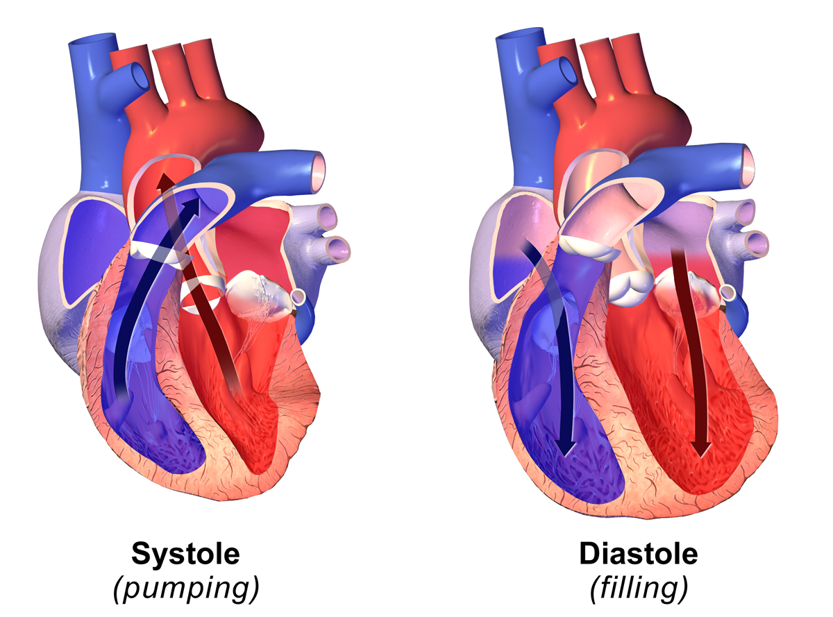
Diastole ( ) is the relaxed phase of the cardiac cycle when the chambers of the heart are refilling with blood. The contrasting phase is systole when the heart chambers are contracting. Atrial diastole is the relaxing of the atria, and ventricular diastole the relaxing of the ventricles. The term originates from the Greek word (''diastolē''), meaning "dilation", from (''diá'', "apart") + (''stéllein'', "to send"). Role in cardiac cycle A typical heart rate is 75 beats per minute (bpm), which means that the cardiac cycle that produces one heartbeat, lasts for less than one second. The cycle requires 0.3 sec in ventricular systole (contraction)—pumping blood to all body systems from the two ventricles; and 0.5 sec in diastole (dilation), re-filling the four chambers of the heart, for a total of 0.8 sec to complete the cycle. Early ventricular diastole During early ventricular diastole, pressure in the two ventricles begins to drop from the peak reached during systole ... [...More Info...] [...Related Items...] OR: [Wikipedia] [Google] [Baidu] |
Supine Position
The supine position () means lying horizontally, with the face and torso facing up, as opposed to the prone position, which is face down. When used in surgical procedures, it grants access to the peritoneal, thoracic, and pericardium, pericardial regions; as well as the head, neck, and extremities. Using anatomical terms of location, the dorsal side is down, and the ventral side is up, when supine. Semi-supine In scientific literature "semi-supine" commonly refers to positions where the upper body is tilted (at 45° or variations) and not completely horizontal. Relation to sudden infant death syndrome The decline in death due to sudden infant death syndrome (SIDS) is said to be attributable to having babies sleep in the supine position. The realization that infants sleeping face down, or in a prone position, had an increased mortality rate re-emerged into medical awareness at the end of the 1980s when two researchers, Susan Beal in Australia and Gus De Jonge in the Nether ... [...More Info...] [...Related Items...] OR: [Wikipedia] [Google] [Baidu] |
Jugular Venous Distension
The jugular venous pressure (JVP, sometimes referred to as ''jugular venous pulse'') is the indirectly observed pressure over the venous system via visualization of the internal jugular vein. It can be useful in the differentiation of different forms of heart and lung disease. Classically three upward deflections and two downward deflections have been described. * The upward deflections are the "a" (atrial contraction), "c" (ventricular contraction and resulting bulging of tricuspid into the right atrium during isovolumetric systole) and "v" (venous filling). * The downward deflections of the wave are the "x" descent (the atrium relaxes and the tricuspid valve moves downward) and the "y" descent (filling of ventricle after tricuspid opening). Method Visualization The patient is positioned at a 45° incline. The head is gently turned to the left, and the right external jugular vein should be identified which may be pulsatile and the filling level noted. If the external jugular ... [...More Info...] [...Related Items...] OR: [Wikipedia] [Google] [Baidu] |


