|
Anterior Tibial Veins
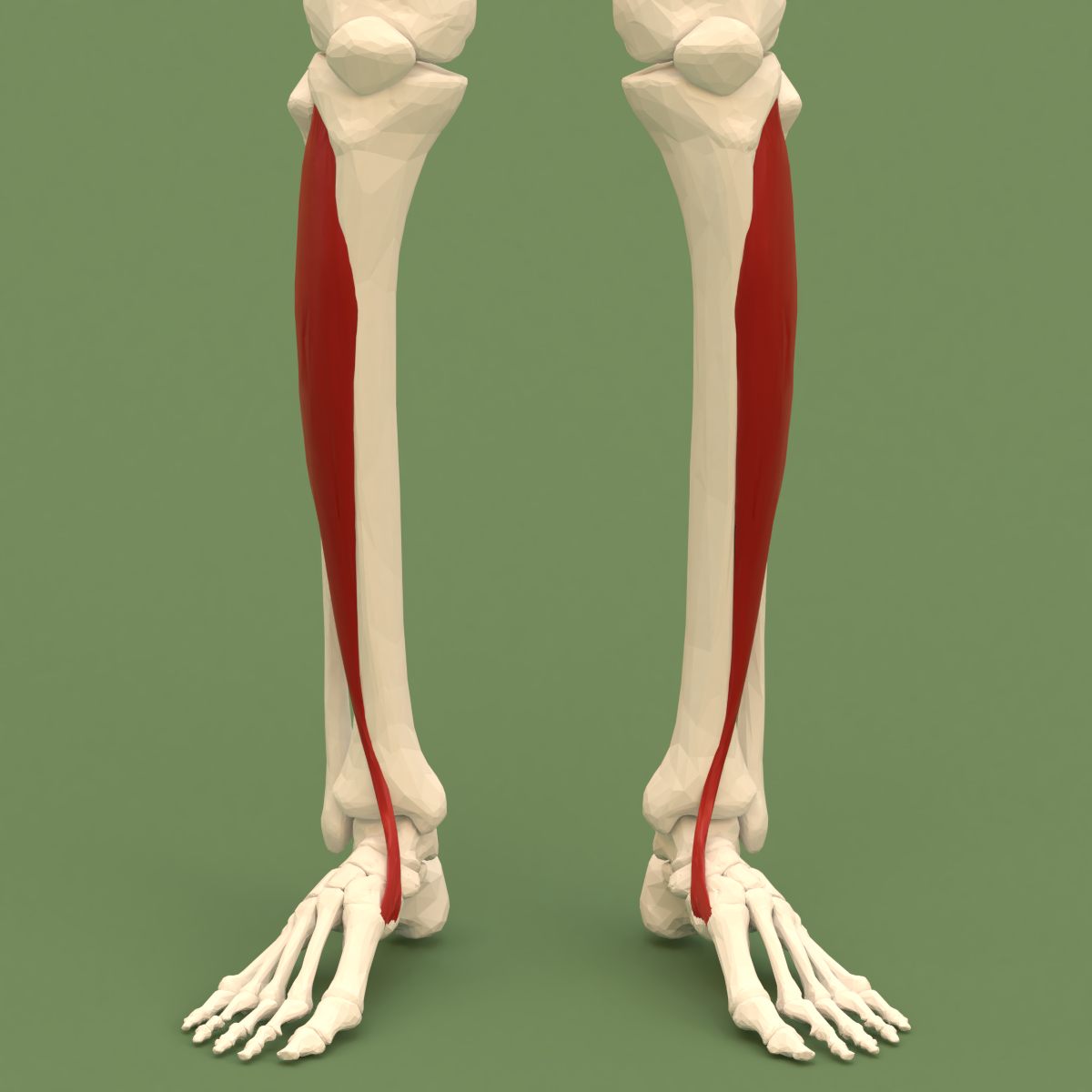
The anterior tibial vein is a vein in the lower leg. In human anatomy, there are two anterior tibial veins. They originate and receive blood from the dorsal venous arch, on the back of the foot and empties into the popliteal vein. The anterior tibial veins drain the ankle joint, knee joint, tibiofibular joint, and the anterior portion of the lower leg. The two anterior tibial veins ascend in the interosseous membrane between the tibia and fibula and unite with the posterior tibial veins to form the popliteal vein. Like most deep veins in legs, anterior tibial veins are accompanied by the homonym artery, the anterior tibial artery The anterior tibial artery is an artery of the leg. It carries blood to the anterior compartment of the leg and dorsum (biology), dorsal surface of the foot, from the popliteal artery. Structure Course The anterior tibial artery is a branch o ..., along its course. References Veins of the lower limb {{Circulatory-stub ... [...More Info...] [...Related Items...] OR: [Wikipedia] [Google] [Baidu] |
Popliteal Vein
The popliteal vein is a vein of the lower limb. It is formed from the anterior tibial vein and the posterior tibial vein. It travels medial to the popliteal artery, and becomes the femoral vein. It drains blood from the leg. It can be assessed using medical ultrasound. It can be affected by popliteal vein entrapment. Structure The popliteal vein is formed by the junction of the venae comitantes of the anterior tibial vein and the posterior tibial vein at the lower border of the popliteus muscle. It travels on the medial side of the popliteal artery. It is superficial to the popliteal artery. As it ascends through the fossa, it crosses behind the popliteal artery so that it comes to lie on its lateral side. It passes through the adductor hiatus (the opening in the adductor magnus muscle) to become the femoral vein.Moore K.L. and Dalley A.F. (2006), Clinically Oriented Anatomy, 5th Edition, Lippincott Williams & Wilkins, Toronto, page 636 Tributaries The tributaries of the p ... [...More Info...] [...Related Items...] OR: [Wikipedia] [Google] [Baidu] |
Tibiofibular Joint (other)
Tibiofibular joint may refer to: * Superior tibiofibular joint * Inferior tibiofibular joint The distal tibiofibular joint (tibiofibular syndesmosis) is formed by the rough, convex surface of the medial side of the distal end of the fibula, and a rough concave surface on the lateral side of the tibia. Below, to the extent of about 4& ... {{disambig ... [...More Info...] [...Related Items...] OR: [Wikipedia] [Google] [Baidu] |
Artery
An artery (plural arteries) () is a blood vessel in humans and most animals that takes blood away from the heart to one or more parts of the body (tissues, lungs, brain etc.). Most arteries carry oxygenated blood; the two exceptions are the pulmonary and the umbilical arteries, which carry deoxygenated blood to the organs that oxygenate it (lungs and placenta, respectively). The effective arterial blood volume is that extracellular fluid which fills the arterial system. The arteries are part of the circulatory system, that is responsible for the delivery of oxygen and nutrients to all cells, as well as the removal of carbon dioxide and waste products, the maintenance of optimum blood pH, and the circulation of proteins and cells of the immune system. Arteries contrast with veins, which carry blood back towards the heart. Structure The anatomy of arteries can be separated into gross anatomy, at the macroscopic level, and microanatomy, which must be studied with a microscop ... [...More Info...] [...Related Items...] OR: [Wikipedia] [Google] [Baidu] |
Deep Vein
A deep vein is a vein that is deep in the body. This contrasts with superficial veins that are close to the body's surface. Deep veins are almost always beside an artery with the same name (e.g. the femoral vein is beside the femoral artery). Collectively, they carry the vast majority of the blood. Occlusion of a deep vein can be life-threatening and is most often caused by thrombosis. Occlusion of a deep vein by thrombosis is called ''deep vein thrombosis''. Because of their location deep within the body, operation on these veins can be difficult. List *Internal jugular vein Upper limb *Brachial vein *Axillary vein *Subclavian vein Lower limb *Common femoral vein *Femoral vein *Profunda femoris vein *Popliteal vein *Peroneal vein *Anterior tibial vein *Posterior tibial vein The posterior tibial veins are veins of the leg in humans. They drain the posterior compartment of the leg and the plantar surface of the foot to the popliteal vein. Structure The posterior tibi ... [...More Info...] [...Related Items...] OR: [Wikipedia] [Google] [Baidu] |
Posterior Tibial Vein
The posterior tibial veins are veins of the leg in humans. They drain the posterior compartment of the leg and the plantar surface of the foot to the popliteal vein. Structure The posterior tibial veins receive blood from the medial and lateral plantar veins. They drain the posterior compartment of the leg and the plantar surface of the foot to the popliteal vein, which it forms when it joins with the anterior tibial vein. The posterior tibial vein is accompanied by an homonym artery, the posterior tibial artery, along its course. It lies posterior to the medial malleolus in the ankle. They receive the most important perforator vein Perforator veins are so called because they perforate the deep fascia of muscles, to connect the superficial veins to the deep veins where they drain. Perforator veins play an essential role in maintaining normal blood draining. They have valves ...s: the Cockett perforators, superior, medial and inferior. Additional images File:Gray440_col ... [...More Info...] [...Related Items...] OR: [Wikipedia] [Google] [Baidu] |
Fibula
The fibula or calf bone is a leg bone on the lateral side of the tibia, to which it is connected above and below. It is the smaller of the two bones and, in proportion to its length, the most slender of all the long bones. Its upper extremity is small, placed toward the back of the head of the tibia, below the knee joint and excluded from the formation of this joint. Its lower extremity inclines a little forward, so as to be on a plane anterior to that of the upper end; it projects below the tibia and forms the lateral part of the ankle joint. Structure The bone has the following components: * Lateral malleolus * Interosseous membrane connecting the fibula to the tibia, forming a syndesmosis joint * The superior tibiofibular articulation is an arthrodial joint between the lateral condyle of the tibia and the head of the fibula. * The inferior tibiofibular articulation (tibiofibular syndesmosis) is formed by the rough, convex surface of the medial side of the lower end of the f ... [...More Info...] [...Related Items...] OR: [Wikipedia] [Google] [Baidu] |
Tibia
The tibia (; ), also known as the shinbone or shankbone, is the larger, stronger, and anterior (frontal) of the two bones in the leg below the knee in vertebrates (the other being the fibula, behind and to the outside of the tibia); it connects the knee with the ankle. The tibia is found on the medial side of the leg next to the fibula and closer to the median plane. The tibia is connected to the fibula by the interosseous membrane of leg, forming a type of fibrous joint called a syndesmosis with very little movement. The tibia is named for the flute ''tibia''. It is the second largest bone in the human body, after the femur. The leg bones are the strongest long bones as they support the rest of the body. Structure In human anatomy, the tibia is the second largest bone next to the femur. As in other vertebrates the tibia is one of two bones in the lower leg, the other being the fibula, and is a component of the knee and ankle joints. The ossification or formation of the bone ... [...More Info...] [...Related Items...] OR: [Wikipedia] [Google] [Baidu] |
Interosseous Membrane Of Leg
The interosseous membrane of the leg (middle tibiofibular ligament) extends between the interosseous crests of the tibia and fibula, helps stabilize the Tib-Fib relationship and separates the muscles on the front from those on the back of the leg. It consists of a thin, aponeurotic joint lamina composed of oblique fibers, which for the most part run downward and lateralward; some few fibers, however, pass in the opposite direction. It is broader above than below. Its upper margin does not quite reach the tibiofibular joint, but presents a free concave border, above which is a large, oval aperture for the passage of the anterior tibial vessels to the front of the leg. In its lower part is an opening for the passage of the anterior peroneal vessels. It is continuous below with the interosseous ligament of the tibiofibular syndesmosis In anatomy, fibrous joints are joints connected by fibrous tissue, consisting mainly of collagen. These are fixed joints where bones are unit ... [...More Info...] [...Related Items...] OR: [Wikipedia] [Google] [Baidu] |
Anterior Compartment Of Leg
The anterior compartment of the leg is a fascial compartments of leg, fascial compartment of the lower leg. It contains muscles that produce Anatomical terms of motion#Flexion and extension of the foot, dorsiflexion and participate in Anatomical terms of motion#Inversion and eversion, inversion and eversion of the foot, as well as vascular and nervous elements, including the anterior tibial artery and anterior tibial vein, veins and the deep fibular nerve. Muscles The muscles of the compartment are: * tibialis anterior muscle, tibialis anterior * extensor hallucis longus muscle, extensor hallucis longus * extensor digitorum longus muscle, extensor digitorum longus * Fibularis tertius, fibularis (peroneus) tertius Function The compartment contains muscles that are Anatomical terms of motion#Flexion and extension of the foot, dorsiflexors and participate in Anatomical terms of motion#Inversion and eversion, inversion and eversion of the foot. Innervation and blood supply The an ... [...More Info...] [...Related Items...] OR: [Wikipedia] [Google] [Baidu] |
Knee Joint
In humans and other primates, the knee joins the thigh with the leg and consists of two joints: one between the femur and tibia (tibiofemoral joint), and one between the femur and patella (patellofemoral joint). It is the largest joint in the human body. The knee is a modified hinge joint, which permits flexion and extension as well as slight internal and external rotation. The knee is vulnerable to injury and to the development of osteoarthritis. It is often termed a ''compound joint'' having tibiofemoral and patellofemoral components. (The fibular collateral ligament is often considered with tibiofemoral components.) Structure The knee is a modified hinge joint, a type of synovial joint, which is composed of three functional compartments: the patellofemoral articulation, consisting of the patella, or "kneecap", and the patellar groove on the front of the femur through which it slides; and the medial and lateral tibiofemoral articulations linking the femur, or thigh bone, ... [...More Info...] [...Related Items...] OR: [Wikipedia] [Google] [Baidu] |
Anterior Tibial Artery
The anterior tibial artery is an artery of the leg. It carries blood to the anterior compartment of the leg and dorsum (biology), dorsal surface of the foot, from the popliteal artery. Structure Course The anterior tibial artery is a branch of the popliteal artery. It originates at the distal end of the popliteus muscle posterior to the tibia. The artery typically passes anterior to the popliteus muscle prior to passing between the tibia and fibula through an oval opening at the superior aspect of the interosseus membrane. The artery then descends between the tibialis anterior and extensor digitorum longus muscles. It is accompanied by the anterior tibial vein, and the deep peroneal nerve, along its course. It crosses the anterior aspect of the ankle joint, at which point it becomes the dorsalis pedis artery. Branches The branches of the anterior tibial artery are: *posterior tibial recurrent artery *anterior tibial recurrent artery *muscular branches *anterior medial malleo ... [...More Info...] [...Related Items...] OR: [Wikipedia] [Google] [Baidu] |
Ankle Joint
The ankle, or the talocrural region, or the jumping bone (informal) is the area where the foot and the leg meet. The ankle includes three joints: the ankle joint proper or talocrural joint, the subtalar joint, and the inferior tibiofibular joint. The movements produced at this joint are dorsiflexion and plantarflexion of the foot. In common usage, the term ankle refers exclusively to the ankle region. In medical terminology, "ankle" (without qualifiers) can refer broadly to the region or specifically to the talocrural joint. The main bones of the ankle region are the talus (in the foot), and the tibia and fibula (in the leg). The talocrural joint is a synovial hinge joint that connects the distal ends of the tibia and fibula in the lower limb with the proximal end of the talus. The articulation between the tibia and the talus bears more weight than that between the smaller fibula and the talus. Structure Region The ankle region is found at the junction of the leg and the f ... [...More Info...] [...Related Items...] OR: [Wikipedia] [Google] [Baidu] |


.jpg)