|
Amphiarthrosis
Amphiarthrosis is a type of continuous, slightly movable joint. Types In amphiarthroses, the contiguous bony surfaces can be: * A symphysis: connected by broad flattened disks of fibrocartilage, of a more or less complex structure, which adhere to the ends of each bone, as in the articulations between the bodies of the vertebrae or the inferior articulation of the two hip bones (aka the pubic symphysis). * An interosseous membrane An interosseous membrane is a thick dense fibrous sheet of connective tissue that spans the space between two bones, forming a type of syndesmosis joint. Interosseous membranes in the human body: * Interosseous membrane of forearm * Interosseous ... - the sheet of connective tissue joining neighboring bones (e.g. tibia and fibula).Principles of Anatomy & Physiology, 12th Edition, Tortora & Derrickson, Pub: Wiley & Sons References External links * Joints {{musculoskeletal-stub ... [...More Info...] [...Related Items...] OR: [Wikipedia] [Google] [Baidu] |
Joint
A joint or articulation (or articular surface) is the connection made between bones, ossicles, or other hard structures in the body which link an animal's skeletal system into a functional whole.Saladin, Ken. Anatomy & Physiology. 7th ed. McGraw-Hill Connect. Webp.274/ref> They are constructed to allow for different degrees and types of movement. Some joints, such as the knee, elbow, and shoulder, are self-lubricating, almost frictionless, and are able to withstand compression and maintain heavy loads while still executing smooth and precise movements. Other joints such as sutures between the bones of the skull permit very little movement (only during birth) in order to protect the brain and the sense organs. The connection between a tooth and the jawbone is also called a joint, and is described as a fibrous joint known as a gomphosis. Joints are classified both structurally and functionally. Classification The number of joints depends on if sesamoids are included, age of t ... [...More Info...] [...Related Items...] OR: [Wikipedia] [Google] [Baidu] |
Symphysis
A symphysis (, pl. symphyses) is a fibrocartilaginous fusion between two bones. It is a type of cartilaginous joint, specifically a secondary cartilaginous joint. # A symphysis is an amphiarthrosis, a slightly movable joint. # A growing together of parts or structures. Unlike synchondroses, symphyses are permanent. Examples The more prominent symphyses are: * the pubic symphysis * sacrococcygeal symphysis * intervertebral disc between two vertebrae * in the sternum, between the manubrium and body * mandibular symphysis In human anatomy, the facial skeleton of the skull the external surface of the mandible is marked in the median line by a faint ridge, indicating the mandibular symphysis (Latin: ''symphysis menti'') or line of junction where the two lateral halve ..., in the jaw Symphysis disorders Pubic symphysis diastasis Pubic symphysis diastasis, is an extremely rare complication that occurs in women who are giving birth. Separation of the two pubic bones during deliv ... [...More Info...] [...Related Items...] OR: [Wikipedia] [Google] [Baidu] |
Fibrocartilage
Fibrocartilage consists of a mixture of white fibrous tissue and cartilaginous tissue in various proportions. It owes its inflexibility and toughness to the former of these constituents, and its elasticity to the latter. It is the only type of cartilage that contains type I collagen in addition to the normal type II. Structure The extracellular matrix of fibrocartilage is mainly made from type I collagen secreted by chondroblasts. Locations of fibrocartilage in the human body * secondary cartilaginous joints: ** pubic symphysis ** annulus fibrosis of intervertebral discs ** manubriosternal joint * glenoid labrum of shoulder joint * acetabular labrum of hip joint * medial and lateral menisci of the knee joint * location where tendons and ligaments attach to bone * Ulnar Triangular Fibrocartilage complex (TFCC) Function Repair If hyaline cartilage Hyaline cartilage is the glass-like (hyaline) and translucent cartilage found on many joint surfaces. It is al ... [...More Info...] [...Related Items...] OR: [Wikipedia] [Google] [Baidu] |
Vertebrae
The spinal column, a defining synapomorphy shared by nearly all vertebrates, Hagfish are believed to have secondarily lost their spinal column is a moderately flexible series of vertebrae (singular vertebra), each constituting a characteristic irregular bone whose complex structure is composed primarily of bone, and secondarily of hyaline cartilage. They show variation in the proportion contributed by these two tissue types; such variations correlate on one hand with the cerebral/caudal rank (i.e., location within the backbone), and on the other with phylogenetic differences among the vertebrate taxa. The basic configuration of a vertebra varies, but the bone is its ''body'', with the central part of the body constituting the ''centrum''. The upper (closer to) and lower (further from), respectively, the cranium and its central nervous system surfaces of the vertebra body support attachment to the intervertebral discs. The posterior part of a vertebra forms a vertebral ar ... [...More Info...] [...Related Items...] OR: [Wikipedia] [Google] [Baidu] |
Hip Bone
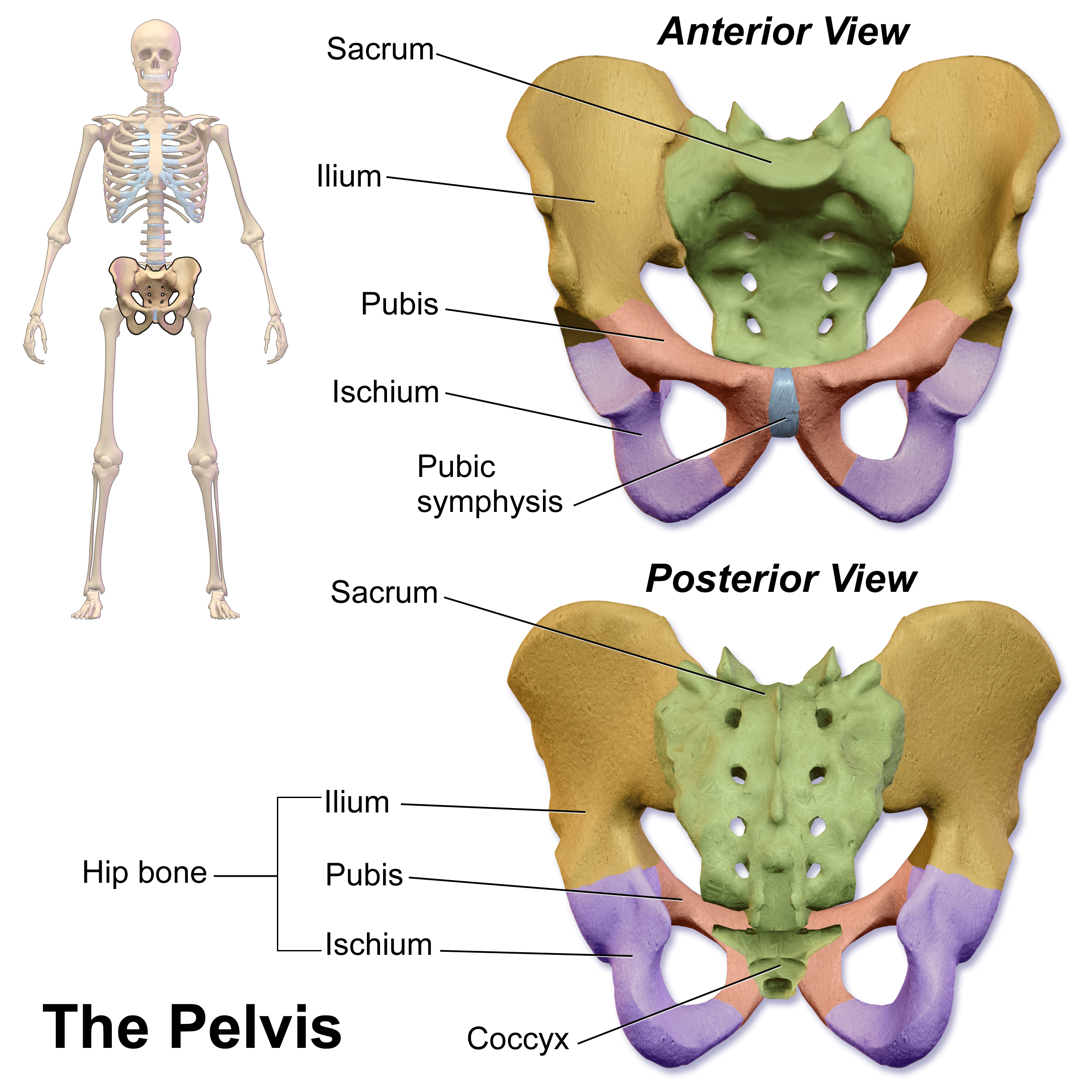
The hip bone (os coxae, innominate bone, pelvic bone or coxal bone) is a large flat bone, constricted in the center and expanded above and below. In some vertebrates (including humans before puberty) it is composed of three parts: the ilium, ischium, and the pubis. The two hip bones join at the pubic symphysis and together with the sacrum and coccyx (the pelvic part of the spine) comprise the skeletal component of the pelvis – the pelvic girdle which surrounds the pelvic cavity. They are connected to the sacrum, which is part of the axial skeleton, at the sacroiliac joint. Each hip bone is connected to the corresponding femur (thigh bone) (forming the primary connection between the bones of the lower limb and the axial skeleton) through the large ball and socket joint of the hip. Structure The hip bone is formed by three parts: the ilium, ischium, and pubis. At birth, these three components are separated by hyaline cartilage. They join each other in a Y-shaped porti ... [...More Info...] [...Related Items...] OR: [Wikipedia] [Google] [Baidu] |
Pubic Symphysis
The pubic symphysis is a secondary cartilaginous joint between the left and right superior rami of the pubis of the hip bones. It is in front of and below the urinary bladder. In males, the suspensory ligament of the penis attaches to the pubic symphysis. In females, the pubic symphysis is close to the clitoris. In most adults it can be moved roughly 2 mm and with 1 degree rotation. This increases for women at the time of childbirth. The name comes from the Greek word ''symphysis'', meaning 'growing together'. Structure The pubic symphysis is a nonsynovial amphiarthrodial joint. The width of the pubic symphysis at the front is 3–5 mm greater than its width at the back. This joint is connected by fibrocartilage and may contain a fluid-filled cavity; the center is avascular, possibly due to the nature of the compressive forces passing through this joint, which may lead to harmful vascular disease. The ends of both pubic bones are covered by a thin layer of hyalin ... [...More Info...] [...Related Items...] OR: [Wikipedia] [Google] [Baidu] |
Interosseous Membrane
An interosseous membrane is a thick dense fibrous sheet of connective tissue that spans the space between two bones, forming a type of syndesmosis joint. Interosseous membranes in the human body: * Interosseous membrane of forearm * Interosseous membrane of leg The interosseous membrane of the leg (middle tibiofibular ligament) extends between the interosseous crests of the tibia and fibula, helps stabilize the Tib-Fib relationship and separates the muscles on the front from those on the back of the leg. ... Gallery File:5 ligaments of interosseous membrane of forearm.png, Five ligaments of interosseous membrane of forearm:* Central band (key portion to be reconstructed in case of injury)* Accessory band * Distal oblique bundle * Proximal oblique cord* Dorsal oblique accessory cord Notes External links * * {{Authority control Skeletal system ... [...More Info...] [...Related Items...] OR: [Wikipedia] [Google] [Baidu] |
Connective Tissue
Connective tissue is one of the four primary types of animal tissue, along with epithelial tissue, muscle tissue, and nervous tissue. It develops from the mesenchyme derived from the mesoderm the middle embryonic germ layer. Connective tissue is found in between other tissues everywhere in the body, including the nervous system. The three meninges, membranes that envelop the brain and spinal cord are composed of connective tissue. Most types of connective tissue consists of three main components: elastic and collagen fibers, ground substance, and cells. Blood, and lymph are classed as specialized fluid connective tissues that do not contain fiber. All are immersed in the body water. The cells of connective tissue include fibroblasts, adipocytes, macrophages, mast cells and leucocytes. The term "connective tissue" (in German, ''Bindegewebe'') was introduced in 1830 by Johannes Peter Müller. The tissue was already recognized as a distinct class in the 18th century. ... [...More Info...] [...Related Items...] OR: [Wikipedia] [Google] [Baidu] |


