|
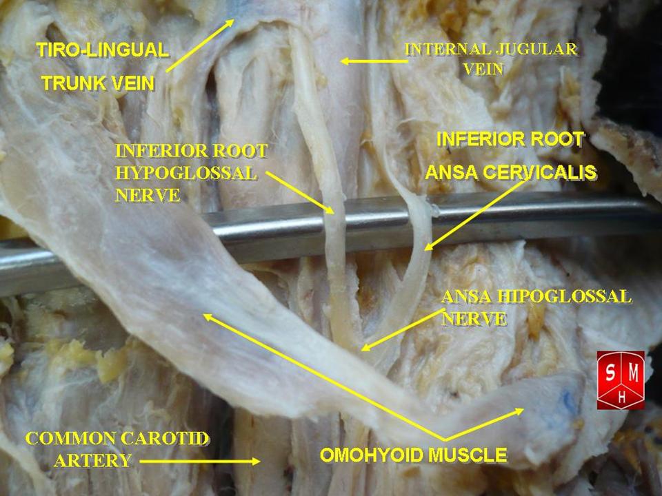
Ansa Cervicalis
The ansa cervicalis (or ansa hypoglossi in older literature) is a loop formed by muscular branches of the cervical plexus formed by branches of cervical spinal nerves C1-C3. The ansa cervicalis has two roots - a superior root (formed by branch of C1) and an inferior root (formed by union of branches of C2 and C3) - that unite distally, forming a loop. It is situated anterior to the carotid sheath. Branches of the ansa cervicalis innervate three of the four infrahyoid muscles: the sternothyroid, sternohyoid, and omohyoid muscles (note that the thyrohyoid muscle is the one infrahyoid muscle not innervated by the ansa cervicalis - it is instead innervated by cervical spinal nerve 1 via a separate thyrohyoid branch). Its name means "handle of the neck" in Latin. Anatomy The ansa cervicalis is typically embedded within the anterior wall of the carotid sheath anterior to the internal jugular vein. Superior root The superior root of the ansa cervicalis (formerly known as d ... [...More Info...] [...Related Items...] OR: [Wikipedia] [Google] [Baidu] |
Sternohyoid Muscle
The sternohyoid muscle is a bilaterally paired, long, thin, narrow strap muscle of the anterior neck. It is one of the infrahyoid muscles. It is innervated by the ansa cervicalis. It acts to depress the hyoid bone. The sternohyoid muscle is a flat muscle located on both sides of the neck, part of the infrahyoid muscle group. It originates from the medial edge of the clavicle, sternoclavicular ligament, and posterior side of the manubrium, and ascends to attach to the body of the hyoid bone. The sternohyoid muscle, along with other infrahyoid muscles, functions to depress the hyoid bone, which is important for activities such as speaking, chewing, and swallowing. Additionally, this muscle group contributes to the protection of the trachea, esophagus, blood vessels, and thyroid gland. The sternohyoid muscle also plays a minor role in head movements. Structure The sternohyoid muscle is one of the paired strap muscles of the infrahyoid muscles. The muscle is directed superom ... [...More Info...] [...Related Items...] OR: [Wikipedia] [Google] [Baidu] |
Cervical Spinal Nerve 2
The cervical spinal nerve 2 (C2) is a spinal nerve of the cervical segment. Nervous System -- Groups of Nerves It is a part of the ansa cervicalis along with the C1 and C3 nerves sometimes forming part of superior root of the ansa cervicalis. it also connects into the inferior root of the ansa cervicalis with the C3. It originates from the spinal column from above the [...More Info...] [...Related Items...] OR: [Wikipedia] [Google] [Baidu] |
Cervical Spinal Nerve 3
The cervical spinal nerve 3 (C3) is a spinal nerve of the cervical segment The spinal cord is a long, thin, tubular structure made up of nervous tissue that extends from the medulla oblongata in the lower brainstem to the lumbar region of the vertebral column (backbone) of vertebrate animals. The center of the spinal c .... Nervous System -- Groups of Nerves It originates from the spinal column from above the cervical vertebra 3 (C3). References Spinal nerves {{neuroanatomy-stub ...[...More Info...] [...Related Items...] OR: [Wikipedia] [Google] [Baidu] |
Cervical Spinal Nerve 2
The cervical spinal nerve 2 (C2) is a spinal nerve of the cervical segment. Nervous System -- Groups of Nerves It is a part of the ansa cervicalis along with the C1 and C3 nerves sometimes forming part of superior root of the ansa cervicalis. it also connects into the inferior root of the ansa cervicalis with the C3. It originates from the spinal column from above the [...More Info...] [...Related Items...] OR: [Wikipedia] [Google] [Baidu] |
Spinal Nerve
A spinal nerve is a mixed nerve, which carries Motor neuron, motor, Sensory neuron, sensory, and Autonomic nervous system, autonomic signals between the spinal cord and the body. In the human body there are 31 pairs of spinal nerves, one on each side of the vertebral column. These are grouped into the corresponding cervical vertebrae, cervical, thoracic vertebrae, thoracic, lumbar vertebrae, lumbar, sacral vertebrae, sacral and coccygeal vertebrae, coccygeal regions of the spine. There are eight pairs of cervical nerves, twelve pairs of thoracic nerves, five pairs of lumbar nerves, five pairs of sacral nerves, and one pair of coccygeal nerves. The spinal nerves are part of the peripheral nervous system. Structure Each spinal nerve is a mixed nerve, formed from the combination of nerve root axon, fibers from its Dorsal root of spinal nerve, dorsal and Ventral root of spinal nerve, ventral roots. The dorsal root is the afferent nerve fiber, afferent sensory root and carries sen ... [...More Info...] [...Related Items...] OR: [Wikipedia] [Google] [Baidu] |
Anterior Rami
The ventral ramus (: rami) (Latin for 'branch') is the anterior division of a spinal nerve. The ventral rami supply the antero-lateral parts of the trunk and the limbs. They are mainly larger than the dorsal rami. Shortly after a spinal nerve exits the intervertebral foramen, it branches into the dorsal ramus, the ventral ramus, and the ramus communicans. Each of these three structures carries both sensory and motor information. Each spinal nerve is a mixed nerve that carries both sensory and motor information, via efferent and afferent nerve fibers—ultimately via the motor cortex in the frontal lobe and to somatosensory cortex in the parietal lobe—but also through the phenomenon of reflex. In the thoracic region they remain distinct from each other and each innervates a narrow strip of muscle and skin along the sides, chest, ribs, and abdominal wall. These rami are called the intercostal nerves. In regions other than the thoracic, ventral rami converge with each oth ... [...More Info...] [...Related Items...] OR: [Wikipedia] [Google] [Baidu] |
Sternohyoid
The sternohyoid muscle is a bilaterally paired, long, thin, narrow strap muscle of the anterior neck. It is one of the infrahyoid muscles. It is innervated by the ansa cervicalis. It acts to depress the hyoid bone. The sternohyoid muscle is a flat muscle located on both sides of the neck, part of the infrahyoid muscle group. It originates from the medial edge of the clavicle, sternoclavicular ligament, and posterior side of the manubrium, and ascends to attach to the body of the hyoid bone. The sternohyoid muscle, along with other infrahyoid muscles, functions to depress the hyoid bone, which is important for activities such as speaking, chewing, and swallowing. Additionally, this muscle group contributes to the protection of the trachea, esophagus, blood vessels, and thyroid gland. The sternohyoid muscle also plays a minor role in head movements. Structure The sternohyoid muscle is one of the paired strap muscles of the infrahyoid muscles. The muscle is directed superome ... [...More Info...] [...Related Items...] OR: [Wikipedia] [Google] [Baidu] |
Sternothyroid
The sternothyroid muscle (or sternothyroideus) is an infrahyoid muscle of the neck. It acts to depress the hyoid bone. Structure The two muscles are in contact with each other proximally (close to their origin), but diverge distally (towards their insertions). Origin The sternothyroid arises from the posterior surface of the manubrium of the sternum from the midline to the notch for the first rib (inferior to the origin of the sternohyoid muscle), and the posterior margin of the first costal cartilage. Insertion It inserts onto the oblique line of the lamina of thyroid cartilage. Innervation The sternothyroid muscle receives motor innervation from branches of the ansa cervicalis (ultimately derived from cervical spinal nerves C1-C3). Relations The sternothyroid muscle is shorter and wider than the sternohyoid muscle and is situated deep to and partially medial to it. Variations The muscle may be absent or doubled. It may issue accessory slips to the thyrohyoid mus ... [...More Info...] [...Related Items...] OR: [Wikipedia] [Google] [Baidu] |
Occipital Artery
The occipital artery is a branch of the external carotid artery that provides arterial supply to the back of the scalp, sternocleidomastoid muscles, and deep muscles of the back and neck. Structure Origin The occipital artery arises from (the posterior aspect of) the external carotid artery (some 2 cm distal to the origin of the external carotid artery). Course and relations At its origin, the hypoglossal nerve (CN XII) crosses artery superficially as the nerve passes posteroanteriorly. The artery passes superoposteriorly deep to the posterior belly of the digastricus muscle. It crosses the internal carotid artery and vein, the vagus nerve (CN X), accessory nerve (CN XI), and hypoglossal nerve (CN XII). It next ascends to the interval between the transverse process of the atlas and the mastoid process of the temporal bone, and passes horizontally backward, grooving the surface of the latter bone, being covered by the sternocleidomastoideus, splenius capitis, longi ... [...More Info...] [...Related Items...] OR: [Wikipedia] [Google] [Baidu] |
Common Carotid Artery
In anatomy, the left and right common carotid arteries (carotids) () are artery, arteries that supply the head and neck with oxygenated blood; they divide in the neck to form the external carotid artery, external and internal carotid artery, internal carotid arteries. Structure The common carotid arteries are present on the left and right sides of the body. These arteries originate from different arteries but follow symmetrical courses. The right common carotid originates in the neck from the brachiocephalic trunk; the left from the aortic arch in the thorax. These split into the external and internal carotid arteries at the upper border of the thyroid cartilage, at around the level of the fourth cervical vertebra. The left common carotid artery can be thought of as having two parts: a thoracic (chest) part and a cervical (neck) part. The right common carotid originates in or close to the neck and contains only a small thoracic portion. There are studies in the bioengineering l ... [...More Info...] [...Related Items...] OR: [Wikipedia] [Google] [Baidu] |
Internal Carotid Artery
The internal carotid artery is an artery in the neck which supplies the anterior cerebral artery, anterior and middle cerebral artery, middle cerebral circulation. In human anatomy, the internal and external carotid artery, external carotid arise from the common carotid artery, where it bifurcates at cervical vertebrae C3 or C4. The internal carotid artery supplies the brain, including the eyes, while the external carotid nourishes other portions of the head, such as the face, scalp, skull, and meninges. Classification Terminologia Anatomica in 1998 subdivided the artery into four parts: "cervical", "petrous", "cavernous", and "cerebral". In clinical settings, however, usually the classification system of the internal carotid artery follows the 1996 recommendations by Bouthillier, describing seven anatomical segments of the internal carotid artery, each with a corresponding alphanumeric identifier: C1 cervical; C2 petrous; C3 lacerum; C4 cavernous; C5 clinoid; C6 ophthalmic; ... [...More Info...] [...Related Items...] OR: [Wikipedia] [Google] [Baidu] |
Carotid Triangle
The carotid triangle (or superior carotid triangle) is a portion of the anterior triangle of the neck. Anatomy Boundaries It is bounded: * Posteriorly by (the anterior border of) the sternocleidomastoid muscle, * Anteroinferiorly by (the superior belly of) the omohyoid muscle. * Superiorly by (the posterior belly of) the digastric muscle. Roof The roof is formed by: * Integument, * Superficial fascia, * Platysma, * Deep fascia. Floor The floor is formed by (parts of) the: * Thyrohyoid membrane, *Hyoglossus, * Constrictor pharyngis medius and constrictor pharyngis inferior muscles. Contents Arteries * Internal carotid artery * External carotid artery and some of its branches: ** Superior thyroid artery, ** Ascending pharyngeal artery, ** Lingual artery, ** Facial artery, ** Occipital artery. Veins * internal jugular vein and its tributaries (correspondng to the branches of the corresponding artery): ** Superior thyroid vein, ** Lingual veins, ** Comm ... [...More Info...] [...Related Items...] OR: [Wikipedia] [Google] [Baidu] |


