|
Anomic Aphasia
Anomic aphasia, also known as dysnomia, nominal aphasia, and amnesic aphasia, is a mild, fluent type of aphasia where individuals have word retrieval failures and cannot express the words they want to say (particularly nouns and verbs). By contrast, ''anomia'' is a deficit of expressive language, and a symptom of all forms of aphasia, but patients whose primary deficit is word retrieval are diagnosed with anomic aphasia. Individuals with aphasia who display anomia can often describe an object in detail and maybe even use hand gestures to demonstrate how the object is used, but cannot find the appropriate word to name the object. Patients with anomic aphasia have relatively preserved speech fluency, repetition, comprehension, and grammatical speech. Types * Word selection anomia is caused by damage to the posterior inferior temporal area. This type of anomia occurs when the patient knows how to use an object and can correctly select the target object from a group of objects ... [...More Info...] [...Related Items...] OR: [Wikipedia] [Google] [Baidu] |
Diffusion Tensor Imaging
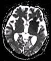
Diffusion-weighted magnetic resonance imaging (DWI or DW-MRI) is the use of specific MRI sequences as well as software that generates images from the resulting data that uses the diffusion of water molecules to generate contrast in MR images. It allows the mapping of the diffusion process of molecules, mainly water, in biological tissues, in vivo and non-invasively. Molecular diffusion in tissues is not random, but reflects interactions with many obstacles, such as macromolecules, fibers, and membranes. Water molecule diffusion patterns can therefore reveal microscopic details about tissue architecture, either normal or in a diseased state. A special kind of DWI, diffusion tensor imaging (DTI), has been used extensively to map white matter tractography in the brain. Introduction In diffusion weighted imaging (DWI), the intensity of each image element (voxel) reflects the best estimate of the rate of water diffusion at that location. Because the mobility of water is driven by ... [...More Info...] [...Related Items...] OR: [Wikipedia] [Google] [Baidu] |
Stroke
Stroke is a medical condition in which poor cerebral circulation, blood flow to a part of the brain causes cell death. There are two main types of stroke: brain ischemia, ischemic, due to lack of blood flow, and intracranial hemorrhage, hemorrhagic, due to bleeding. Both cause parts of the brain to stop functioning properly. Signs and symptoms of stroke may include an hemiplegia, inability to move or feel on one side of the body, receptive aphasia, problems understanding or expressive aphasia, speaking, dizziness, or homonymous hemianopsia, loss of vision to one side. Signs and symptoms often appear soon after the stroke has occurred. If symptoms last less than 24 hours, the stroke is a transient ischemic attack (TIA), also called a mini-stroke. subarachnoid hemorrhage, Hemorrhagic stroke may also be associated with a thunderclap headache, severe headache. The symptoms of stroke can be permanent. Long-term complications may include pneumonia and Urinary incontinence, loss of b ... [...More Info...] [...Related Items...] OR: [Wikipedia] [Google] [Baidu] |
Progressive Aphasia
In neurology, primary progressive aphasia (PPA) is a type of neurological syndrome in which language capabilities slowly and progressively become impaired. As with other types of aphasia, the symptoms that accompany PPA depend on what parts of the brain's left hemisphere are significantly damaged. However, unlike most other aphasias, PPA results from continuous deterioration in brain tissue, which leads to early symptoms being far less detrimental than later symptoms. Those with PPA slowly lose the ability to speak, write, read, and generally comprehend language. Eventually, almost every patient becomes mute and completely loses the ability to understand both written and spoken language. Although it was first described as solely impairment of language capabilities while other mental functions remain intact, it is now recognized that many, if not most of those with PPA experience impairment of memory, short-term memory formation and loss of executive functions. It was firs ... [...More Info...] [...Related Items...] OR: [Wikipedia] [Google] [Baidu] |
Dementia
Dementia is a syndrome associated with many neurodegenerative diseases, characterized by a general decline in cognitive abilities that affects a person's ability to perform activities of daily living, everyday activities. This typically involves problems with memory, thinking, behavior, and motor control. Aside from memory impairment and a thought disorder, disruption in thought patterns, the most common symptoms of dementia include emotional problems, difficulties with language, and decreased motivation. The symptoms may be described as occurring in a continuum (measurement), continuum over several stages. Dementia is a life-limiting condition, having a significant effect on the individual, their caregivers, and their social relationships in general. A diagnosis of dementia requires the observation of a change from a person's usual mental functioning and a greater cognitive decline than might be caused by the normal aging process. Several diseases and injuries to the brain, ... [...More Info...] [...Related Items...] OR: [Wikipedia] [Google] [Baidu] |
Posterior Cerebral Artery Syndrome
Posterior cerebral artery syndrome is a condition whereby the blood supply from the posterior cerebral artery (PCA) is restricted, leading to a reduction of the function of the portions of the brain supplied by that vessel: the occipital lobe, the inferomedial temporal lobe, a large portion of the thalamus, and the upper brainstem and midbrain. This event restricts the flow of blood to the brain in a near-immediate fashion. The blood hammer is analogous to the water hammer in hydrology, and consists of a sudden increase of the upstream blood pressure in a blood vessel when the bloodstream is abruptly blocked by vessel obstruction. Complete understanding of the relationship between mechanical parameters in vascular occlusions is a critical issue, which can play an important role in the future diagnosis, understanding and treatment of vascular diseases. Depending upon the location and severity of the occlusion, signs and symptoms may vary within the population affected with PCA sy ... [...More Info...] [...Related Items...] OR: [Wikipedia] [Google] [Baidu] |
Arcuate Fasciculus
In neuroanatomy, the arcuate fasciculus (AF; ) is a bundle of axons that generally connects Broca's area and Wernicke's area in the brain. It is an association fiber tract connecting caudal temporal lobe and inferior frontal lobe. Structure The arcuate fasciculus is a white matter tract that runs parallel to the superior longitudinal fasciculus. Due to their proximity, they are sometimes referred to interchangeably. They can be distinguished by the location and function of their endpoints in the frontal cortex. The arcuate fasciculus terminates in Broca's area (specifically BA 44) which is linked to processing complex syntax. However, the superior longitudinal fasciculus ends in the premotor cortex which is implicated in acoustic-motor mapping. Connection Historically, the arcuate fasciculus has been understood to connect two important areas for language use: Broca's area in the inferior frontal gyrus and Wernicke's area in the posterior superior temporal gyrus. It is mo ... [...More Info...] [...Related Items...] OR: [Wikipedia] [Google] [Baidu] |
White Matter
White matter refers to areas of the central nervous system that are mainly made up of myelinated axons, also called Nerve tract, tracts. Long thought to be passive tissue, white matter affects learning and brain functions, modulating the distribution of action potentials, acting as a relay and coordinating communication between different brain regions. White matter is named for its relatively light appearance resulting from the lipid content of myelin. Its white color in prepared specimens is due to its usual preservation in formaldehyde. It appears pinkish-white to the naked eye otherwise, because myelin is composed largely of lipid tissue veined with capillaries. Structure White matter White matter is composed of bundles, which connect various grey matter areas (the locations of nerve cell bodies) of the brain to each other, and carry nerve impulses between neurons. Myelin acts as an insulator, which allows saltatory conduction, electrical signals to jump, rather than cour ... [...More Info...] [...Related Items...] OR: [Wikipedia] [Google] [Baidu] |
Grey Matter
Grey matter, or gray matter in American English, is a major component of the central nervous system, consisting of neuronal cell bodies, neuropil ( dendrites and unmyelinated axons), glial cells ( astrocytes and oligodendrocytes), synapses, and capillaries. Grey matter is distinguished from white matter in that it contains numerous cell bodies and relatively few myelinated axons, while white matter contains relatively few cell bodies and is composed chiefly of long-range myelinated axons. The colour difference arises mainly from the whiteness of myelin. In living tissue, grey matter actually has a very light grey colour with yellowish or pinkish hues, which come from capillary blood vessels and neuronal cell bodies. Structure Grey matter refers to unmyelinated neurons and other cells of the central nervous system. It is present in the brain, brainstem and cerebellum, and present throughout the spinal cord. Grey matter is distributed at the surface of the cerebral hemisp ... [...More Info...] [...Related Items...] OR: [Wikipedia] [Google] [Baidu] |
Lesion
A lesion is any damage or abnormal change in the tissue of an organism, usually caused by injury or diseases. The term ''Lesion'' is derived from the Latin meaning "injury". Lesions may occur in both plants and animals. Types There is no designated classification or naming convention for lesions. Because lesions can occur anywhere in the body and their definition is so broad, the varieties of lesions are virtually endless. Generally, lesions may be classified by their patterns, sizes, locations, or causes. They can also be named after the person who discovered them. For example, Ghon lesions, which are found in the lungs of those with tuberculosis, are named after the lesion's discoverer, Anton Ghon. The characteristic skin lesions of a varicella zoster virus infection are called '' chickenpox''. Lesions of the teeth are usually called dental caries, or "cavities". Location Lesions are often classified by their tissue types or locations. For example, "skin lesions" or ... [...More Info...] [...Related Items...] OR: [Wikipedia] [Google] [Baidu] |
Wernicke's Area
Wernicke's area (; ), also called Wernicke's speech area, is one of the two parts of the cerebral cortex that are linked to speech, the other being Broca's area. It is involved in the comprehension of written and spoken language, in contrast to Broca's area, which is primarily involved in the production of language. It is traditionally thought to reside in Brodmann area 22, located in the superior temporal gyrus in the dominant cerebral hemisphere, which is the left hemisphere in about 95% of right-handed individuals and 70% of left-handed individuals. Damage caused to Wernicke's area results in receptive, fluent aphasia. This means that the person with aphasia will be able to fluently connect words, but the phrases will lack meaning. This is unlike non-fluent aphasia, in which the person will use meaningful words, but in a non-fluent, telegraphic manner. Emerging research on the developmental trajectory of Wernicke's area highlights its evolving role in language acquisiti ... [...More Info...] [...Related Items...] OR: [Wikipedia] [Google] [Baidu] |
Functional Magnetic Resonance Imaging
Functional magnetic resonance imaging or functional MRI (fMRI) measures brain activity by detecting changes associated with blood flow. This technique relies on the fact that cerebral blood flow and neuronal activation are coupled. When an area of the brain is in use, blood flow to that region also increases. The primary form of fMRI uses the blood-oxygen-level dependent (BOLD) contrast, discovered by Seiji Ogawa in 1990. This is a type of specialized brain and body scan used to map neuron, neural activity in the brain or spinal cord of humans or other animals by imaging the change in blood flow (hemodynamic response) related to energy use by brain cells. Since the early 1990s, fMRI has come to dominate brain mapping research because it does not involve the use of injections, surgery, the ingestion of substances, or exposure to ionizing radiation. This measure is frequently corrupted by noise from various sources; hence, statistical procedures are used to extract the underlying si ... [...More Info...] [...Related Items...] OR: [Wikipedia] [Google] [Baidu] |
Broca's Area
Broca's area, or the Broca area (, also , ), is a region in the frontal lobe of the dominant Cerebral hemisphere, hemisphere, usually the left, of the Human brain, brain with functions linked to speech production. Language processing in the brain, Language processing has been linked to Broca's area since Pierre Paul Broca reported impairments in two patients. They had lost the ability to speak after injury to the posterior inferior frontal gyrus (pars triangularis) (BA45) of the brain. Since then, the approximate region he identified has become known as Broca's area, and the deficit in language production as Broca's aphasia, also called expressive aphasia. Broca's area is now typically defined in terms of the pars opercularis and pars triangularis of the inferior frontal gyrus, represented in Korbinian Brodmann, Brodmann's Brodmann area, cytoarchitectonic map as Brodmann area 44 and Brodmann area 45 of the dominant hemisphere. Functional magnetic resonance imaging (fMRI) has show ... [...More Info...] [...Related Items...] OR: [Wikipedia] [Google] [Baidu] |


