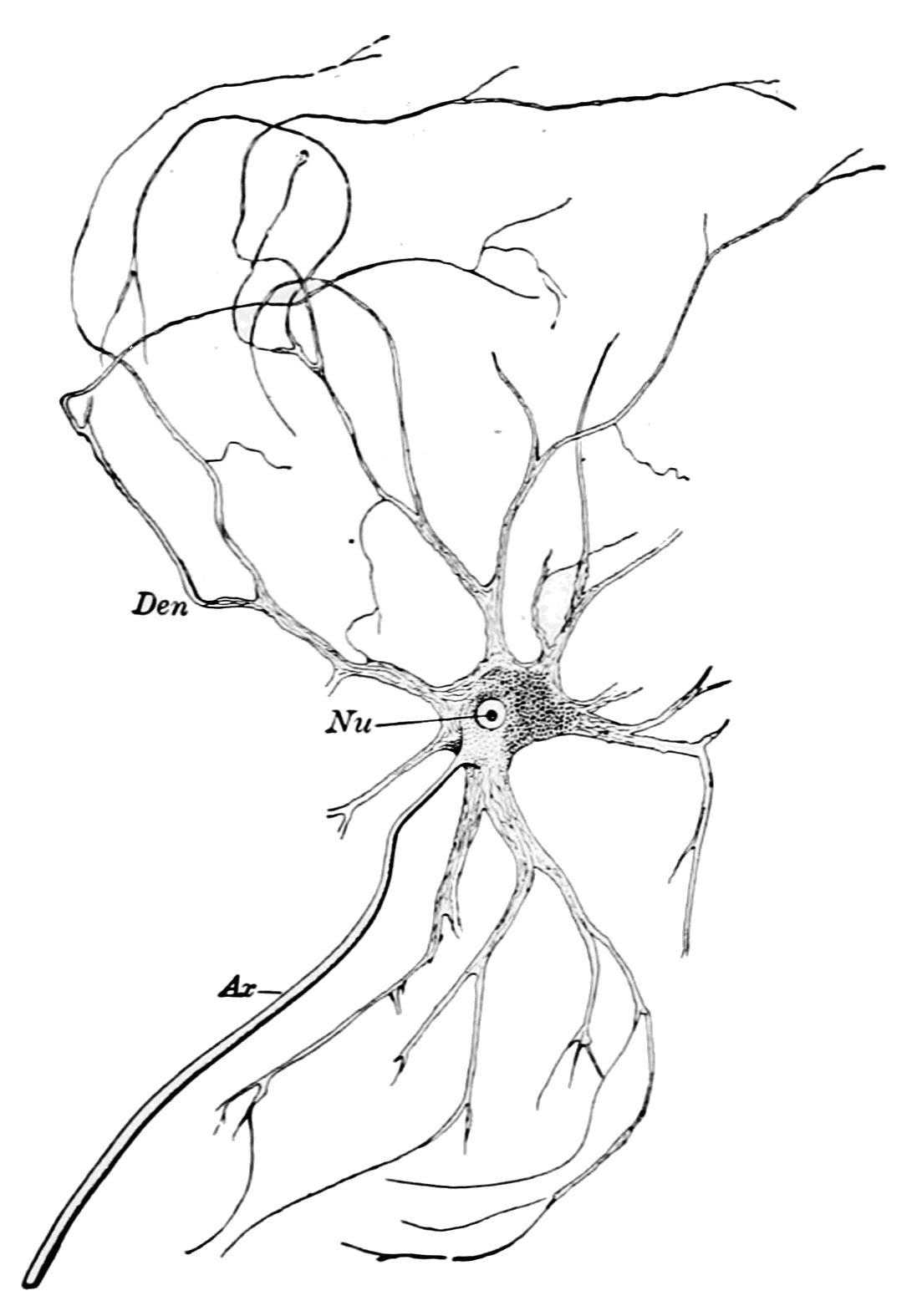
Motor nerve on:
[Wikipedia]
[Google]
[Amazon]
 A motor nerve is a

 A motor nerve is a
 nerve
A nerve is an enclosed, cable-like bundle of nerve fibers (called axons) in the peripheral nervous system.
A nerve transmits electrical impulses. It is the basic unit of the peripheral nervous system. A nerve provides a common pathway for the e ...
that transmits motor signals from the central nervous system
The central nervous system (CNS) is the part of the nervous system consisting primarily of the brain and spinal cord. The CNS is so named because the brain integrates the received information and coordinates and influences the activity of all par ...
(CNS) to the muscles of the body. This is different from the motor neuron
A motor neuron (or motoneuron or efferent neuron) is a neuron whose cell body is located in the motor cortex, brainstem or the spinal cord, and whose axon (fiber) projects to the spinal cord or outside of the spinal cord to directly or indirectl ...
, which includes a cell body and branching of dendrites, while the nerve is made up of a bundle of axons. Motor nerves act as efferent nerves
Efferent nerve fibers refer to axonal projections that ''exit'' a particular region; as opposed to afferent projections that ''arrive'' at the region. These terms have a slightly different meaning in the context of the peripheral nervous syste ...
which carry information out from the CNS to muscles, as opposed to afferent nerve
A sensory nerve, or afferent nerve, is a general anatomic term for a nerve which contains predominantly somatic afferent nerve fibers. Afferent nerve fibers in a sensory nerve carry sensory information toward the central nervous system (CNS) fr ...
s (also called sensory nerve
A sensory nerve, or afferent nerve, is a general anatomic term for a nerve which contains predominantly somatic afferent nerve fibers. Afferent nerve fibers in a sensory nerve carry sensory information toward the central nervous system (CNS) fr ...
s), which transfer signals from sensory receptors in the periphery to the CNS. Efferent nerves can also connect to gland
In animals, a gland is a group of cells in an animal's body that synthesizes substances (such as hormones) for release into the bloodstream (endocrine gland) or into cavities inside the body or its outer surface (exocrine gland).
Structure
De ...
s or other organs/issues instead of muscles (and so motor nerves are not equivalent to efferent nerves). In addition, there are nerves that serve as both sensory and motor nerves called mixed nerves.
Structure and function
Motor nerve fibers transduce signals from the CNS to peripheral neurons of proximal muscle tissue. Motor nerve axon terminals innervateskeletal
A skeleton is the structural frame that supports the body of an animal. There are several types of skeletons, including the exoskeleton, which is the stable outer shell of an organism, the endoskeleton, which forms the support structure inside ...
and smooth muscle
Smooth muscle is an involuntary non-striated muscle, so-called because it has no sarcomeres and therefore no striations (''bands'' or ''stripes''). It is divided into two subgroups, single-unit and multiunit smooth muscle. Within single-unit mus ...
, as they are heavily involved in muscle
Skeletal muscles (commonly referred to as muscles) are organs of the vertebrate muscular system and typically are attached by tendons to bones of a skeleton. The muscle cells of skeletal muscles are much longer than in the other types of muscl ...
control. Motor nerves tend to be rich in acetylcholine
Acetylcholine (ACh) is an organic chemical that functions in the brain and body of many types of animals (including humans) as a neurotransmitter. Its name is derived from its chemical structure: it is an ester of acetic acid and choline. Part ...
vesicles because the motor nerve, a bundle of motor nerve axons that deliver motor signals and signal for movement and motor control. Calcium vesicles
Vesicle may refer to:
; In cellular biology or chemistry
* Vesicle (biology and chemistry), a supramolecular assembly of lipid molecules, like a cell membrane
* Synaptic vesicle
; In human embryology
* Vesicle (embryology), bulge-like features o ...
reside in the axon terminal
Axon terminals (also called synaptic boutons, terminal boutons, or end-feet) are distal terminations of the telodendria (branches) of an axon. An axon, also called a nerve fiber, is a long, slender projection of a nerve cell, or neuron, that condu ...
s of the motor nerve bundles. The high calcium concentration outside of presynaptic motor nerves increases the size of end-plate potential
End plate potentials (EPPs) are the voltages which cause depolarization of skeletal muscle fibers caused by neurotransmitters binding to the postsynaptic membrane in the neuromuscular junction. They are called "end plates" because the postsynapt ...
s (EPPs).
Protective tissues
Within motor nerves, each axon is wrapped by theendoneurium
The endoneurium (also called endoneurial channel, endoneurial sheath, endoneurial tube, or Henle's sheath) is a layer of delicate connective tissue around the myelin sheath of each myelinated nerve fiber in the peripheral nervous system. Its comp ...
, which is a layer of connective tissue that surrounds the myelin sheath
Myelin is a lipid-rich material that surrounds nerve cell axons (the nervous system's "wires") to insulate them and increase the rate at which electrical impulses (called action potentials) are passed along the axon. The myelinated axon can be l ...
. Bundles of axons are called fascicles, which are wrapped in perineurium
The perineurium is a protective sheath that surrounds a nerve fascicle. This bundles together axons targeting the same anatomical location. The perineurium is composed from fibroblasts.
In the peripheral nervous system, the myelin sheath of each ...
. All of the fascicles wrapped in the perineurium
The perineurium is a protective sheath that surrounds a nerve fascicle. This bundles together axons targeting the same anatomical location. The perineurium is composed from fibroblasts.
In the peripheral nervous system, the myelin sheath of each ...
are wound together and wrapped by a final layer of connective tissue known as the epineurium
The epineurium is the outermost layer of dense irregular connective tissue surrounding a peripheral nerve. It usually surrounds multiple nerve fascicles as well as blood vessels which supply the nerve. Smaller branches of these blood vessels p ...
. These protective tissues defend nerves from injury, pathogens and help to maintain nerve function. Layers of connective tissue maintain the rate at which nerves conduct action potentials
An action potential occurs when the membrane potential of a specific cell location rapidly rises and falls. This depolarization then causes adjacent locations to similarly depolarize. Action potentials occur in several types of animal cells, c ...
.

Spinal cord exit
Most motor pathways originate in themotor cortex
The motor cortex is the region of the cerebral cortex believed to be involved in the planning, control, and execution of voluntary movements.
The motor cortex is an area of the frontal lobe located in the posterior precentral gyrus immediately a ...
of the brain. Signals run down the brainstem and spinal cord ipsilaterally, on the same side, and exit the spinal cord at the ventral horn of the spinal cord on either side. Motor nerves communicate with the muscle cells they innervate through motor neuron
A motor neuron (or motoneuron or efferent neuron) is a neuron whose cell body is located in the motor cortex, brainstem or the spinal cord, and whose axon (fiber) projects to the spinal cord or outside of the spinal cord to directly or indirectl ...
s once they exit the spinal cord.
Motor nerve types
Motor nerves can vary based on the subtype ofmotor neuron
A motor neuron (or motoneuron or efferent neuron) is a neuron whose cell body is located in the motor cortex, brainstem or the spinal cord, and whose axon (fiber) projects to the spinal cord or outside of the spinal cord to directly or indirectl ...
they are associate with.
Alpha
Alpha motor neuron
Alpha (α) motor neurons (also called alpha motoneurons), are large, multipolar lower motor neurons of the brainstem and spinal cord. They innervate extrafusal muscle fibers of skeletal muscle and are directly responsible for initiating their con ...
s target extrafusal muscle fiber
Extrafusal muscle fibers are the standard skeletal muscle fibers that are innervated by alpha motor neurons and generate tension by contracting, thereby allowing for skeletal movement. They make up the large mass of skeletal striated muscle ti ...
s. The motor nerves associated with these neurons innervate extrafusal fibers and are responsible for muscle contraction. These nerve fibers have the largest diameter of the motor neurons and require the highest conduction velocity of the three types.
Beta
Beta motor neurons
Beta motor neurons (β motor neurons), also called beta motoneurons, are a kind of lower motor neuron, along with alpha motor neurons and gamma motor neurons. Beta motor neurons innervate intrafusal fibers of muscle spindles with collaterals to ex ...
innervate intrafusal fibers
Intrafusal muscle fibers are skeletal muscle fibers that serve as specialized sensory organs ( proprioceptors). They detect the amount and rate of change in length of a muscle.Casagrand, Janet (2008) ''Action and Movement: Spinal Control of ...
of muscle spindles
Muscle spindles are stretch receptors within the body of a skeletal muscle that primarily detect changes in the length of the muscle. They convey length information to the central nervous system via afferent nerve fibers. This information can be ...
. These nerves are responsible for signaling slow twitch muscle fibers.
Gamma
Gamma motor neuron
A gamma motor neuron (γ motor neuron), also called gamma motoneuron, or fusimotor neuron, is a type of lower motor neuron that takes part in the process of muscle contraction, and represents about 30% of ( Aγ) fibers going to the muscle. Like al ...
s, unlike alpha motor neurons, are not directly involved in muscle contraction. The nerves associated with these neurons do not send signals that directly adjust the shortening or lengthening of muscle fibers. However, these nerves are important in keeping muscle spindles taut.
Neurodegeneration
Motor neural degeneration is the progressive weakening of neural tissues and connections in the nervous system. Muscles begin to weaken as there are no longer any motor nerves or pathways that allows for muscle innervation. Motor neuron diseases can be viral, genetic or be a result of environmental factors. The exact causes remain unclear, however many experts believe that toxic and environmental factors play a large role.Neuroregeneration
There are problems withneuroregeneration
Neuroregeneration refers to the regrowth or repair of nervous tissues, cells or cell products. Such mechanisms may include generation of new neurons, glia, axons, myelin, or synapses. Neuroregeneration differs between the peripheral nervous system ...
due to many sources, both internal and external. There is a weak regenerative ability of nerves and new nerve cells cannot simply be made. The outside environment can also play a role in nerve regeneration. Neural stem cell
Neural stem cells (NSCs) are self-renewing, multipotent cells that firstly generate the radial glial progenitor cells that generate the neurons and glia of the nervous system of all animals during embryonic development. Some neural progenitor ste ...
s (NSCs), however, are able to differentiate into many different types of nerve cells. This is one way that nerves can "repair" themselves. NSC transplant into damaged areas usually leads to the cells differentiating into astrocyte
Astrocytes (from Ancient Greek , , "star" + , , "cavity", "cell"), also known collectively as astroglia, are characteristic star-shaped glial cells in the brain and spinal cord. They perform many functions, including biochemical control of endo ...
s which assists the surrounding neurons. Schwann cell
Schwann cells or neurolemmocytes (named after German physiologist Theodor Schwann) are the principal glia of the peripheral nervous system (PNS). Glial cells function to support neurons and in the PNS, also include satellite cells, olfactory ensh ...
s have the ability to regenerate, but the capacity that these cells can repair nerve cells declines as time goes on as well as distance the Schwann cells are from site of damage.
References
{{Nervous tissue Nervous system