|
Sphincter
A sphincter is a circular muscle that normally maintains constriction of a natural body passage or orifice and relaxes as required by normal physiological functioning. Sphincters are found in many animals. There are over 60 types in the human body, some microscopically small, in particular the millions of precapillary sphincters. Sphincters relax at death, often releasing Body fluid, fluids and faeces. Functioning Each sphincter is associated with the lumen (opening) it surrounds. As long as the sphincter muscle is contracted, its length is shortened and the lumen is constricted (closed). Relaxation of the muscle causes it to lengthen, opening the lumen and allowing the passage of liquids, solids, or gases. This is evident, for example, in the Blowhole (biology), blowholes of numerous marine mammals. Many sphincters are used every day in the normal course of digestion. For example, the lower oesophageal sphincter (or cardiac sphincter), which resides at the top of the stomach, ... [...More Info...] [...Related Items...] OR: [Wikipedia] [Google] [Baidu] [Amazon] |
Oesophageal
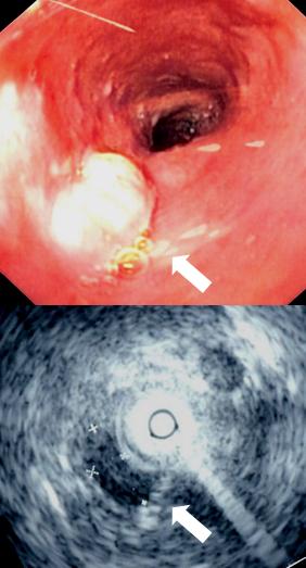
The esophagus (American English), oesophagus (British English), or œsophagus (Œ, archaic spelling) (American and British English spelling differences#ae and oe, see spelling difference) all ; : ((o)e)(œ)sophagi or ((o)e)(œ)sophaguses), colloquially known also as the food pipe, food tube, or gullet, is an Organ (anatomy), organ in vertebrates through which food passes, aided by Peristalsis, peristaltic contractions, from the Human pharynx, pharynx to the stomach. The esophagus is a :wiktionary:fibromuscular, fibromuscular tube, about long in adults, that travels behind the trachea and human heart, heart, passes through the Thoracic diaphragm, diaphragm, and empties into the uppermost region of the stomach. During swallowing, the epiglottis tilts backwards to prevent food from going down the larynx and lungs. The word ''esophagus'' is from Ancient Greek οἰσοφάγος (oisophágos), from οἴσω (oísō), future form of φέρω (phérō, "I carry") + ἔφαγον ( ... [...More Info...] [...Related Items...] OR: [Wikipedia] [Google] [Baidu] [Amazon] |
Oesophagus
The esophagus (American English), oesophagus (British English), or œsophagus ( archaic spelling) ( see spelling difference) all ; : ((o)e)(œ)sophagi or ((o)e)(œ)sophaguses), colloquially known also as the food pipe, food tube, or gullet, is an organ in vertebrates through which food passes, aided by peristaltic contractions, from the pharynx to the stomach. The esophagus is a fibromuscular tube, about long in adults, that travels behind the trachea and heart, passes through the diaphragm, and empties into the uppermost region of the stomach. During swallowing, the epiglottis tilts backwards to prevent food from going down the larynx and lungs. The word ''esophagus'' is from Ancient Greek οἰσοφάγος (oisophágos), from οἴσω (oísō), future form of φέρω (phérō, "I carry") + ἔφαγον (éphagon, "I ate"). The wall of the esophagus from the lumen outwards consists of mucosa, submucosa (connective tissue), layers of muscle fibers between lay ... [...More Info...] [...Related Items...] OR: [Wikipedia] [Google] [Baidu] [Amazon] |
Esophagus
The esophagus (American English), oesophagus (British English), or œsophagus (Œ, archaic spelling) (American and British English spelling differences#ae and oe, see spelling difference) all ; : ((o)e)(œ)sophagi or ((o)e)(œ)sophaguses), colloquially known also as the food pipe, food tube, or gullet, is an Organ (anatomy), organ in vertebrates through which food passes, aided by Peristalsis, peristaltic contractions, from the Human pharynx, pharynx to the stomach. The esophagus is a :wiktionary:fibromuscular, fibromuscular tube, about long in adults, that travels behind the trachea and human heart, heart, passes through the Thoracic diaphragm, diaphragm, and empties into the uppermost region of the stomach. During swallowing, the epiglottis tilts backwards to prevent food from going down the larynx and lungs. The word ''esophagus'' is from Ancient Greek οἰσοφάγος (oisophágos), from οἴσω (oísō), future form of φέρω (phérō, "I carry") + ἔφαγον ( ... [...More Info...] [...Related Items...] OR: [Wikipedia] [Google] [Baidu] [Amazon] |
Precapillary Sphincter
A precapillary sphincter is a band of contractile mural cells either classified as smooth muscle or pericytes that adjusts blood flow into capillaries. They were originally described in the mesenteric microcirculation, and were thought to only reside there. At the point where each of the capillaries originates from an arteriole, contractile mural cells encircle the capillary. This is called the precapillary sphincter. The precapillary sphincter has now also been found in the brain, where it regulates blood flow to the capillary bed. The sphincter can open and close the entrance to the capillary, by which contraction causes blood flow in a capillary to change as vasomotion occurs. In some tissues, the entire capillary bed may be bypassed by blood flow through arteriovenous anastomoses or through preferential flow through metarterioles. If the sphincter is damaged or cannot contract, blood can flow into the capillary bed at high pressures. When capillary pressures are high (as p ... [...More Info...] [...Related Items...] OR: [Wikipedia] [Google] [Baidu] [Amazon] |
Sphincter Of Oddi
The sphincter of Oddi (SO) (also hepatopancreatic sphincter or Glisson's sphincter), is a sphincter, a muscular valve that, in humans and some animals, controls the flow of bile and pancreatic juice out of the gallbladder and pancreas respectively through the ampulla of Vater into the second part of the duodenum. It is named after Ruggero Oddi. Structure The sphincter of Oddi is a circular muscle band, or sphincter that surrounds the major duodenal papilla. Function The sphincter regulates the secretion of pancreatic juice and bile into the duodenum. It also prevents reflux of duodenal contents into the ampulla of Vater. By preventing reflux of the contents of the duodenum, the sphincter of Oddi prevents the accumulation of particulate matter and sludge in the bile ducts, reducing the risk of cholangitis. The sphincter of Oddi also allows retrograde filling of the gallbladder. The sphincter of Oddi is relaxed by the hormone cholecystokinin via vasoactive intestinal pep ... [...More Info...] [...Related Items...] OR: [Wikipedia] [Google] [Baidu] [Amazon] |
Cardia
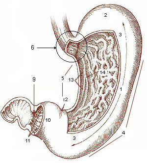
The stomach is a muscular, hollow organ in the upper gastrointestinal tract of humans and many other animals, including several invertebrates. The Ancient Greek name for the stomach is ''gaster'' which is used as ''gastric'' in medical terms related to the stomach. The stomach has a dilated structure and functions as a vital organ in the digestive system. The stomach is involved in the gastric phase of digestion, following the cephalic phase in which the sight and smell of food and the act of chewing are stimuli. In the stomach a chemical breakdown of food takes place by means of secreted digestive enzymes and gastric acid. It also plays a role in regulating gut microbiota, influencing digestion and overall health. The stomach is located between the esophagus and the small intestine. The pyloric sphincter controls the passage of partially digested food (chyme) from the stomach into the duodenum, the first and shortest part of the small intestine, where peristalsis takes over ... [...More Info...] [...Related Items...] OR: [Wikipedia] [Google] [Baidu] [Amazon] |
Stomach
The stomach is a muscular, hollow organ in the upper gastrointestinal tract of Human, humans and many other animals, including several invertebrates. The Ancient Greek name for the stomach is ''gaster'' which is used as ''gastric'' in medical terms related to the stomach. The stomach has a dilated structure and functions as a vital organ in the digestive system. The stomach is involved in the gastric phase, gastric phase of digestion, following the cephalic phase in which the sight and smell of food and the act of chewing are stimuli. In the stomach a chemical breakdown of food takes place by means of secreted digestive enzymes and gastric acid. It also plays a role in regulating gut microbiota, influencing digestion and overall health. The stomach is located between the esophagus and the small intestine. The pyloric sphincter controls the passage of partially digested food (chyme) from the stomach into the duodenum, the first and shortest part of the small intestine, where p ... [...More Info...] [...Related Items...] OR: [Wikipedia] [Google] [Baidu] [Amazon] |
Pyloric Sphincter
The pylorus ( or ) connects the stomach to the duodenum. The pylorus is considered as having two parts, the ''pyloric antrum'' (opening to the body of the stomach) and the ''pyloric canal'' (opening to the duodenum). The ''pyloric canal'' ends as the ''pyloric orifice'', which marks the junction between the stomach and the duodenum. The orifice is surrounded by a sphincter, a band of muscle, called the ''pyloric sphincter''. The word ''pylorus'' comes from Greek language, Greek πυλωρός, via Latin. The word ''pylorus'' in Greek means "gatekeeper", related to "gate" () and is thus linguistically related to the word "pylon (architecture), pylon". Structure The pylorus is the furthest part of the stomach that connects to the duodenum. It is divided into two parts, the ''antrum'', which connects to the body of the stomach, and the ''pyloric canal'', which connects to the duodenum. Antrum The antrum also called the ''gastric antrum'' or the ''pyloric antrum'' is the in ... [...More Info...] [...Related Items...] OR: [Wikipedia] [Google] [Baidu] [Amazon] |
Iris (anatomy)
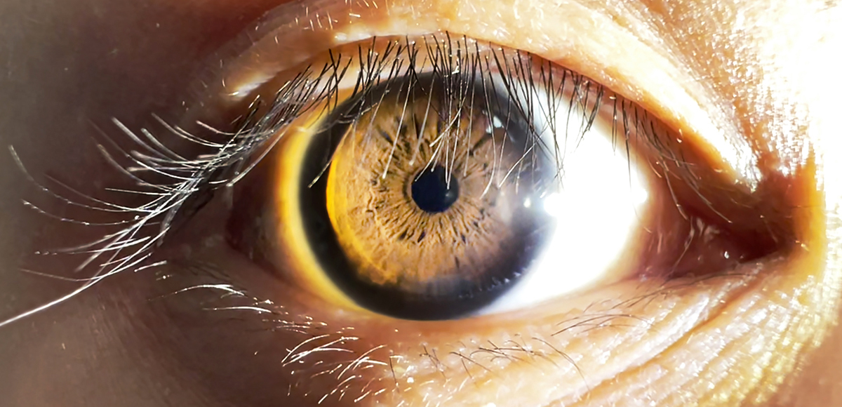
The iris (: irides or irises) is a thin, annular structure in the eye in most mammals and birds that is responsible for controlling the diameter and size of the pupil, and thus the amount of light reaching the retina. In optical terms, the pupil is the eye's aperture, while the iris is the diaphragm (optics), diaphragm. Eye color is defined by the iris. Etymology The word "iris" is derived from the Greek word for "rainbow", also Iris (mythology), its goddess plus messenger of the gods in the ''Iliad'', because of the many eye color, colours of this eye part. Structure The iris consists of two layers: the front pigmented Wikt:fibrovascular, fibrovascular layer known as a stroma of iris, stroma and, behind the stroma, pigmented epithelial cells. The stroma is connected to a sphincter muscle (sphincter pupillae), which contracts the pupil in a circular motion, and a set of dilator muscles (dilator pupillae), which pull the iris radially to enlarge the pupil, pulling it in folds. ... [...More Info...] [...Related Items...] OR: [Wikipedia] [Google] [Baidu] [Amazon] |
Pupillary Sphincter
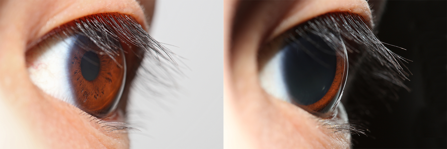
The iris sphincter muscle (pupillary sphincter, pupillary constrictor, circular muscle of iris, circular fibers) is a muscle in the part of the eye called the iris. It encircles the pupil of the iris, appropriate to its function as a constrictor of the pupil. The ciliary muscle, pupillary sphincter muscle and pupillary dilator muscle sometimes are called intrinsic ocular muscles or intraocular muscles. Comparative anatomy This structure is found in vertebrates and in some cephalopods. General structure All the myocytes are of the smooth muscle type. Its dimensions are about 0.75 mm wide by 0.15 mm thick. Mode of action In humans, it functions to constrict the pupil in bright light (pupillary light reflex) or during accommodation. In , the muscle cells themselves are photosensitive causing iris action without brain input. Innervation It is controlled by parasympathetic postganglionic fibers releasing acetylcholine acting primarily on the muscarinic acetylcholine re ... [...More Info...] [...Related Items...] OR: [Wikipedia] [Google] [Baidu] [Amazon] |
Autonomic Nervous System
The autonomic nervous system (ANS), sometimes called the visceral nervous system and formerly the vegetative nervous system, is a division of the nervous system that operates viscera, internal organs, smooth muscle and glands. The autonomic nervous system is a control system that acts largely unconsciously and regulates bodily functions, such as the heart rate, its Myocardial contractility, force of contraction, digestion, respiratory rate, pupillary dilation, pupillary response, Micturition, urination, and Animal sexual behaviour, sexual arousal. The fight-or-flight response, also known as the acute stress response, is set into action by the autonomic nervous system. The autonomic nervous system is regulated by integrated reflexes through the brainstem to the spinal cord and organ (anatomy), organs. Autonomic functions include control of respiration, heart rate, cardiac regulation (the cardiac control center), vasomotor activity (the vasomotor center), and certain reflex, reflex ... [...More Info...] [...Related Items...] OR: [Wikipedia] [Google] [Baidu] [Amazon] |
Digestion
Digestion is the breakdown of large insoluble food compounds into small water-soluble components so that they can be absorbed into the blood plasma. In certain organisms, these smaller substances are absorbed through the small intestine into the blood stream. Digestion is a form of catabolism that is often divided into two processes based on how food is broken down: mechanical and chemical digestion. The term mechanical digestion refers to the physical breakdown of large pieces of food into smaller pieces which can subsequently be accessed by digestive enzymes. Mechanical digestion takes place in the mouth through Chewing, mastication and in the small intestine through segmentation contractions. In chemical digestion, enzymes break down food into the small compounds that the body can use. In the human digestive system, food enters the mouth and mechanical digestion of the food starts by the action of mastication (chewing), a form of mechanical digestion, and the wetting contact ... [...More Info...] [...Related Items...] OR: [Wikipedia] [Google] [Baidu] [Amazon] |


