serous membrane on:
[Wikipedia]
[Google]
[Amazon]
 The serous membrane (or serosa) is a smooth tissue membrane of mesothelium lining the contents and inner walls of
 The
The
Image:Illu stomach layers.jpg, Layers of stomach wall
Image:Gray1058.png, Section of duodenum of cat
edition of Anatomy & Physiolog
text-book by OpenStax College''
 The serous membrane (or serosa) is a smooth tissue membrane of mesothelium lining the contents and inner walls of
 body cavities
A body cavity is any space or compartment, or potential space, in an animal body. Cavities accommodate organs and other structures; cavities as potential spaces contain fluid.
The two largest human body cavities are the ventral body cavity, and ...
, which secrete serous fluid
In physiology, serous fluid or serosal fluid (originating from the Medieval Latin word ''serosus'', from Latin ''serum'') is any of various body fluids resembling Serum (blood), serum, that are typically pale yellow or transparent and of a benign ...
to allow lubricated sliding movements between opposing surfaces. The serous membrane that covers internal organs
In biology, an organ is a collection of tissues joined in a structural unit to serve a common function. In the hierarchy of life, an organ lies between tissue and an organ system. Tissues are formed from same type cells to act together in a ...
is called a ''visceral'' membrane; while the one that covers the cavity wall is called the ''parietal'' membrane. Between the two opposing serosal surfaces is often a potential space
In anatomy, a potential space is a space between two adjacent structures that are normally pressed together (directly apposed). Many anatomic spaces are potential spaces, which means that they are potential rather than realized (with their realiz ...
, mostly empty except for the small amount of serous fluid.
The Latin anatomical name is '' tunica serosa''. Serous membranes line and enclose several body cavities
A body cavity is any space or compartment, or potential space, in an animal body. Cavities accommodate organs and other structures; cavities as potential spaces contain fluid.
The two largest human body cavities are the ventral body cavity, and ...
, also known as serous cavities, where they secrete a lubricating fluid which reduces friction from movements. Serosa is entirely different from the adventitia
The adventitia () is the outer layer of fibrous connective tissue surrounding an organ.
The outer layer of connective tissue that surrounds an artery, or vein – the tunica externa, is also called the ''tunica adventitia''.
To some degree, its ...
, a connective tissue
Connective tissue is one of the four primary types of animal tissue, along with epithelial tissue, muscle tissue, and nervous tissue. It develops from the mesenchyme derived from the mesoderm the middle embryonic germ layer. Connective tiss ...
layer which binds together structures rather than reducing friction between them. The serous membrane covering the heart
The heart is a muscular organ in most animals. This organ pumps blood through the blood vessels of the circulatory system. The pumped blood carries oxygen and nutrients to the body, while carrying metabolic waste such as carbon dioxide t ...
and lining the mediastinum
The mediastinum (from ) is the central compartment of the thoracic cavity. Surrounded by loose connective tissue, it is an undelineated region that contains a group of structures within the thorax, namely the heart and its vessels, the esophagu ...
is referred to as the pericardium
The pericardium, also called pericardial sac, is a double-walled sac containing the heart and the roots of the great vessels. It has two layers, an outer layer made of strong connective tissue (fibrous pericardium), and an inner layer made of ...
, the serous membrane lining the thoracic cavity
The thoracic cavity (or chest cavity) is the chamber of the body of vertebrates that is protected by the thoracic wall (rib cage and associated skin, muscle, and fascia). The central compartment of the thoracic cavity is the mediastinum. There ...
and surrounding the lungs
The lungs are the primary organs of the respiratory system in humans and most other animals, including some snails and a small number of fish. In mammals and most other vertebrates, two lungs are located near the backbone on either side ...
is referred to as the pleura
The pulmonary pleurae (''sing.'' pleura) are the two opposing layers of serous membrane overlying the lungs and the inside of the surrounding chest walls.
The inner pleura, called the visceral pleura, covers the surface of each lung and dips b ...
, and that lining the abdominopelvic cavity
The abdominopelvic cavity is a body cavity that consists of the abdominal cavity and the pelvic cavity. The upper portion is the abdominal cavity, and it contains the stomach, liver, pancreas, spleen, gallbladder, kidneys, small intestine, and ...
and the viscera
In biology, an organ is a collection of tissues joined in a structural unit to serve a common function. In the hierarchy of life, an organ lies between tissue and an organ system. Tissues are formed from same type cells to act together in a ...
is referred to as the peritoneum
The peritoneum is the serous membrane forming the lining of the abdominal cavity or coelom in amniotes and some invertebrates, such as annelids. It covers most of the intra-abdominal (or coelomic) organs, and is composed of a layer of mesoth ...
.
Structure
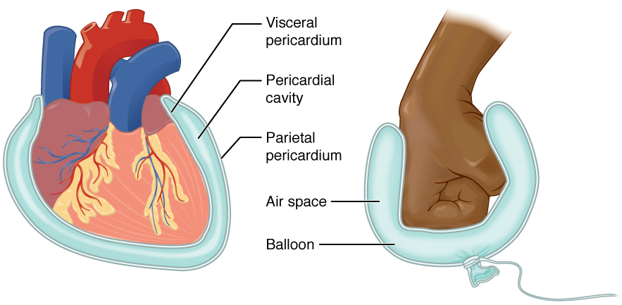
Serous membranes have two layers. The parietal layers of the membranes line the walls of the body cavity (pariet- refers to a cavity wall). The visceral layer of the membrane covers the organs (the viscera). Between the parietal and visceral layers is a very thin, fluid-filled serous space, or cavity.Visceral and parietal layers
Each serous membrane is composed of a secretoryepithelial
Epithelium or epithelial tissue is one of the four basic types of animal tissue, along with connective tissue, muscle tissue and nervous tissue. It is a thin, continuous, protective layer of compactly packed cells with a little intercellula ...
layer and a connective tissue
Connective tissue is one of the four primary types of animal tissue, along with epithelial tissue, muscle tissue, and nervous tissue. It develops from the mesenchyme derived from the mesoderm the middle embryonic germ layer. Connective tiss ...
layer underneath.
* The ''epithelial layer'', known as mesothelium, consists of a single layer of avascular flat nucleated
The cell nucleus (pl. nuclei; from Latin or , meaning ''kernel'' or ''seed'') is a membrane-bound organelle found in eukaryotic cells. Eukaryotic cells usually have a single nucleus, but a few cell types, such as mammalian red blood cells, ...
cells (simple squamous epithelium
A simple squamous epithelium, also known as pavement epithelium, and tessellated epithelium is a single layer of flattened, polygonal cells in contact with the basal lamina (one of the two layers of the basement membrane) of the epithelium. This ...
) which produce the lubricating serous fluid. This fluid has a consistency similar to thin mucus
Mucus ( ) is a slippery aqueous secretion produced by, and covering, mucous membranes. It is typically produced from cells found in mucous glands, although it may also originate from mixed glands, which contain both serous and mucous cells. It ...
. These cells are bound tightly to the underlying connective tissue.
* The ''connective tissue layer'' provides the blood vessels
The blood vessels are the components of the circulatory system that transport blood throughout the human body. These vessels transport blood cells, nutrients, and oxygen to the tissues of the body. They also take waste and carbon dioxide away f ...
and nerves
A nerve is an enclosed, cable-like bundle of nerve fibers (called axons) in the peripheral nervous system.
A nerve transmits electrical impulses. It is the basic unit of the peripheral nervous system. A nerve provides a common pathway for the e ...
for the overlying secretory cells, and also serves as the binding layer which allows the whole serous membrane to adhere to organs and other structures.
For the heart, the layers of the serous membrane are called the parietal pericardium
The pericardium, also called pericardial sac, is a double-walled sac containing the heart and the roots of the great vessels. It has two layers, an outer layer made of strong connective tissue (fibrous pericardium), and an inner layer made of ...
, and the visceral pericardium (sometimes called the epicardium
The pericardium, also called pericardial sac, is a double-walled sac containing the heart and the roots of the great vessels. It has two layers, an outer layer made of strong connective tissue (fibrous pericardium), and an inner layer made o ...
). Other parts of the body may also have specific names for these structures. For example, the serosa of the uterus
The uterus (from Latin ''uterus'', plural ''uteri'') or womb () is the organ in the reproductive system of most female mammals, including humans that accommodates the embryonic and fetal development of one or more embryos until birth. The uter ...
is called the perimetrium.
 The
The pericardial cavity
The pericardium, also called pericardial sac, is a double-walled sac containing the heart and the roots of the great vessels. It has two layers, an outer layer made of strong connective tissue (fibrous pericardium), and an inner layer made of ...
(surrounding the heart
The heart is a muscular organ in most animals. This organ pumps blood through the blood vessels of the circulatory system. The pumped blood carries oxygen and nutrients to the body, while carrying metabolic waste such as carbon dioxide t ...
), pleural cavity (surrounding the lung
The lungs are the primary organs of the respiratory system in humans and most other animals, including some snails and a small number of fish. In mammals and most other vertebrates, two lungs are located near the backbone on either side of t ...
s) and peritoneal cavity
The peritoneal cavity is a potential space between the parietal peritoneum (the peritoneum that surrounds the abdominal wall) and visceral peritoneum (the peritoneum that surrounds the internal organs). The parietal and visceral peritonea are la ...
(surrounding most organs of the abdomen
The abdomen (colloquially called the belly, tummy, midriff, tucky or stomach) is the part of the body between the thorax (chest) and pelvis, in humans and in other vertebrates. The abdomen is the front part of the abdominal segment of the torso. ...
) are the three serous cavities within the human body. While serous membranes have a lubricative role to play in all three cavities, in the pleural cavity it has a greater role to play in the function of breathing.
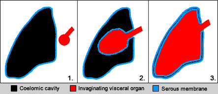
The serous cavities are formed from the intraembryonic coelom and are basically an empty space within the body surrounded by serous membrane. Early in embryonic life visceral organs develop adjacent to a cavity and invaginate into the bag-like coelom. Therefore, each organ becomes surrounded by serous membrane - they ''do not'' lie within the serous cavity. The layer in contact with the organ is known as the visceral layer, while the parietal layer is in contact with the body wall.
Examples
In the human body, there are three serous cavities with associated serous membranes: *A serous membrane lines thepericardial cavity
The pericardium, also called pericardial sac, is a double-walled sac containing the heart and the roots of the great vessels. It has two layers, an outer layer made of strong connective tissue (fibrous pericardium), and an inner layer made of ...
of the heart, and reflects back to cover the heart, much like an under-inflated balloon would form two layers surrounding a fist. Called the pericardium
The pericardium, also called pericardial sac, is a double-walled sac containing the heart and the roots of the great vessels. It has two layers, an outer layer made of strong connective tissue (fibrous pericardium), and an inner layer made of ...
, this serous membrane is a two-layered sac that surrounds the entire heart except where blood vessels emerge on the heart’s superior side;
*The pleura
The pulmonary pleurae (''sing.'' pleura) are the two opposing layers of serous membrane overlying the lungs and the inside of the surrounding chest walls.
The inner pleura, called the visceral pleura, covers the surface of each lung and dips b ...
is the serous membrane that surrounds the lungs in the pleural cavity;
*The peritoneum
The peritoneum is the serous membrane forming the lining of the abdominal cavity or coelom in amniotes and some invertebrates, such as annelids. It covers most of the intra-abdominal (or coelomic) organs, and is composed of a layer of mesoth ...
is the serous membrane that surrounds several organs in the abdominopelvic cavity.
*The tunica vaginalis
The tunica vaginalis is the pouch of serous membrane that covers the testes. It is derived from the vaginal process of the peritoneum, which in the fetus precedes the descent of the testes from the abdomen into the scrotum.
After its descent, ...
is the serous membrane, which surrounds the male gonad, the testis.
The two layers of serous membranes are named parietal and visceral. Between the two layers is a thin fluid filled space. The fluid is produced by the serous membranes and stays between the two layers to reduce friction between the walls of the cavities and the internal organs when they move with respect to one another, such as when the lungs inflate or the heart beats. Such movement could otherwise lead to inflammation of the organs.
Development
All serous membranes found in the human body formed ultimately from themesoderm
The mesoderm is the middle layer of the three germ layers that develops during gastrulation in the very early development of the embryo of most animals. The outer layer is the ectoderm, and the inner layer is the endoderm.Langman's Medical E ...
of the trilaminar embryo. The trilaminar embryo consists of three relatively flat layers of ectoderm
The ectoderm is one of the three primary germ layers formed in early embryonic development. It is the outermost layer, and is superficial to the mesoderm (the middle layer) and endoderm (the innermost layer). It emerges and originates from t ...
, endoderm
Endoderm is the innermost of the three primary germ layers in the very early embryo. The other two layers are the ectoderm (outside layer) and mesoderm (middle layer). Cells migrating inward along the archenteron form the inner layer of the gast ...
(also known as "entoderm") and mesoderm
The mesoderm is the middle layer of the three germ layers that develops during gastrulation in the very early development of the embryo of most animals. The outer layer is the ectoderm, and the inner layer is the endoderm.Langman's Medical E ...
.
As the embryo develops, the mesoderm starts to segment into three main regions: the paraxial mesoderm
Paraxial mesoderm, also known as presomitic or somitic mesoderm is the area of mesoderm in the neurulating embryo that flanks and forms simultaneously with the neural tube. The cells of this region give rise to somites, blocks of tissue running ...
, the intermediate mesoderm
Intermediate mesoderm or intermediate mesenchyme is a narrow section of the mesoderm (one of the three primary germ layers) located between the paraxial mesoderm and the lateral plate of the developing embryo. The intermediate mesoderm develop ...
and the lateral plate mesoderm
The lateral plate mesoderm is the mesoderm that is found at the periphery of the embryo. It is to the side of the paraxial mesoderm, and further to the axial mesoderm. The lateral plate mesoderm is separated from the paraxial mesoderm by a narrow ...
.
The lateral plate mesoderm later splits in half to form two layers bounding a cavity known as the intraembryonic coelom
In the development of the human embryo the intraembryonic coelom (or somatic coelom) is a portion of the conceptus forming in the mesoderm during the third week of development. During the third week of development, the lateral plate mesoderm split ...
. Individually, each layer is known as splanchnopleure
In the anatomy of an embryo, the splanchnopleuric mesenchyme is a structure created during embryogenesis when the lateral mesodermal germ layer splits into two layers. The inner (or splanchnic) layer adheres to the endoderm, and with it forms the ...
and somatopleure
In the anatomy of an embryo, the somatopleure is a structure created during embryogenesis when the lateral plate mesoderm splits into two layers. The outer (or somatic) layer becomes applied to the inner surface of the ectoderm, and with it (part ...
.
* The ''splanchnopleure'' is associated with the underlying endoderm with which it is in contact, and later becomes the serous membrane in contact with visceral organs within the body.
* The ''somatopleure'' is associated with the overlying ectoderm and later becomes the serous membrane in contact with the body wall.
The intraembryonic coelom
In the development of the human embryo the intraembryonic coelom (or somatic coelom) is a portion of the conceptus forming in the mesoderm during the third week of development. During the third week of development, the lateral plate mesoderm split ...
can now be seen as a cavity within the body which is covered with serous membrane derived from the splanchnopleure. This cavity is divided and demarcated by the folding and development of the embryo, ultimately forming the serous cavities which house many different organs within the thorax
The thorax or chest is a part of the anatomy of humans, mammals, and other tetrapod animals located between the neck and the abdomen. In insects, crustaceans, and the extinct trilobites, the thorax is one of the three main divisions of the cre ...
and abdomen
The abdomen (colloquially called the belly, tummy, midriff, tucky or stomach) is the part of the body between the thorax (chest) and pelvis, in humans and in other vertebrates. The abdomen is the front part of the abdominal segment of the torso. ...
.
Diseases
Mesothelioma
Mesothelioma is a type of cancer that develops from the thin layer of tissue that covers many of the internal organs (known as the mesothelium). The most common area affected is the lining of the lungs and chest wall. Less commonly the lining ...
s are neoplasia
A neoplasm () is a type of abnormal and excessive growth of tissue. The process that occurs to form or produce a neoplasm is called neoplasia. The growth of a neoplasm is uncoordinated with that of the normal surrounding tissue, and persists ...
s that are relatively specific for serous membranes. The modified Mullerian-derived serous membranes that surrounds the ovaries
The ovary is an organ in the female reproductive system that produces an ovum. When released, this travels down the fallopian tube into the uterus, where it may become fertilized by a sperm. There is an ovary () found on each side of the body. T ...
in females can give rise to serous tumor
A serous tumour is a neoplasm that typically has papillary to solid formations of tumor cells with crowded nuclei, and which typically arises on the modified Mullerian-derived serous membranes that surround the ovaries in females. Such ovarian t ...
s, a solid to papillary tumor type that may also arise within the uterus
The uterus (from Latin ''uterus'', plural ''uteri'') or womb () is the organ in the reproductive system of most female mammals, including humans that accommodates the embryonic and fetal development of one or more embryos until birth. The uter ...
.
Anatomical images
See also
*Adventitia
The adventitia () is the outer layer of fibrous connective tissue surrounding an organ.
The outer layer of connective tissue that surrounds an artery, or vein – the tunica externa, is also called the ''tunica adventitia''.
To some degree, its ...
References
''This Wikipedia entry incorporates text from the freely licensed Connexionedition of Anatomy & Physiolog
text-book by OpenStax College''
External links
* * {{Abdominopelvic cavity Membrane biology Tissues (biology)