Functional near-infrared spectroscopy (fNIRS) is an optical brain monitoring technique which uses
near-infrared spectroscopy for the purpose of
functional neuroimaging.
Using fNIRS, brain activity is measured by using near-infrared light to estimate cortical
hemodynamic activity which occur in response to neural activity. Alongside
EEG, fNIRS is one of the most common non-invasive neuroimaging techniques which can be used in portable contexts. The signal is often compared with the
BOLD
In typography, emphasis is the strengthening of words in a text with a font in a different style from the rest of the text, to highlight them. It is the equivalent of prosody stress in speech.
Methods and use
The most common methods in W ...
signal measured by
fMRI and is capable of measuring changes both in oxy- and deoxyhemoglobin concentration,
but can only measure from regions near the cortical surface. fNIRS may also be referred to as Optical Topography (OT) and is sometimes referred to simply as NIRS.
Description

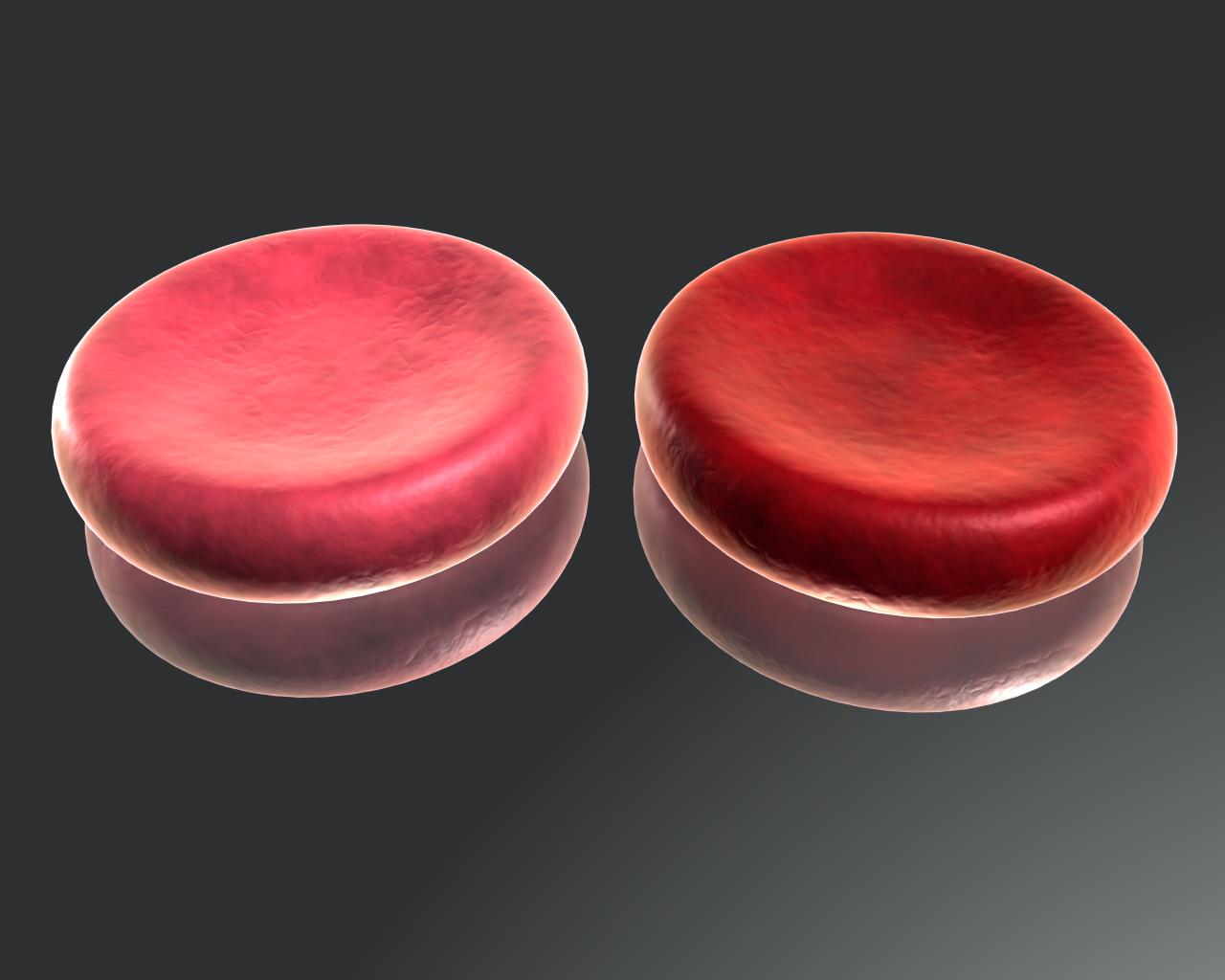
fNIRS estimates the concentration of hemoglobin from changes in absorption of near infrared light. As light moves or propagates through the head, it is alternately scattered or absorbed by the tissue through which it travels. Because hemoglobin is a significant absorber of near-infrared light, changes in absorbed light can be used to reliably measure changes in hemoglobin concentration. Different fNIRS techniques can also use the way in which light propagates to estimate blood volume and oxygenation. The technique is safe, non-invasive, and can be used with other imaging modalities.
fNIRS is a non-invasive imaging method involving the quantification of
chromophore
A chromophore is the part of a molecule responsible for its color.
The color that is seen by our eyes is the one not absorbed by the reflecting object within a certain wavelength spectrum of visible light. The chromophore is a region in the molec ...
concentration resolved from the measurement of near infrared (NIR)
light attenuation or temporal or phasic changes. The technique takes advantage of the
optical window in which (a) skin, tissue, and bone are mostly transparent to NIR light (700–900 nm spectral interval) and (b)
hemoglobin (Hb) and deoxygenated-hemoglobin (deoxy-Hb) are strong absorbers of light.

There are six different ways for infrared light to interact with the brain tissue: direct transmission, diffuse transmission, specular reflection, diffuse reflection, scattering, and absorption. fNIRS focuses primarily on absorption: differences in the absorption spectra of deoxy-Hb and oxy-Hb allow the measurement of relative changes in hemoglobin concentration through the use of light attenuation at multiple
wavelengths. Two or more wavelengths are selected, with one wavelength above and one below the
isosbestic point of 810 nm—at which deoxy-Hb and oxy-Hb have identical absorption
coefficient
In mathematics, a coefficient is a multiplicative factor in some term of a polynomial, a series, or an expression; it is usually a number, but may be any expression (including variables such as , and ). When the coefficients are themselves var ...
s. Using the modified
Beer-Lambert law (mBLL), relative changes in concentration can be calculated as a function of total photon path length.
Typically, the light emitter and detector are placed ipsilaterally (each emitter/detector pair on the same side) on the subject's skull so recorded measurements are due to back-scattered (reflected) light following elliptical pathways.
fNIRS is most sensitive to hemodynamic changes which occur nearest to the scalp
and these superficial artifacts are often addressed using additional light detectors located closer to the light source (short-separation detectors).
Modified Beer–Lambert law
Changes in light intensity can be related to changes in relative concentrations of hemoglobin through the modified
Beer–Lambert law (mBLL). The Beer lambert-law has to deal with concentration of hemoglobin. This technique also measures relative changes in light attenuation as well as using mBLL to quantify hemoglobin concentration changes.
History
US & UK
In 1977, Jöbsis reported that brain tissue transparency to NIR light allowed a non-invasive and continuous method of tissue oxygen saturation using
transillumination. Transillumination (forward-scattering) was of limited utility in adults because of light attenuation and was quickly replaced by reflectance-mode based techniques - resulting in development of NIRS systems proceeding rapidly. Then, by 1985, the first studies on cerebral oxygenation were conducted by M. Ferrari. Later, in 1989, following work with David Delpy at University College London, Hamamatsu developed the first commercial NIRS system: NIR-1000 cerebral oxygenation monitor. NIRS methods were initially used for cerebral oximetry in the 1990s. In 1993, four publications by Chance et al. ''PNAS'', Hoshi & Tamura ''J Appl Physiol'', Kato et al. ''JCBFM,'' Villringer ''et al'' ''Neuros. Lett.'' demonstrated the feasibility of fNIRS in adult humans. NIRS techniques were further expanded on by the work of Randall Barbour,
Britton Chance, Arno Villringer, M. Cope, D. T. Delpy, Enrico Gratton, and others. Currently, wearable fNIRS are being developed.

Japan
Meanwhile, in the mid-80's, Japanese researchers at the central research laboratory of Hitachi Ltd set out to build a NIRS-based brain monitoring system using a pulse of 70-picosecond rays. This effort came into light when the team, along with their leading expert, Dr Hideaki Koizumi (小泉 英明), held an open symposium to announce the principle of "Optical Topography" in January 1995. In fact, the term "Optical Topography" derives from the concept of using light on "2-Dimensional mapping combined with 1-Dimensional information", or ''topography''. The idea had been successfully implemented in launching their first fNIRS (or Optical Topography, as they call it) device based on Frequency Domain in 2001: Hitachi ETG-100. Later, Harumi Oishi (大石 晴美), a PhD-to-be at Nagoya University, published her doctoral dissertation in 2003 with the subject of "language learners' cortical activation patterns measured by ETG-100" under the supervision of Professor Toru Kinoshita (木下 微)—presenting a new prospect on the use of fNIRS. The company has been advancing the ETG series ever since.
Spectroscopic techniques
Currently, there are three modalities of fNIR spectroscopy:
1. Continuous wave
2. Frequency domain
3. Time-domain
Continuous wave
Continuous wave (CW) system uses light sources with constant frequency and amplitude. In fact, to measure absolute changes in HbO concentration with the mBLL, we need to know photon path-length. However, CW-fNIRS does not provide any knowledge of photon path-length, so changes in HbO concentration are relative to an unknown path-length. Many CW-fNIRS commercial systems use estimations of photon path-length derived from computerized
Monte-Carlo simulations and physical models, to approximate absolute quantification of hemoglobin concentrations.
Where
is the optical density or attenuation,
is emitted light intensity,
is measured light intensity,
is the
attenuation coefficient,
 Functional near-infrared spectroscopy (fNIRS) is an optical brain monitoring technique which uses near-infrared spectroscopy for the purpose of functional neuroimaging. Using fNIRS, brain activity is measured by using near-infrared light to estimate cortical hemodynamic activity which occur in response to neural activity. Alongside EEG, fNIRS is one of the most common non-invasive neuroimaging techniques which can be used in portable contexts. The signal is often compared with the
  fNIRS estimates the concentration of hemoglobin from changes in absorption of near infrared light. As light moves or propagates through the head, it is alternately scattered or absorbed by the tissue through which it travels. Because hemoglobin is a significant absorber of near-infrared light, changes in absorbed light can be used to reliably measure changes in hemoglobin concentration. Different fNIRS techniques can also use the way in which light propagates to estimate blood volume and oxygenation. The technique is safe, non-invasive, and can be used with other imaging modalities.
fNIRS is a non-invasive imaging method involving the quantification of
  There are six different ways for infrared light to interact with the brain tissue: direct transmission, diffuse transmission, specular reflection, diffuse reflection, scattering, and absorption. fNIRS focuses primarily on absorption: differences in the absorption spectra of deoxy-Hb and oxy-Hb allow the measurement of relative changes in hemoglobin concentration through the use of light attenuation at multiple wavelengths. Two or more wavelengths are selected, with one wavelength above and one below the isosbestic point of 810 nm—at which deoxy-Hb and oxy-Hb have identical absorption
 Functional near-infrared spectroscopy (fNIRS) is an optical brain monitoring technique which uses near-infrared spectroscopy for the purpose of functional neuroimaging. Using fNIRS, brain activity is measured by using near-infrared light to estimate cortical hemodynamic activity which occur in response to neural activity. Alongside EEG, fNIRS is one of the most common non-invasive neuroimaging techniques which can be used in portable contexts. The signal is often compared with the
  fNIRS estimates the concentration of hemoglobin from changes in absorption of near infrared light. As light moves or propagates through the head, it is alternately scattered or absorbed by the tissue through which it travels. Because hemoglobin is a significant absorber of near-infrared light, changes in absorbed light can be used to reliably measure changes in hemoglobin concentration. Different fNIRS techniques can also use the way in which light propagates to estimate blood volume and oxygenation. The technique is safe, non-invasive, and can be used with other imaging modalities.
fNIRS is a non-invasive imaging method involving the quantification of
  There are six different ways for infrared light to interact with the brain tissue: direct transmission, diffuse transmission, specular reflection, diffuse reflection, scattering, and absorption. fNIRS focuses primarily on absorption: differences in the absorption spectra of deoxy-Hb and oxy-Hb allow the measurement of relative changes in hemoglobin concentration through the use of light attenuation at multiple wavelengths. Two or more wavelengths are selected, with one wavelength above and one below the isosbestic point of 810 nm—at which deoxy-Hb and oxy-Hb have identical absorption