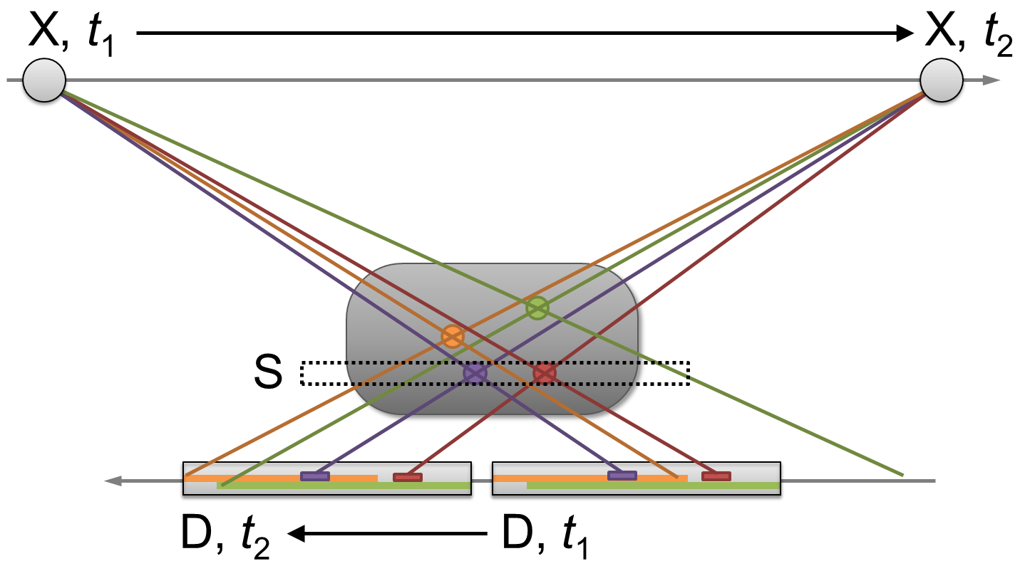
focal plane tomography on:
[Wikipedia]
[Google]
[Amazon]
In
 This is the most basic form of conventional tomography. The
radiography
Radiography is an imaging technique using X-rays, gamma rays, or similar ionizing radiation and non-ionizing radiation to view the internal form of an object. Applications of radiography include medical radiography ("diagnostic" and "therapeut ...
, focal plane tomography is tomography
Tomography is imaging by sections or sectioning that uses any kind of penetrating wave. The method is used in radiology, archaeology, biology, atmospheric science, geophysics, oceanography, plasma physics, materials science, astrophysics, quantu ...
(imaging a single plane, or slice, of an object) by simultaneously moving the X-ray generator
An X-ray generator is a device that produces X-rays. Together with an X-ray detector, it is commonly used in a variety of applications including medicine, X-ray fluorescence, electronic assembly inspection, and measurement of material thickness ...
and X-ray detector
X-ray detectors are devices used to measure the flux, spatial distribution, spectrum, and/or other properties of X-rays.
Detectors can be divided into two major categories: imaging detectors (such as photographic plates and X-ray film (photograp ...
so as to keep a consistent exposure of only the plane of interest during image acquisition. This was the main method of obtaining tomographs in medical imaging
Medical imaging is the technique and process of imaging the interior of a body for clinical analysis and medical intervention, as well as visual representation of the function of some organs or tissues (physiology). Medical imaging seeks to rev ...
until the late-1970s. It has since been largely replaced by more advanced imaging techniques such as CT and MRI
Magnetic resonance imaging (MRI) is a medical imaging technique used in radiology to form pictures of the anatomy and the physiological processes of the body. MRI scanners use strong magnetic fields, magnetic field gradients, and radio waves ...
. It remains in use today in a few specialized applications, such as for acquiring orthopantomographs of the jaw in dental radiography
Dental radiographs, commonly known as X-rays, are radiographs used to diagnose hidden dental structures, malignant or benign masses, bone loss, and cavities.
A radiographic image is formed by a controlled burst of X-ray radiation which penet ...
.
Focal plane tomography’s development began in the 1930s as a means of reducing the problem of superimposition
Superimposition is the placement of one thing over another, typically so that both are still evident.
Graphics
In graphics, superimposition is the placement of an image or video on top of an already-existing image or video, usually to add to t ...
of structures which is inherent to projectional radiography
Projectional radiography, also known as conventional radiography, is a form of radiography and medical imaging that produces two-dimensional images by x-ray radiation. The image acquisition is generally performed by radiographers, and the images ...
. It was invented in parallel by, among others, by the French physician Bocage
Bocage (, ) is a terrain of mixed woodland and pasture characteristic of parts of Northern France, Southern England, Ireland, the Netherlands and Northern Germany, in regions where pastoral farming is the dominant land use.
''Bocage'' may als ...
, the Italian radiologist Alessandro Vallebona
Alessandro is both a given name and a surname, the Italian form of the name Alexander. Notable people with the name include:
People with the given name Alessandro
* Alessandro Allori (1535–1607), Italian portrait painter
* Alessandro Baricco ...
and the Dutch radiologist Bernard George Ziedses des Plantes.
Technique
Focal plane tomography generally uses mechanical movement of anX-ray
An X-ray, or, much less commonly, X-radiation, is a penetrating form of high-energy electromagnetic radiation. Most X-rays have a wavelength ranging from 10 picometers to 10 nanometers, corresponding to frequencies in the range 30&nb ...
source and film in unison to generate a tomogram using the principles of projective geometry
In mathematics, projective geometry is the study of geometric properties that are invariant with respect to projective transformations. This means that, compared to elementary Euclidean geometry, projective geometry has a different setting, pro ...
. Synchronizing the movement of the radiation source and detector which are situated in the opposite direction from each other causes structures which are not in the focal plane being studied to blur out.
Limitations
The blurring provided by focal plane tomography is only marginally effective, since it only occurs in the ''X''plane
Plane(s) most often refers to:
* Aero- or airplane, a powered, fixed-wing aircraft
* Plane (geometry), a flat, 2-dimensional surface
Plane or planes may also refer to:
Biology
* Plane (tree) or ''Platanus'', wetland native plant
* ''Planes' ...
. Moreover, since focal plane tomography uses plain X-rays, it is not particularly effective at resolving soft tissues.
The increased availability and power of computers in the 1960s and 70s gave rise to new imaging techniques such as CT and MRI which use computational (in addition to or in lieu of mechanical) methods to acquire and process tomographic image data, and which do not suffer from the limitations of focal plane tomography.
Variants
Initially focal plane tomography used simple linear movements. The technique advanced through the mid-twentieth century however, steadily producing sharper images, and with a greater ability to vary the thickness of the cross-section being examined. This was achieved through the introduction of more complex, pluridirectional devices that can move in more than one plane and perform more effective blurring.Linear tomography
 This is the most basic form of conventional tomography. The
 X-ray tube
An X-ray tube is a vacuum tube that converts electrical input power into X-rays. The availability of this controllable source of X-rays created the field of radiography, the imaging of partly opaque objects with penetrating radiation. In contrast ...
moved from point "A" to point "B" above the patient, while the detector (such as cassette holder or "bucky") moves simultaneously under the patient from point "B" to point "A". The fulcrum
A fulcrum is the support about which a lever pivots.
Fulcrum may also refer to:
Companies and organizations
* Fulcrum (Anglican think tank), a Church of England think tank
* Fulcrum Press, a British publisher of poetry
* Fulcrum Wheels, a bicy ...
, or pivot point, is set to the area of interest. In this manner, the points above and below the focal plane are blurred out, just as the background is blurred when panning a camera during exposure. Rarely used, and has largely been replaced by computed tomography (CT).
Poly tomography
This was achieved using a more advanced X-ray apparatus that allows for more sophisticated and continuous movements of the X-ray tube and film. With this technique, a number of complex synchronous geometrical movements could be programmed, such as hypocycloidic, circular, figure 8, andelliptical
Elliptical may mean:
* having the shape of an ellipse, or more broadly, any oval shape
** in botany, having an elliptic leaf shape
** of aircraft wings, having an elliptical planform
* characterised by ellipsis (the omission of words), or by conc ...
. Philips
Koninklijke Philips N.V. (), commonly shortened to Philips, is a Dutch multinational conglomerate corporation that was founded in Eindhoven in 1891. Since 1997, it has been mostly headquartered in Amsterdam, though the Benelux headquarters i ...
Medical Systems for example produced one such device called the 'Polytome'. This pluridirectional unit was still in use into the 1990s, as its resulting images for small or difficult physiology, such as the inner ear, were still difficult to image with CTs at that time. As the resolution of CT scanners got better, this procedure was taken over by CT.
Zonography
This is a variant of linear tomography, where a limited arc of movement is used, resulting in less blurring than linear tomography. It is still used in some centres for visualising thekidney
The kidneys are two reddish-brown bean-shaped organs found in vertebrates. They are located on the left and right in the retroperitoneal space, and in adult humans are about in length. They receive blood from the paired renal arteries; blood ...
during an intravenous urogram
Pyelogram (or pyelography or urography) is a form of imaging of the renal pelvis and ureter.
Types include:
* Intravenous pyelogram – In which a contrast solution is introduced through a vein into the circulatory system.
* Retrograde pyelogram ...
(IVU), though it too is being supplanted by CT.
Panoramic radiograph
Panoramic radiograph
A panoramic radiograph is a panoramic scanning dental X-ray of the upper and lower jaw. It shows a two-dimensional view of a half-circle from ear to ear. Panoramic radiography is a form of focal plane tomography; thus, images of multiple pl ...
y is the only common tomographic examination still in use. This makes use of a complex movement to allow the radiographic examination of the mandible
In anatomy, the mandible, lower jaw or jawbone is the largest, strongest and lowest bone in the human facial skeleton. It forms the lower jaw and holds the lower tooth, teeth in place. The mandible sits beneath the maxilla. It is the only movabl ...
, as if it were a flat bone. It is commonly performed in dental practices and is often referred to as a "Panorex", though this is a trademark of a specific company and not a generic term.
See also
*Tomography
Tomography is imaging by sections or sectioning that uses any kind of penetrating wave. The method is used in radiology, archaeology, biology, atmospheric science, geophysics, oceanography, plasma physics, materials science, astrophysics, quantu ...
References
External links
*{{Commonscatinline Tomography