|
Intravenous Pyelogram
Pyelogram (or pyelography or urography) is a form of imaging of the renal pelvis and ureter. Types include: * Intravenous pyelogram – In which a contrast solution is introduced through a vein into the circulatory system. * Retrograde pyelogram – Any pyelogram in which contrast medium is introduced from the lower urinary tract and flows toward the kidney (i.e. in a "retrograde" direction, against the normal flow of urine). * Anterograde pyelogram (also antegrade pyelogram) – A pyelogram where a contrast medium passes from the kidneys toward the bladder, mimicking the normal flow of urine. * Gas pyelogram – A pyelogram that uses a gaseous rather than liquid contrast medium. It may also form without the injection of a gas, when gas producing micro-organisms infect the most upper parts of urinary system. Intravenous pyelogram An intravenous pyelogram (IVP), also called an intravenous urogram (IVU), is a radiological procedure used to visualize abnormalities of the urinary sy ... [...More Info...] [...Related Items...] OR: [Wikipedia] [Google] [Baidu] |
Renal Pelvis
The renal pelvis or pelvis of the kidney is the funnel-like dilated part of the ureter in the kidney. It is formed by the covnvergence of the major calyces, acting as a funnel for urine flowing from the major calyces to the ureter. It has a mucous membrane and is covered with transitional epithelium and an underlying lamina propria of loose-to-dense connective tissue. The renal pelvis is situated within the renal sinus alongside the other structures of the renal sinus. The renal pelvis is the location of several kinds of kidney cancer and is affected by infection in pyelonephritis. Clinical significance The renal pelvis is the location of several kinds of kidney cancer and is affected by infection in pyelonephritis. A large "staghorn" kidney stone may block all or part of the renal pelvis. The size of the renal pelvis plays a major role in the grading of hydronephrosis. Normally, the anteroposterior diameter of the renal pelvis is less than 4 mm in fetuses up to 32 ... [...More Info...] [...Related Items...] OR: [Wikipedia] [Google] [Baidu] |
Retrograde Pyelogram
Pyelogram (or pyelography or urography) is a form of imaging of the renal pelvis and ureter. Types include: * Intravenous pyelogram – In which a contrast solution is introduced through a vein into the circulatory system. * Retrograde pyelogram – Any pyelogram in which contrast medium is introduced from the lower urinary tract and flows toward the kidney (i.e. in a "retrograde" direction, against the normal flow of urine). * Anterograde pyelogram (also antegrade pyelogram) – A pyelogram where a contrast medium passes from the kidneys toward the bladder, mimicking the normal flow of urine. * Gas pyelogram – A pyelogram that uses a gaseous rather than liquid contrast medium. It may also form without the injection of a gas, when gas producing micro-organisms infect the most upper parts of urinary system. Intravenous pyelogram An intravenous pyelogram (IVP), also called an intravenous urogram (IVU), is a radiological procedure used to visualize abnormalities of the urinary sy ... [...More Info...] [...Related Items...] OR: [Wikipedia] [Google] [Baidu] |
Kidneys, Ureters, And Bladder X-ray
An abdominal x-ray is an x-ray of the abdomen. It is sometimes abbreviated to AXR, or KUB (for kidneys, ureters, and urinary bladder). Indications In children, abdominal x-ray is indicated in the acute setting: *Suspected bowel obstruction or gastrointestinal perforation; Abdominal x-ray will demonstrate most cases of bowel obstruction, by showing dilated bowel loops. * Foreign body in the alimentary tract; can be identified if it is radiodense. *Suspected abdominal mass *In suspected intussusception, an abdominal x-ray does not exclude intussusception but is useful in the differential diagnosis to exclude perforation or obstruction. Yet, CT scan is the best alternative for diagnosing intra-abdominal injury. Computed tomography provides an overall better surgical strategy planning, and possibly less unnecessary laparotomies. Abdominal x-ray is therefore not recommended for adults with acute abdominal pain presenting in the emergency department. Projections The standard abd ... [...More Info...] [...Related Items...] OR: [Wikipedia] [Google] [Baidu] |
Kidney Stones
Kidney stone disease, also known as nephrolithiasis or urolithiasis, is a crystallopathy where a calculus (medicine), solid piece of material (kidney stone) develops in the urinary tract. Kidney stones typically form in the kidney and leave the body in the urine stream. A small stone may pass without causing symptoms. If a stone grows to more than , it can cause blockage of the ureter, resulting in renal colic, sharp and severe pain in the lower back or abdomen. A stone may also result in Hematuria, blood in the urine, vomiting, or Dysuria, painful urination. About half of people who have had a kidney stone will have another within ten years. Most stones form by a combination of genetics and environmental factors. Risk factors include hypercalciuria, high urine calcium levels, obesity, certain foods, some medications, calcium supplements, hyperparathyroidism, gout and not drinking enough fluids. Stones form in the kidney when minerals in urine are at high concentration. The Med ... [...More Info...] [...Related Items...] OR: [Wikipedia] [Google] [Baidu] |
Hematuria
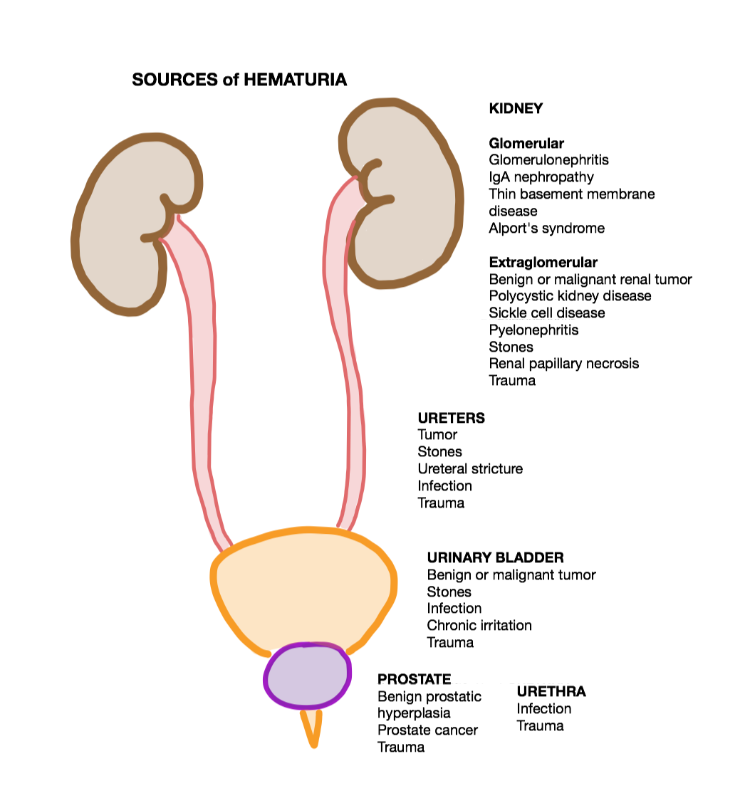
Hematuria or haematuria is defined as the presence of blood or red blood cells in the urine. “Gross hematuria” occurs when urine appears red, brown, or tea-colored due to the presence of blood. Hematuria may also be subtle and only detectable with a microscope or laboratory test. Blood that enters and mixes with the urine can come from any location within the urinary system, including the kidney, ureter, urinary bladder, urethra, and in men, the prostate. Common causes of hematuria include urinary tract infection (UTI), kidney stones, viral illness, trauma, bladder cancer, and exercise. These causes are grouped into glomerular and non-glomerular causes, depending on the involvement of the glomerulus of the kidney. But not all red urine is hematuria. Other substances such as certain medications and foods (e.g. blackberries, beets, food dyes) can cause urine to appear red. Menstruation in women may also cause the appearance of hematuria and may result in a positive urine dipstic ... [...More Info...] [...Related Items...] OR: [Wikipedia] [Google] [Baidu] |
Renal Colic
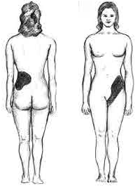
Renal colic is a type of abdominal pain commonly caused by obstruction of ureter from dislodged kidney stones. The most frequent site of obstruction is the vesico-ureteric junction (VUJ), the narrowest point of the upper urinary tract. Acute obstruction and the resultant urinary stasis (disruption of urine flow) can distend the ureter ( hydroureter) and cause a reflexive peristaltic smooth muscle spasm, which leads to a very intense visceral pain transmitted via the ureteric plexus. Signs and symptoms Renal colic typically begins in the flank and often radiates to below the ribs or the groin. It typically comes in waves due to ureteric peristalsis, but may be constant. It is often described as one of the most severe pains. Although this condition can be very painful, most ureteric stones under 5 mm size will eventually pass into the bladder without needing treatments, and cause no permanent physical damage. The experience is said to be traumatizing due to the severe ... [...More Info...] [...Related Items...] OR: [Wikipedia] [Google] [Baidu] |
Ureters
The ureters are tubes made of smooth muscle that propel urine from the kidneys to the urinary bladder. In a human adult, the ureters are usually long and around in diameter. The ureter is lined by urothelial cells, a type of transitional epithelium, and has an additional smooth muscle layer that assists with peristalsis in its lowest third. The ureters can be affected by a number of diseases, including urinary tract infections and kidney stone. is when a ureter is narrowed, due to for example chronic inflammation. Congenital abnormalities that affect the ureters can include the development of two ureters on the same side or abnormally placed ureters. Additionally, reflux of urine from the bladder back up the ureters is a condition commonly seen in children. The ureters have been identified for at least two thousand years, with the word "ureter" stemming from the stem relating to urinating and seen in written records since at least the time of Hippocrates. It is, however, on ... [...More Info...] [...Related Items...] OR: [Wikipedia] [Google] [Baidu] |
X-rays
An X-ray, or, much less commonly, X-radiation, is a penetrating form of high-energy electromagnetic radiation. Most X-rays have a wavelength ranging from 10 picometers to 10 nanometers, corresponding to frequencies in the range 30 petahertz to 30 exahertz ( to ) and energies in the range 145 eV to 124 keV. X-ray wavelengths are shorter than those of UV rays and typically longer than those of gamma rays. In many languages, X-radiation is referred to as Röntgen radiation, after the German scientist Wilhelm Conrad Röntgen, who discovered it on November 8, 1895. He named it ''X-radiation'' to signify an unknown type of radiation.Novelline, Robert (1997). ''Squire's Fundamentals of Radiology''. Harvard University Press. 5th edition. . Spellings of ''X-ray(s)'' in English include the variants ''x-ray(s)'', ''xray(s)'', and ''X ray(s)''. The most familiar use of X-rays is checking for fractures (broken bones), but X-rays are also used in other ways. ... [...More Info...] [...Related Items...] OR: [Wikipedia] [Google] [Baidu] |
Vein
Veins are blood vessels in humans and most other animals that carry blood towards the heart. Most veins carry deoxygenated blood from the tissues back to the heart; exceptions are the pulmonary and umbilical veins, both of which carry oxygenated blood to the heart. In contrast to veins, arteries carry blood away from the heart. Veins are less muscular than arteries and are often closer to the skin. There are valves (called ''pocket valves'') in most veins to prevent backflow. Structure Veins are present throughout the body as tubes that carry blood back to the heart. Veins are classified in a number of ways, including superficial vs. deep, pulmonary vs. systemic, and large vs. small. *Superficial veins are those closer to the surface of the body, and have no corresponding arteries. *Deep veins are deeper in the body and have corresponding arteries. * Perforator veins drain from the superficial to the deep veins. These are usually referred to in the lower limbs and feet. *Comm ... [...More Info...] [...Related Items...] OR: [Wikipedia] [Google] [Baidu] |
Cannula
A cannula (; Latin meaning 'little reed'; plural or ) is a tube that can be inserted into the body, often for the delivery or removal of fluid or for the gathering of samples. In simple terms, a cannula can surround the inner or outer surfaces of a trocar needle thus extending the effective needle length by at least half the length of the original needle. Its size mainly ranges from 14 to 24 gauge. Different-sized cannula have different colours as coded. Decannulation is the permanent removal of a cannula ( extubation), especially of a tracheostomy cannula, once a physician determines it is no longer needed for breathing. Medicine Cannulas normally come with a trocar inside. The trocar is a needle, which punctures the body in order to get into the intended space. Many types of cannulas exist: Intravenous cannulas are the most common in hospital use. A variety of cannulas are used to establish cardiopulmonary bypass in cardiac surgery. A nasal cannula is a piece of plastic ... [...More Info...] [...Related Items...] OR: [Wikipedia] [Google] [Baidu] |
Contrast Medium
A contrast agent (or contrast medium) is a substance used to increase the contrast of structures or fluids within the body in medical imaging. Contrast agents absorb or alter external electromagnetism or ultrasound, which is different from radiopharmaceuticals, which emit radiation themselves. In x-rays, contrast agents enhance the radiodensity in a target tissue or structure. In MRIs, contrast agents shorten (or in some instances increase) the relaxation times of nuclei within body tissues in order to alter the contrast in the image. Contrast agents are commonly used to improve the visibility of blood vessels and the gastrointestinal tract. Several types of contrast agent are in use in medical imaging and they can roughly be classified based on the imaging modalities where they are used. Most common contrast agents work based on X-ray attenuation and magnetic resonance signal enhancement. Radiocontrast media For radiography, which is based on X-rays, iodine and barium are ... [...More Info...] [...Related Items...] OR: [Wikipedia] [Google] [Baidu] |
Medullary Sponge Kidney
Medullary sponge kidney is a congenital disorder of the kidneys characterized by cystic dilatation of the collecting tubules in one or both kidneys. Individuals with medullary sponge kidney are at increased risk for kidney stones and urinary tract infection (UTI). Patients with MSK typically pass twice as many stones per year as do other stone formers without MSK. While having a low morbidity rate, as many as 10% of patients with MSK have an increased risk of morbidity associated with frequent stones and UTIs. While many patients report increased chronic kidney pain, the source of the pain, when a UTI or blockage is not present, is unclear at this time. Renal colic (flank and back pain) is present in 55% of patients. Women with MSK experience more stones, UTIs, and complications than men. MSK was previously believed not to be hereditary but there is more evidence coming forth that may indicate otherwise. Signs and symptoms Most cases are asymptomatic or are discovered during an i ... [...More Info...] [...Related Items...] OR: [Wikipedia] [Google] [Baidu] |


