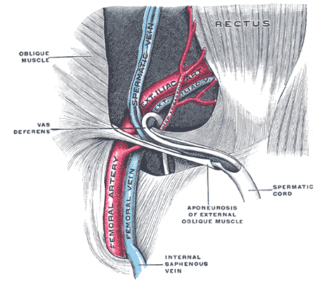
Femoral Vessel on:
[Wikipedia]
[Google]
[Amazon]
 The femoral vessels are those blood vessels passing through the
 The femoral vessels are those blood vessels passing through the
 femoral ring
The femoral ring is the opening at the proximal, abdominal end of the femoral canal, and represents the (superiorly directed/oriented) base of the conically-shaped femoral canal. The femoral ring is oval-shaped, with its long diameter being direc ...
into the femoral canal thereby passing down the length of the thigh until behind the knee. These large vessel are the:
* Femoral artery
The femoral artery is a large artery in the thigh and the main arterial supply to the thigh and leg. The femoral artery gives off the deep femoral artery or profunda femoris artery and descends along the anteromedial part of the thigh in the f ...
(also known in this location as the common femoral artery) and
* Femoral vein
In the human body, the femoral vein is a blood vessel that accompanies the femoral artery in the femoral sheath. It begins at the adductor hiatus (an opening in the adductor magnus muscle) as the continuation of the popliteal vein. It ends ...
Lymphatic vessels
The lymphatic vessels (or lymph vessels or lymphatics) are thin-walled vessels (tubes), structured like blood vessels, that carry lymph. As part of the lymphatic system, lymph vessels are complementary to the cardiovascular system. Lymph vessel ...
found in the thigh aren’t usually included in this collective noun.
As the blood vessels pass along the thigh, they branch, with their main branches remaining closely associated, where they are still referred to collectively as femoral vessels.
The adjective femoral, in this case, relates to the thigh, which contains the femur
The femur (; ), or thigh bone, is the proximal bone of the hindlimb in tetrapod vertebrates. The head of the femur articulates with the acetabulum in the pelvic bone forming the hip joint, while the distal part of the femur articulates wit ...
.
The relative position of these two large vessels is very important in medicine and surgery, because several medical interventions involve puncturing one or the other of them. Reliably distinguishing between them is therefore important. The location of the vessel is also used as an anatomical landmark for the femoral nerve
The femoral nerve is a nerve in the thigh that supplies skin on the upper thigh and inner leg, and the muscles that extend the knee.
Structure
The femoral nerve is the major nerve supplying the anterior compartment of the thigh. It is the largest ...
.
References
{{Reflist Arteries of the lower limb Veins of the lower limb