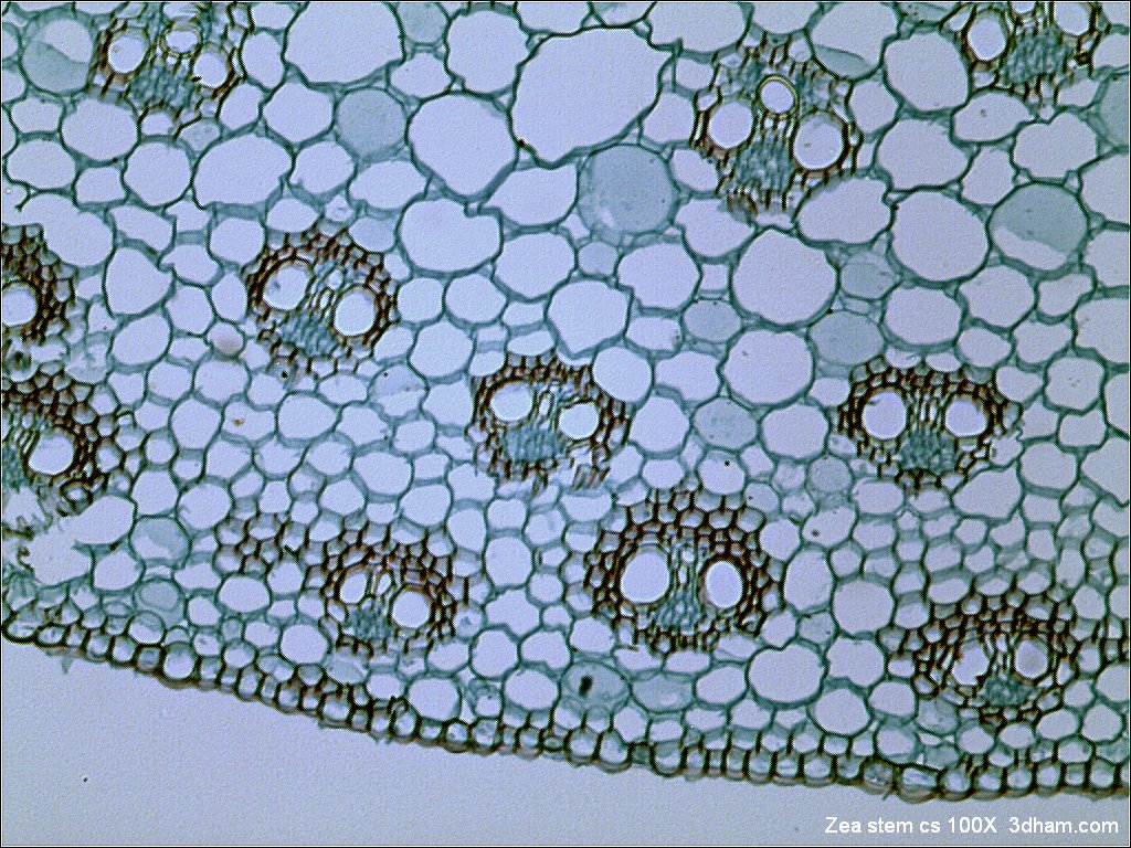
Bright field microscopy on:
[Wikipedia]
[Google]
[Amazon]
 Bright-field microscopy (BF) is the simplest of all the
tissue paper
Tissue paper or simply tissue is a lightweight paper or, light crêpe paper. Tissue can be made from recycled paper pulp on a paper machine.
Tissue paper is very versatile, and different kinds of tissue are made to best serve these purposes, w ...
(1.559 μm/pixel)" align="center">
Image:Paper_Micrograph_Bright.png, Bright-field illumination, sample contrast comes from
Microscopy and Imaging Literature
h2>
 Bright-field microscopy (BF) is the simplest of all the
 optical microscopy
Optics is the branch of physics that studies the behaviour and properties of light, including its interactions with matter and the construction of instruments that use or detect it. Optics usually describes the behaviour of visible, ultravio ...
illumination techniques. Sample illumination is transmitted (i.e., illuminated from below and observed from above) white light
White is the lightest color and is achromatic (having no hue). It is the color of objects such as snow, chalk, and milk, and is the opposite of black. White objects fully reflect and scatter all the visible wavelengths of light. White on ...
, and contrast in the sample is caused by attenuation
In physics, attenuation (in some contexts, extinction) is the gradual loss of flux intensity through a medium. For instance, dark glasses attenuate sunlight, lead attenuates X-rays, and water and air attenuate both light and sound at var ...
of the transmitted light in dense areas of the sample. Bright-field microscopy is the simplest of a range of techniques used for illumination of samples in light microscopes, and its simplicity makes it a popular technique. The typical appearance of a bright-field microscopy image is a dark sample on a bright background, hence the name.
Light path
The light path of a bright-field microscope is extremely simple, no additional components are required beyond the normal light-microscope setup. The light path therefore consists of: * a transillumination light source, commonly ahalogen lamp
A halogen lamp (also called tungsten halogen, quartz-halogen, and quartz iodine lamp) is an incandescent lamp consisting of a tungsten filament sealed in a compact transparent envelope that is filled with a mixture of an inert gas and a small ...
in the microscope stand;
* a condenser lens, which focuses light from the light source onto the sample;
* an objective lens
In optical engineering, the objective is the optical element that gathers light from the object being observed and focuses the light rays to produce a real image. Objectives can be a single lens or mirror, or combinations of several optical elem ...
, which collects light from the sample and magnifies the image;
* oculars and/or a camera
A camera is an optical instrument that can capture an image. Most cameras can capture 2D images, with some more advanced models being able to capture 3D images. At a basic level, most cameras consist of sealed boxes (the camera body), with ...
to view the sample image.
Bright-field microscopy may use critical
Critical or Critically may refer to:
*Critical, or critical but stable, medical states
**Critical, or intensive care medicine
* Critical juncture, a discontinuous change studied in the social sciences.
*Critical Software, a company specializing ...
or Köhler illumination Köhler illumination is a method of specimen illumination used for transmitted and reflected light (trans- and epi-illuminated) optical microscopy. Köhler illumination acts to generate an even illumination of the sample and ensures that an image o ...
to illuminate the sample.
Performance
Bright-field microscopy typically has low contrast with most biological samples, as few absorb light to a great extent.Staining
Staining is a technique used to enhance contrast in samples, generally at the microscopic level. Stains and dyes are frequently used in histology (microscopic study of biological tissues), in cytology (microscopic study of cells), and in th ...
is often required to increase contrast, which prevents use on live cells in many situations. Bright-field illumination is useful for samples that have an intrinsic color, for example chloroplasts
A chloroplast () is a type of membrane-bound organelle known as a plastid that conducts photosynthesis mostly in plant and algal cells. The photosynthetic pigment chlorophyll captures the energy from sunlight, converts it, and stores it in ...
in plant cells.
absorbance
Absorbance is defined as "the logarithm of the ratio of incident to transmitted radiant power through a sample (excluding the effects on cell walls)". Alternatively, for samples which scatter light, absorbance may be defined as "the negative lo ...
of light in the sample
Image:Paper_Micrograph_Cross-Polarised.png, Cross-polarized light
Polarized light microscopy can mean any of a number of optical microscopy techniques involving polarized light. Simple techniques include illumination of the sample with polarized light. Directly transmitted light can, optionally, be blocked with ...
illumination, sample contrast comes from the rotation of polarized light through the sample
Image:Paper_Micrograph_Dark.png, Dark-field illumination, sample contrast comes from light scattered
Scattered may refer to:
Music
* ''Scattered'' (album), a 2010 album by The Handsome Family
* "Scattered" (The Kinks song), 1993
* "Scattered", a song by Ace Young
* "Scattered", a song by Lauren Jauregui
* "Scattered", a song by Green Day from ' ...
by the sample
Image:Paper_Micrograph_Phase.png, Phase-contrast
__NOTOC__
Phase-contrast microscopy (PCM) is an optical microscopy technique that converts phase shifts in light passing through a transparent specimen to brightness changes in the image. Phase shifts themselves are invisible, but become visibl ...
illumination, sample contrast comes from interference of different path lengths of light through the sample
Bright-field microscopy is a standard light-microscopy technique, and therefore magnification
Magnification is the process of enlarging the apparent size, not physical size, of something. This enlargement is quantified by a calculated number also called "magnification". When this number is less than one, it refers to a reduction in si ...
is limited by the resolving power possible with the wavelength
In physics, the wavelength is the spatial period of a periodic wave—the distance over which the wave's shape repeats.
It is the distance between consecutive corresponding points of the same phase on the wave, such as two adjacent crests, tr ...
of visible light
Light or visible light is electromagnetic radiation that can be perceived by the human eye. Visible light is usually defined as having wavelengths in the range of 400–700 nanometres (nm), corresponding to frequencies of 750–420 t ...
.
Advantages
* Simplicity of setup with only basic equipment required. * Living cells can be seen with bright-field microscopes.Limitations
* Very low contrast of most biological samples. * The practical limit to magnification with a light microscope is around 1300X. Although higher magnifications are possible, it becomes increasingly difficult to maintain image clarity as the magnification increases. * Low apparentoptical resolution
Optical resolution describes the ability of an imaging system to resolve detail, in the object that is being imaged.
An imaging system may have many individual components, including one or more lenses, and/or recording and display components. ...
due to the blur of out-of-focus material.
* Samples that are naturally colorless and transparent cannot be seen well, e.g. many types of mammalian cells. These samples often have to be stained before viewing. Samples that do have their own color can be seen without preparation, e.g. the observation of cytoplasmic streaming
Cytoplasmic streaming, also called protoplasmic streaming and cyclosis, is the flow of the cytoplasm inside the cell, driven by forces from the cytoskeleton. It is likely that its function is, at least in part, to speed up the transport of mol ...
in Chara cells.
Enhancements
* Reducing or increasing the amount of the light source by theiris diaphragm
In optics, a diaphragm is a thin opaque structure with an opening (aperture) at its center. The role of the diaphragm is to ''stop'' the passage of light, except for the light passing through the ''aperture''. Thus it is also called a stop (an ...
.
* Use of an oil-immersion objective lens and a special immersion oil placed on a glass cover over the specimen. Immersion oil has the same refraction
In physics, refraction is the redirection of a wave as it passes from one medium to another. The redirection can be caused by the wave's change in speed or by a change in the medium. Refraction of light is the most commonly observed phenomen ...
as glass and improves the resolution of the observed specimen.
* Use of sample-staining methods for use in microbiology
Microbiology () is the scientific study of microorganisms, those being unicellular (single cell), multicellular (cell colony), or acellular (lacking cells). Microbiology encompasses numerous sub-disciplines including virology, bacteriology, ...
, such as simple stains (methylene blue
Methylthioninium chloride, commonly called methylene blue, is a salt used as a dye and as a medication. Methylene blue is a thiazine dye. As a medication, it is mainly used to treat methemoglobinemia by converting the ferric iron in hemoglobin ...
, safranin, crystal violet
Crystal violet or gentian violet, also known as methyl violet 10B or hexamethyl pararosaniline chloride, is a triarylmethane dye used as a histological stain and in Gram's method of classifying bacteria. Crystal violet has antibacterial, antif ...
) and differential stains (negative stains, flagellar stains, endospore stains).
* Use of a colored (usually blue) or polarizing filter on the light source to highlight features not visible under white light. The use of filters is especially useful with mineral
In geology and mineralogy, a mineral or mineral species is, broadly speaking, a solid chemical compound with a fairly well-defined chemical composition and a specific crystal structure that occurs naturally in pure form.John P. Rafferty, ed. (2 ...
samples.
References
# Advanced Light Microscopy vol. 1 Principles and Basic Properties by Maksymilian Pluta, Elsevier (1988) # Advanced Light Microscopy vol. 2 Specialised Methods by Maksymilian Pluta, Elsevier (1989) # Introduction to Light Microscopy by S. Bradbury, B. Bracegirdle, BIOS Scientific Publishers (1998) # Microbiology: Principles and Explorations by Jacquelyn G. Black, John Wiley & Sons, Inc. (2005)Microscopy and Imaging Literature
h2>
Notes
{{Optical microscopy Optical microscopy techniques