|
Zona Incerta
The zona incerta (ZI) is a horizontally elongated region of gray matter in the subthalamus below the thalamus. Its connections project extensively over the brain from the cerebral cortex down into the spinal cord. Its function is unknown, though several potential functions related to "limbic–motor integration" have been proposed, such as controlling visceral activity and pain; gating sensory input and synchronizing cortical and subcortical brain rhythms. Its dysfunction may play a role in central pain syndrome. It has also been identified as a promising deep brain stimulation therapy target for treating Parkinson's disease. Its existence was first described by Auguste Forel in 1877 as a "region of which nothing certain can be said". A hundred and thirty years later in 2007, Nadia Urbain and Martin Deschênes of Université Laval noted that the "zona incerta is among the least studied regions of the brain; its name does not even appear in the index of many textbooks." Structure ... [...More Info...] [...Related Items...] OR: [Wikipedia] [Google] [Baidu] |
Gray Matter
Grey matter is a major component of the central nervous system, consisting of neuronal cell bodies, neuropil (dendrites and unmyelinated axons), glial cells (astrocytes and oligodendrocytes), synapses, and capillaries. Grey matter is distinguished from white matter in that it contains numerous cell bodies and relatively few myelinated axons, while white matter contains relatively few cell bodies and is composed chiefly of long-range myelinated axons. The colour difference arises mainly from the whiteness of myelin. In living tissue, grey matter actually has a very light grey colour with yellowish or pinkish hues, which come from capillary blood vessels and neuronal cell bodies. Structure Grey matter refers to unmyelinated neurons and other cells of the central nervous system. It is present in the brain, brainstem and cerebellum, and present throughout the spinal cord. Grey matter is distributed at the surface of the cerebral hemispheres (cerebral cortex) and of the cerebellum ... [...More Info...] [...Related Items...] OR: [Wikipedia] [Google] [Baidu] |
Lateral Inhibition
In neurobiology, lateral inhibition is the capacity of an excited neuron to reduce the activity of its neighbors. Lateral inhibition disables the spreading of action potentials from excited neurons to neighboring neurons in the lateral direction. This creates a contrast in stimulation that allows increased sensory perception. It is also referred to as lateral antagonism and occurs primarily in visual processes, but also in tactile, auditory, and even olfactory processing. Cells that utilize lateral inhibition appear primarily in the cerebral cortex and thalamus and make up lateral inhibitory networks (LINs). Artificial lateral inhibition has been incorporated into artificial sensory systems, such as vision chips, hearing systems, and optical mice. An often under-appreciated point is that although lateral inhibition is visualised in a spatial sense, it is also thought to exist in what is known as "lateral inhibition across abstract dimensions." This refers to lateral inhibition ... [...More Info...] [...Related Items...] OR: [Wikipedia] [Google] [Baidu] |
Parietal Lobe
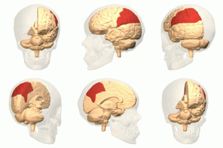
The parietal lobe is one of the four major lobes of the cerebral cortex in the brain of mammals. The parietal lobe is positioned above the temporal lobe and behind the frontal lobe and central sulcus. The parietal lobe integrates sensory information among various modalities, including spatial sense and navigation (proprioception), the main sensory receptive area for the sense of touch in the somatosensory cortex which is just posterior to the central sulcus in the postcentral gyrus, and the dorsal stream of the visual system. The major sensory inputs from the skin (touch, temperature, and pain receptors), relay through the thalamus to the parietal lobe. Several areas of the parietal lobe are important in language processing. The somatosensory cortex can be illustrated as a distorted figure – the cortical homunculus (Latin: "little man") in which the body parts are rendered according to how much of the somatosensory cortex is devoted to them. The superior parietal lobule and in ... [...More Info...] [...Related Items...] OR: [Wikipedia] [Google] [Baidu] |
Cingulate Cortex
The cingulate cortex is a part of the brain situated in the medial aspect of the cerebral cortex. The cingulate cortex includes the entire cingulate gyrus, which lies immediately above the corpus callosum, and the continuation of this in the cingulate sulcus. The cingulate cortex is usually considered part of the limbic lobe. It receives inputs from the thalamus and the neocortex, and projects to the entorhinal cortex via the cingulum. It is an integral part of the limbic system, which is involved with emotion formation and processing, learning, and memory. The combination of these three functions makes the cingulate gyrus highly influential in linking motivational outcomes to behavior (e.g. a certain action induced a positive emotional response, which results in learning). This role makes the cingulate cortex highly important in disorders such as depression and schizophrenia. It also plays a role in executive function and respiratory control. Etymology The term ''cingul ... [...More Info...] [...Related Items...] OR: [Wikipedia] [Google] [Baidu] |
Occipital Lobe
The occipital lobe is one of the four major lobes of the cerebral cortex in the brain of mammals. The name derives from its position at the back of the head, from the Latin ''ob'', "behind", and ''caput'', "head". The occipital lobe is the visual processing center of the mammalian brain containing most of the anatomical region of the visual cortex. The primary visual cortex is Brodmann area 17, commonly called V1 (visual one). Human V1 is located on the medial side of the occipital lobe within the calcarine sulcus; the full extent of V1 often continues onto the occipital pole. V1 is often also called striate cortex because it can be identified by a large stripe of myelin, the Stria of Gennari. Visually driven regions outside V1 are called extrastriate cortex. There are many extrastriate regions, and these are specialized for different visual tasks, such as visuospatial processing, color differentiation, and motion perception. Bilateral lesions of the occipital lobe can lead ... [...More Info...] [...Related Items...] OR: [Wikipedia] [Google] [Baidu] |
Frontal Lobe
The frontal lobe is the largest of the four major lobes of the brain in mammals, and is located at the front of each cerebral hemisphere (in front of the parietal lobe and the temporal lobe). It is parted from the parietal lobe by a groove between tissues called the central sulcus and from the temporal lobe by a deeper groove called the lateral sulcus (Sylvian fissure). The most anterior rounded part of the frontal lobe (though not well-defined) is known as the frontal pole, one of the three poles of the cerebrum. The frontal lobe is covered by the frontal cortex. The frontal cortex includes the premotor cortex, and the primary motor cortex – parts of the motor cortex. The front part of the frontal cortex is covered by the prefrontal cortex. There are four principal gyri in the frontal lobe. The precentral gyrus is directly anterior to the central sulcus, running parallel to it and contains the primary motor cortex, which controls voluntary movements of specific body parts ... [...More Info...] [...Related Items...] OR: [Wikipedia] [Google] [Baidu] |
Brainstem
The brainstem (or brain stem) is the posterior stalk-like part of the brain that connects the cerebrum with the spinal cord. In the human brain the brainstem is composed of the midbrain, the pons, and the medulla oblongata. The midbrain is continuous with the thalamus of the diencephalon through the tentorial notch, and sometimes the diencephalon is included in the brainstem. The brainstem is very small, making up around only 2.6 percent of the brain's total weight. It has the critical roles of regulating cardiac, and respiratory function, helping to control heart rate and breathing rate. It also provides the main motor and sensory nerve supply to the face and neck via the cranial nerves. Ten pairs of cranial nerves come from the brainstem. Other roles include the regulation of the central nervous system and the body's sleep cycle. It is also of prime importance in the conveyance of motor and sensory pathways from the rest of the brain to the body, and from the body back to t ... [...More Info...] [...Related Items...] OR: [Wikipedia] [Google] [Baidu] |
Basal Ganglia
The basal ganglia (BG), or basal nuclei, are a group of subcortical nuclei, of varied origin, in the brains of vertebrates. In humans, and some primates, there are some differences, mainly in the division of the globus pallidus into an external and internal region, and in the division of the striatum. The basal ganglia are situated at the base of the forebrain and top of the midbrain. Basal ganglia are strongly interconnected with the cerebral cortex, thalamus, and brainstem, as well as several other brain areas. The basal ganglia are associated with a variety of functions, including control of voluntary motor movements, procedural learning, habit learning, conditional learning, eye movements, cognition, and emotion. The main components of the basal ganglia – as defined functionally – are the striatum, consisting of both the dorsal striatum (caudate nucleus and putamen) and the ventral striatum (nucleus accumbens and olfactory tubercle), the globus pallidus, ... [...More Info...] [...Related Items...] OR: [Wikipedia] [Google] [Baidu] |
Diencephalon
The diencephalon (or interbrain) is a division of the forebrain (embryonic ''prosencephalon''). It is situated between the telencephalon and the midbrain (embryonic ''mesencephalon''). The diencephalon has also been known as the 'tweenbrain in older literature. It consists of structures that are on either side of the third ventricle, including the thalamus, the hypothalamus, the epithalamus and the subthalamus. The diencephalon is one of the main vesicles of the brain formed during embryogenesis. During the third week of development a neural tube is created from the ectoderm, one of the three primary germ layers. The tube forms three main vesicles during the third week of development: the prosencephalon, the mesencephalon and the rhombencephalon. The prosencephalon gradually divides into the telencephalon and the diencephalon. Structure The diencephalon consists of the following structures: *Thalamus *Hypothalamus including the posterior pituitary *Epithalamus which consists of: ... [...More Info...] [...Related Items...] OR: [Wikipedia] [Google] [Baidu] |
Melanocyte Stimulating Hormone
The melanocyte-stimulating hormones, known collectively as MSH, also known as melanotropins or intermedins, are a family of peptide hormones and neuropeptides consisting of α-melanocyte-stimulating hormone (α-MSH), β-melanocyte-stimulating hormone (β-MSH), and γ-melanocyte-stimulating hormone (γ-MSH) that are produced by cells in the pars intermedia of the anterior lobe of the pituitary gland. Synthetic analogues of α-MSH, such as afamelanotide (melanotan I; Scenesse), melanotan II, and bremelanotide (PT-141), have been developed and researched. Biosynthesis The various forms of MSH are generated from different cleavages of the proopiomelanocortin protein, which also yields other important neuropeptides like adrenocorticotropic hormone. Melanocytes in skin make and secrete MSH in response to ultraviolet light, where it increases synthesis of melanin. Some neurons in arcuate nucleus of the hypothalamus make and secrete α-MSH in response to leptin; α-MSH is also ... [...More Info...] [...Related Items...] OR: [Wikipedia] [Google] [Baidu] |
Angiotensin II
Angiotensin is a peptide hormone that causes vasoconstriction and an increase in blood pressure. It is part of the renin–angiotensin system, which regulates blood pressure. Angiotensin also stimulates the release of aldosterone from the adrenal cortex to promote sodium retention by the kidneys. An oligopeptide, angiotensin is a hormone and a dipsogen. It is derived from the precursor molecule angiotensinogen, a serum globulin produced in the liver. Angiotensin was isolated in the late 1930s (first named 'angiotonin' or 'hypertensin') and subsequently characterized and synthesized by groups at the Cleveland Clinic and Ciba laboratories. Precursor and types Angiotensinogen Angiotensinogen is an α-2-globulin synthesized in the liver and is a precursor for angiotensin, but has also been indicated as having many other roles not related to angiotensin peptides. It is a member of the serpin family of proteins, leading to another name: Serpin A8, although it is not known ... [...More Info...] [...Related Items...] OR: [Wikipedia] [Google] [Baidu] |
Somatostatin
Somatostatin, also known as growth hormone-inhibiting hormone (GHIH) or by several other names, is a peptide hormone that regulates the endocrine system and affects neurotransmission and cell proliferation via interaction with G protein-coupled somatostatin receptors and inhibition of the release of numerous secondary hormones. Somatostatin inhibits insulin and glucagon secretion. Somatostatin has two active forms produced by the alternative cleavage of a single preproprotein: one consisting of 14 amino acids (shown in infobox to right), the other consisting of 28 amino acids. Among the vertebrates, there exist six different somatostatin genes that have been named ''SS1, SS2, SS3, SS4, SS5'' and ''SS6''. Zebrafish have all six. The six different genes, along with the five different somatostatin receptors, allow somatostatin to possess a large range of functions. Humans have only one somatostatin gene, ''SST''. Nomenclature Synonyms of "somatostatin" include: * growth hormoneâ ... [...More Info...] [...Related Items...] OR: [Wikipedia] [Google] [Baidu] |


