|
Cingulate Cortex
The cingulate cortex is a part of the brain situated in the medial aspect of the cerebral cortex. The cingulate cortex includes the entire cingulate gyrus, which lies immediately above the corpus callosum, and the continuation of this in the cingulate sulcus. The cingulate cortex is usually considered part of the limbic lobe. It receives inputs from the thalamus and the neocortex, and projects to the entorhinal cortex via the cingulum. It is an integral part of the limbic system, which is involved with emotion formation and processing, learning, and memory. The combination of these three functions makes the cingulate gyrus highly influential in linking motivational outcomes to behavior (e.g. a certain action induced a positive emotional response, which results in learning). This role makes the cingulate cortex highly important in disorders such as depression and schizophrenia. It also plays a role in executive function and respiratory control. Etymology The term ''cingul ... [...More Info...] [...Related Items...] OR: [Wikipedia] [Google] [Baidu] |
Cerebral Cortex
The cerebral cortex, also known as the cerebral mantle, is the outer layer of neural tissue of the cerebrum of the brain in humans and other mammals. The cerebral cortex mostly consists of the six-layered neocortex, with just 10% consisting of allocortex. It is separated into two cortices, by the longitudinal fissure that divides the cerebrum into the left and right cerebral hemispheres. The two hemispheres are joined beneath the cortex by the corpus callosum. The cerebral cortex is the largest site of neural integration in the central nervous system. It plays a key role in attention, perception, awareness, thought, memory, language, and consciousness. The cerebral cortex is part of the brain responsible for cognition. In most mammals, apart from small mammals that have small brains, the cerebral cortex is folded, providing a greater surface area in the confined volume of the cranium. Apart from minimising brain and cranial volume, cortical folding is crucial for the brain ... [...More Info...] [...Related Items...] OR: [Wikipedia] [Google] [Baidu] |
Executive Function
In cognitive science and neuropsychology, executive functions (collectively referred to as executive function and cognitive control) are a set of cognitive processes that are necessary for the cognitive control of behavior: selecting and successfully monitoring behaviors that facilitate the attainment of chosen goals. Executive functions include basic cognitive processes such as attentional control, cognitive inhibition, inhibitory control, working memory, and cognitive flexibility. Higher-order executive functions require the simultaneous use of multiple basic executive functions and include planning and fluid intelligence (e.g., reasoning and problem-solving). Executive functions gradually develop and change across the lifespan of an individual and can be improved at any time over the course of a person's life. Similarly, these cognitive processes can be adversely affected by a variety of events which affect an individual. Both neuropsychological tests (e.g., the Stroop test ... [...More Info...] [...Related Items...] OR: [Wikipedia] [Google] [Baidu] |
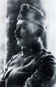
Constantin Von Economo
Constantin Freiherr von Economo ( gr, Κωνσταντίνος Οικονόμου; 21 August 1876 – 21 October 1931) was an Austrian psychiatrist and neurologist of Greek descent, born in modern-day Romania (then Ottoman Empire). He is mostly known for his discovery of encephalitis lethargica and his atlas of cytoarchitectonics of the cerebral cortex. Biography Youth and schooling Constantin Economo von San Serff was born in Brăila, Romania, to Johannes and Helene Economo, a wealthy family with large holdings in Thessaly and Macedonia. The Economo (Οικονόμου, '' Oikonomou'') family originated from Edessa, in the Ottoman Sanjak of Salonica (modern Edessa, Central Macedonia, Greece) where some of Constantin's ancestors were notables, and his family included many bishops. In 1877, the family moved to Trieste, Austria-Hungary,Economo, K. (1932). ''Constantin Freiherr von Economo''. Wien: Mayer & Co. and Constantin spent his childhood and youth in Trieste. He wa ... [...More Info...] [...Related Items...] OR: [Wikipedia] [Google] [Baidu] |
Spindle Neurons - Very High Mag
Spindle may refer to: Textiles and manufacturing * Spindle (textiles), a straight spike to spin fibers into yarn * Spindle (tool), a rotating axis of a machine tool Biology * Common spindle and other species of shrubs and trees in genus ''Euonymus'' whose hard wood was used to make spindles * Spindle apparatus or mitotic spindle, a cellular structure in cell biology * Muscle spindle, stretch receptors within the body of a muscle * Spindle neuron, a specific class of neuron * Sleep spindle, bursts of neural oscillatory activity during sleep * Spindle transfer, an ''in vitro'' fertilization the technique Computing * Spindle (hard disk drive), the axis of a hard disk drive * Spindle (disc packaging), a plastic case for bulk optical disks Vehicles * Spindle (automobile), a part of a car's suspension system * Spindle (vehicle), an autonomous ice-penetrating vehicle Other uses * Spindle (furniture), cylindrically symmetric shaft, usually made of wood * ''Spindle'' (sculpture), ... [...More Info...] [...Related Items...] OR: [Wikipedia] [Google] [Baidu] |
Brodmann Area 33
Brodmann area 33, also known as pregenual area 33, is a subdivision of the cytoarchitecturally defined cingulate region of cerebral cortex. It is a narrow band located in the anterior cingulate gyrus adjacent to the supracallosal gyrus in the depth of the callosal sulcus, near the genu of the corpus callosum. Cytoarchitecturally it is bounded by the ventral anterior cingulate area 24 and the supracallosal gyrus (Brodmann-1909). The pregenual area 33 is heavily involved in emotions, especially happy emotions. Image File:Brodmann area 33 animation small.gif, Animation. File:Brodmann area 33 medial.jpg, Medial view. See also * Brodmann area A Brodmann area is a region of the cerebral cortex, in the human or other primate brain, defined by its cytoarchitecture, or histological structure and organization of cells. History Brodmann areas were originally defined and numbered by th ... References 33 Medial surface of cerebral hemisphere {{Neuroanatomy-stub ... [...More Info...] [...Related Items...] OR: [Wikipedia] [Google] [Baidu] |
Brodmann Area 32
The Brodmann area 32, also known in the human brain as the dorsal anterior cingulate area 32, refers to a subdivision of the cytoarchitecturally defined cingulate cortex. In the human it forms an outer arc around the anterior cingulate gyrus. The cingulate sulcus defines approximately its inner boundary and the superior rostral sulcus (H) its ventral boundary; rostrally it extends almost to the margin of the frontal lobe. Cytoarchitecturally it is bounded internally by the ventral anterior cingulate area 24, externally by medial margins of the agranular frontal area 6, intermediate frontal area 8, granular frontal area 9, frontopolar area 10, and prefrontal area 11-1909. (Brodmann19-09). The dorsal region of the anterior cingulate gyrus is associated with rational thought processes, most notably active during the Stroop task. Guenon In the guenon, Brodmann area 32 is a subdivision of the cytoarchitecturally defined cingulate region of cerebral cortex. This area was named ... [...More Info...] [...Related Items...] OR: [Wikipedia] [Google] [Baidu] |
Brodmann Area 31
Brodmann area 31, also known as dorsal posterior cingulate area 31, is a subdivision of the cytoarchitecturally defined cingulate region of the cerebral cortex. In the human, it occupies portions of the posterior cingulate gyrus and medial aspect of the parietal lobe. Approximate boundaries are the cingulate sulcus dorsally and the parieto-occipital sulcus caudally. It partially surrounds the subparietal sulcus, the ventral continuation of the cingulate sulcus in the parietal lobe. Cytoarchitecturally it is bounded rostrally by the ventral anterior cingulate area 24, ventrally by the ventral posterior cingulate area 23, dorsally by the gigantopyramidal area 4 and preparietal area 5 and caudally by the superior parietal area 7 (H) (Brodmann-1909). See also * Brodmann area A Brodmann area is a region of the cerebral cortex, in the human or other primate brain, defined by its cytoarchitecture, or histological structure and organization of cells. History Brodmann areas wer ... [...More Info...] [...Related Items...] OR: [Wikipedia] [Google] [Baidu] |
Brodmann Area 30
Brodmann area 30, also known as agranular retrolimbic area 30, is a subdivision of the cytoarchitecturally defined retrosplenial region of the cerebral cortex. In the human it is located in the isthmus of cingulate gyrus. Cytoarchitecturally it is bounded internally by the granular retrolimbic area 29, dorsally by the ventral posterior cingulate area 23 and ventrolaterally by the ectorhinal area 36 (Brodmann-1909). See also * Brodmann area A Brodmann area is a region of the cerebral cortex, in the human or other primate brain, defined by its cytoarchitecture, or histological structure and organization of cells. History Brodmann areas were originally defined and numbered by th ... 30 Medial surface of cerebral hemisphere {{Neuroanatomy-stub ... [...More Info...] [...Related Items...] OR: [Wikipedia] [Google] [Baidu] |
Brodmann Area 29
Brodmann area 29, also known as granular retrolimbic area 29 or granular retrosplenial cortex, is a cytoarchitecturally defined portion of the retrosplenial region of the cerebral cortex. In the human it is a narrow band located in the isthmus of cingulate gyrus. Cytoarchitecturally it is bounded internally by the ectosplenial area 26 and externally by the agranular retrolimbic area 30 (Brodmann-1909). Brodmann has this to say about area 29, amongst his other comments on it: "An unusual regression of layer II together with layer III is found in the granular retrosplenial cortex, illustrated in Figures 38 to 41 for four different animals. Here, in addition to the regression of layers II and III, there is fusion of the original layers V and VI and at the same time an isolated massive increase of the inner granular layer, that is particularly prominent in Figures 38 and 39;..." See also * Brodmann area * Retrosplenial region The retrosplenial cortex (RSC) is a cortical area ... [...More Info...] [...Related Items...] OR: [Wikipedia] [Google] [Baidu] |
Brodmann Area 26
Brodmann area 26 is the name for a small part of the brain. Human In the human this area is called ectosplenial area 26. It is a cytoarchitecturally defined portion of the retrosplenial region of the cerebral cortex. It is a narrow band located in the isthmus of cingulate gyrus adjacent to the fasciolar gyrus internally. It is bounded externally by the granular retrolimbic area 29 (Brodmann-1909). Guenon In the guenon Brodmann area 26 is a subdivision of the cerebral cortex defined on the basis of cytoarchitecture. The smallest of Brodmann's cortical areas in the monkey, it represents cortex that is less differentiated and smaller in monkey and human than in other species. Brodmann regarded it as topographically and cytoarchitecturally homologous to the combined human ectosplenial area 26, granular retrolimbic area 29 and agranular retrolimbic area 30 (Brodmann-1909). Distinctive features (Brodmann-1905): thin cortex; distinct but narrow layers. See also * Brodmann area * ... [...More Info...] [...Related Items...] OR: [Wikipedia] [Google] [Baidu] |
Brodmann Area 24
Brodmann area 24 is part of the anterior cingulate in the human brain. Human In the human this area is known as ventral anterior cingulate area 24, and it refers to a subdivision of the cytoarchitecturally defined cingulate cortex region of cerebral cortex (area cingularis anterior ventralis). It occupies most of the anterior cingulate gyrus in an arc around the genu of the corpus callosum. Its outer border corresponds approximately to the cingulate sulcus. Cytoarchitecturally it is bounded internally by the pregenual area 33, externally by the dorsal anterior cingulate area 32, and caudally by the ventral posterior cingulate area 23 and the dorsal posterior cingulate area 31. Guenon In the guenon this area is referred to as area 24 of Brodmann-1905. It includes portions of the cingulate gyrus and the frontal lobe. The cortex is thin; it lacks the internal granular layer (IV) so that the densely distributed, plump pyramidal cells of sublayer 3b of the external pyramidal layer ... [...More Info...] [...Related Items...] OR: [Wikipedia] [Google] [Baidu] |
Brodmann Area 23
Brodmann area 23 (BA23) is a region in the brain that lies inside the posterior cingulate cortex. It lies between Brodmann area 30 and Brodmann area 31 and is located on the medial wall of the cingulate gyrus between the callosal sulcus and the cingulate sulcus. Human This area is also known as ventral posterior cingulate area 23. It is a subdivision of the cytoarchitecturally defined cingulate region of cerebral cortex. In the human it occupies most of the posterior cingulate gyrus adjacent to the corpus callosum. At the caudal extreme it is bounded approximately by the parieto-occipital sulcus. Cytoarchitecturally it is bounded dorsally by the dorsal posterior cingulate area 31, rostrally by the ventral anterior cingulate area 24, and ventrorostrally in its caudal half by the retrosplenial region (Brodmann-1909). Guenon Brodmann area 23 is a subdivision of the cerebral cortex of the guenon defined on the basis of cytoarchitecture. Brodmann regarded it as topographically and cyt ... [...More Info...] [...Related Items...] OR: [Wikipedia] [Google] [Baidu] |


