|
Ventricular Outflow Tract
A ventricular outflow tract is a portion of either the left ventricle or right ventricle of the heart through which blood passes in order to enter the great arteries. The right ventricular outflow tract (RVOT) is an infundibular extension of the ventricular cavity that connects to the pulmonary artery. The left ventricular outflow tract (LVOT), which connects to the aorta, is nearly indistinguishable from the rest of the ventricle. The outflow tract is derived from the secondary heart field, during cardiogenesis. Both the left and right outflow tract have their own term. The right outflow tract is called "conus arteriosus" from the outside, and infundibulum from the inside. In the left ventricle the outflow tract is the "aortic vestibule". They both possess smooth walls, and are derived from the embryonic bulbus cordis In both left and right ventricle there are specific structures separating the inflow and outflow of blood. In the right ventricle, the inflow and outflow is separ ... [...More Info...] [...Related Items...] OR: [Wikipedia] [Google] [Baidu] |
Circulatory System
The blood circulatory system is a system of organs that includes the heart, blood vessels, and blood which is circulated throughout the entire body of a human or other vertebrate. It includes the cardiovascular system, or vascular system, that consists of the heart and blood vessels (from Greek ''kardia'' meaning ''heart'', and from Latin ''vascula'' meaning ''vessels''). The circulatory system has two divisions, a systemic circulation or circuit, and a pulmonary circulation or circuit. Some sources use the terms ''cardiovascular system'' and ''vascular system'' interchangeably with the ''circulatory system''. The network of blood vessels are the great vessels of the heart including large elastic arteries, and large veins; other arteries, smaller arterioles, capillaries that join with venules (small veins), and other veins. The Closed circulatory system, circulatory system is closed in vertebrates, which means that the blood never leaves the network of blood vessels. Some in ... [...More Info...] [...Related Items...] OR: [Wikipedia] [Google] [Baidu] |
Left Ventricle
A ventricle is one of two large chambers toward the bottom of the heart that collect and expel blood towards the peripheral beds within the body and lungs. The blood pumped by a ventricle is supplied by an atrium, an adjacent chamber in the upper heart that is smaller than a ventricle. Interventricular means between the ventricles (for example the interventricular septum), while intraventricular means within one ventricle (for example an intraventricular block). In a four-chambered heart, such as that in humans, there are two ventricles that operate in a double circulatory system: the right ventricle pumps blood into the pulmonary circulation to the lungs, and the left ventricle pumps blood into the systemic circulation through the aorta. Structure Ventricles have thicker walls than atria and generate higher blood pressures. The physiological load on the ventricles requiring pumping of blood throughout the body and lungs is much greater than the pressure generated by the atria ... [...More Info...] [...Related Items...] OR: [Wikipedia] [Google] [Baidu] |
Right Ventricle
A ventricle is one of two large chambers toward the bottom of the heart that collect and expel blood towards the peripheral beds within the body and lungs. The blood pumped by a ventricle is supplied by an atrium, an adjacent chamber in the upper heart that is smaller than a ventricle. Interventricular means between the ventricles (for example the interventricular septum), while intraventricular means within one ventricle (for example an intraventricular block). In a four-chambered heart, such as that in humans, there are two ventricles that operate in a double circulatory system: the right ventricle pumps blood into the pulmonary circulation to the lungs, and the left ventricle pumps blood into the systemic circulation through the aorta. Structure Ventricles have thicker walls than atria and generate higher blood pressures. The physiological load on the ventricles requiring pumping of blood throughout the body and lungs is much greater than the pressure generated by the atria t ... [...More Info...] [...Related Items...] OR: [Wikipedia] [Google] [Baidu] |
Heart
The heart is a muscular organ in most animals. This organ pumps blood through the blood vessels of the circulatory system. The pumped blood carries oxygen and nutrients to the body, while carrying metabolic waste such as carbon dioxide to the lungs. In humans, the heart is approximately the size of a closed fist and is located between the lungs, in the middle compartment of the chest. In humans, other mammals, and birds, the heart is divided into four chambers: upper left and right atria and lower left and right ventricles. Commonly the right atrium and ventricle are referred together as the right heart and their left counterparts as the left heart. Fish, in contrast, have two chambers, an atrium and a ventricle, while most reptiles have three chambers. In a healthy heart blood flows one way through the heart due to heart valves, which prevent backflow. The heart is enclosed in a protective sac, the pericardium, which also contains a small amount of fluid. The wall ... [...More Info...] [...Related Items...] OR: [Wikipedia] [Google] [Baidu] |
Blood
Blood is a body fluid in the circulatory system of humans and other vertebrates that delivers necessary substances such as nutrients and oxygen to the cells, and transports metabolic waste products away from those same cells. Blood in the circulatory system is also known as ''peripheral blood'', and the blood cells it carries, ''peripheral blood cells''. Blood is composed of blood cells suspended in blood plasma. Plasma, which constitutes 55% of blood fluid, is mostly water (92% by volume), and contains proteins, glucose, mineral ions, hormones, carbon dioxide (plasma being the main medium for excretory product transportation), and blood cells themselves. Albumin is the main protein in plasma, and it functions to regulate the colloidal osmotic pressure of blood. The blood cells are mainly red blood cells (also called RBCs or erythrocytes), white blood cells (also called WBCs or leukocytes) and platelets (also called thrombocytes). The most abundant cells in vertebrate blo ... [...More Info...] [...Related Items...] OR: [Wikipedia] [Google] [Baidu] |
Great Arteries
The great arteries are the primary arteries that carry blood away from the heart, which include: * ''Pulmonary artery'': the vessel that carries oxygen-depleted blood from the right ventricle to the lungs. * ''Aorta'': the blood vessel through which oxygenated blood from the left ventricle enters the systemic circulation. Development The great arteries originate from the aortic arches during embryonic development. The aortic arches start as five pairs of symmetrical vessels connecting the heart with the dorsal aorta but then undergo a significant remodelling, in which some of these vessels regress (aortic arches 1 and 2), the 3rd pair of arches contribute to form the common carotids, the right 4th will contribute to the base and central part of the right subclavian artery, while the left 4th will form the central portion of the aortic arch. The 5th pair of vessels only form in some cases without any known contribution to the final structure of the great arteries. The right 6th ... [...More Info...] [...Related Items...] OR: [Wikipedia] [Google] [Baidu] |
Conus Arteriosus
The infundibulum (also known as ''conus arteriosus'') is a conical pouch formed from the upper and left angle of the right ventricle in the chordate heart, from which the pulmonary trunk arises. It develops from the bulbus cordis. Typically, the infundibulum refers to the corresponding internal structure, whereas the conus arteriosus refers to the external structure. Defects in infundibulum development can result in a heart condition known as tetralogy of Fallot. A tendinous band extends upward from the right atrioventricular fibrous ring and connects the posterior surface of the infundibulum to the aorta. The infundibulum is the entrance from the right ventricle into the pulmonary artery and pulmonary trunk. The wall of the infundibulum is smooth. Additional images File:Gray490.png, Front view of human Humans (''Homo sapiens'') are the most abundant and widespread species of primate, characterized by bipedalism and exceptional cognitive skills due to a large and compl ... [...More Info...] [...Related Items...] OR: [Wikipedia] [Google] [Baidu] |
Pulmonary Artery
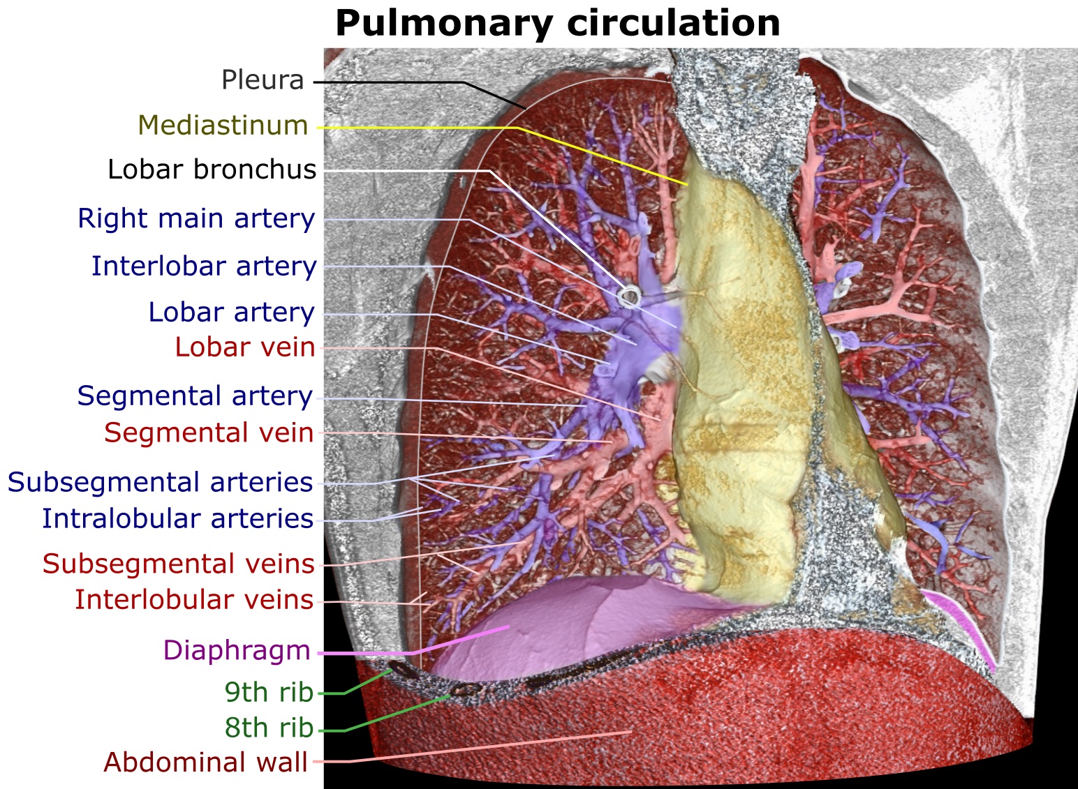
A pulmonary artery is an artery in the pulmonary circulation that carries deoxygenated blood from the right side of the heart to the lungs. The largest pulmonary artery is the ''main pulmonary artery'' or ''pulmonary trunk'' from the heart, and the smallest ones are the arterioles, which lead to the capillaries that surround the pulmonary alveoli. Structure The pulmonary arteries are blood vessels that carry systemic venous blood from the right ventricle of the heart to the microcirculation of the lungs. Unlike in other organs where arteries supply oxygenated blood, the blood carried by the pulmonary arteries is deoxygenated, as it is venous blood returning to the heart. The main pulmonary arteries emerge from the right side of the heart, and then split into smaller arteries that progressively divide and become arterioles, eventually narrowing into the capillary microcirculation of the lungs where gas exchange occurs. Pulmonary trunk In order of blood flow, the pulmonary art ... [...More Info...] [...Related Items...] OR: [Wikipedia] [Google] [Baidu] |
Aorta
The aorta ( ) is the main and largest artery in the human body, originating from the left ventricle of the heart and extending down to the abdomen, where it splits into two smaller arteries (the common iliac arteries). The aorta distributes oxygenated blood to all parts of the body through the systemic circulation. Structure Sections In anatomical sources, the aorta is usually divided into sections. One way of classifying a part of the aorta is by anatomical compartment, where the thoracic aorta (or thoracic portion of the aorta) runs from the heart to the diaphragm. The aorta then continues downward as the abdominal aorta (or abdominal portion of the aorta) from the diaphragm to the aortic bifurcation. Another system divides the aorta with respect to its course and the direction of blood flow. In this system, the aorta starts as the ascending aorta, travels superiorly from the heart, and then makes a hairpin turn known as the aortic arch. Following the aortic arch ... [...More Info...] [...Related Items...] OR: [Wikipedia] [Google] [Baidu] |
Ventricular Tachycardia
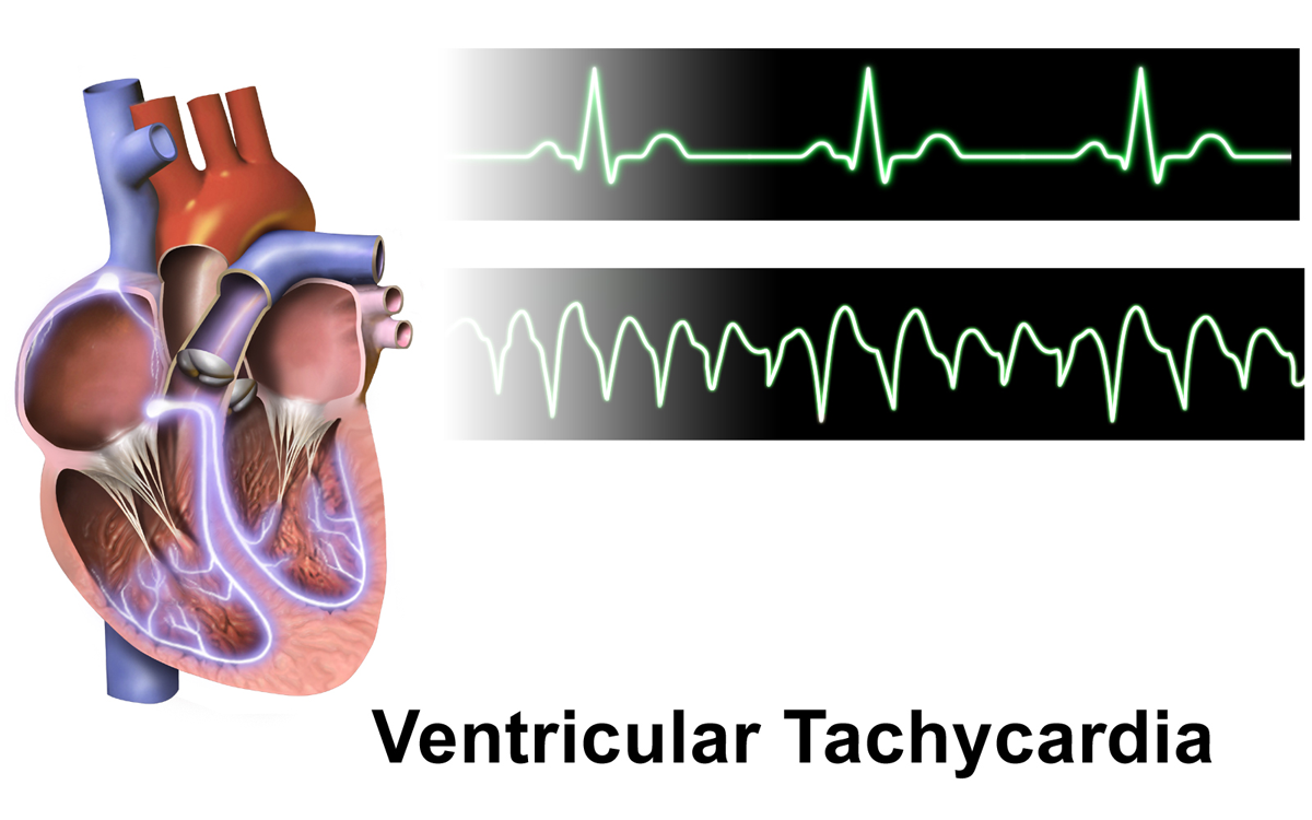
Ventricular tachycardia (V-tach or VT) is a fast heart rate arising from the lower chambers of the heart. Although a few seconds of VT may not result in permanent problems, longer periods are dangerous; and multiple episodes over a short period of time are referred to as an electrical storm. Short periods may occur without symptoms, or present with lightheadedness, palpitations, or chest pain. Ventricular tachycardia may result in ventricular fibrillation (VF) and turn into cardiac arrest. This conversion of the VT into VF is called the degeneration of the VT. It is found initially in about 7% of people in cardiac arrest. Ventricular tachycardia can occur due to coronary heart disease, aortic stenosis, cardiomyopathy, electrolyte problems, or a heart attack. Diagnosis is by an electrocardiogram (ECG) showing a rate of greater than 120 beats per minute and at least three wide QRS complexes in a row. It is classified as non-sustained versus sustained based on whether it lasts le ... [...More Info...] [...Related Items...] OR: [Wikipedia] [Google] [Baidu] |
Brugada Syndrome
Brugada syndrome (BrS) is a genetic disorder in which the electrical activity of the heart is abnormal due to channelopathy. It increases the risk of abnormal heart rhythms and sudden cardiac death. Those affected may have episodes of syncope. The abnormal heart rhythms seen in those with Brugada syndrome often occur at rest. They may be triggered by a fever. About a quarter of those with Brugada syndrome have a family member who also has the condition. Some cases may be due to a new genetic mutation or certain medications. The most commonly involved gene is SCN5A which encodes the cardiac sodium channel. Diagnosis is typically by electrocardiogram (ECG), however, the abnormalities may not be consistently present. Medications such as ajmaline may be used to reveal the ECG changes. Similar ECG patterns may be seen in certain electrolyte disturbances or when the blood supply to the heart has been reduced. There is no cure for Brugada syndrome. Those at higher risk of sudden ... [...More Info...] [...Related Items...] OR: [Wikipedia] [Google] [Baidu] |
Velocity Time Integral
Velocity Time Integral is a clinical Doppler ultrasound measurement of blood flow, equivalent to the area under the velocity time curve. The product of VTI (cm/stroke) and the cross sectional area of a valve (cm2) yields a stroke volume (cm3/stroke), which can be used to calculate cardiac output. VTI can be performed across the left ventricular outflow tract (LVOT), carotid artery, or other blood vessels. __TOC__ LVOT VTI can be incorporated into a POCUS examination to determine the etiology of shock or to predict fluid responsiveness. LVOT VTI can also be used to monitor cardiac output intra-operatively,{{Cite journal, last1=Perrino, first1=Albert C., last2=Harris, first2=Stephen N., last3=Luther, first3=Martha A., date=1998-08-01, title=Intraoperative Determination of Cardiac Output Using Multiplane Transesophageal Echocardiography A Comparison to Thermodilution, url=https://anesthesiology.pubs.asahq.org/article.aspx?articleid=2027702, journal=Anesthesiology: The Journal of the Am ... [...More Info...] [...Related Items...] OR: [Wikipedia] [Google] [Baidu] |


