|
Ventricular Drain
An external ventricular drain (EVD), also known as a ventriculostomy or extraventricular drain, is a device used in neurosurgery to treat hydrocephalus and relieve elevated intracranial pressure when the normal flow of cerebrospinal fluid (CSF) inside the brain is obstructed. An EVD is a flexible plastic catheter placed by a neurosurgeon or neurointensivist and managed by intensive care unit (ICU) physicians and nurses. The purpose of external ventricular drainage is to divert fluid from the ventricles of the brain and allow for monitoring of intracranial pressure. An EVD must be placed in a center with full neurosurgical capabilities, because immediate neurosurgical intervention can be needed if a complication of EVD placement, such as bleeding, is encountered. EVDs are a short-term solution to hydrocephalus, and if the underlying hydrocephalus does not eventually resolve, it may be necessary to convert the EVD to a cerebral shunt, which is a fully internalized, long-term treatm ... [...More Info...] [...Related Items...] OR: [Wikipedia] [Google] [Baidu] |
Ventriculostomy
Ventriculostomy is a neurosurgical procedure that involves creating a hole (stoma) within a cerebral ventricle for drainage. It is most commonly performed on those with hydrocephalus. It is done by surgically penetrating the skull, dura mater, and brain such that the ventricular system ventricle of the brain is accessed. When catheter drainage is temporary, it is commonly referred to as an external ventricular drain (EVD). When catheter drainage is permanent, it is usually referred to as a shunt. There are many catheter-based ventricular shunts that are named for where they terminate, for example, a ventriculi-peritoneal shunt terminates in the peritoneal cavity, a ventriculoarterial shunt terminates within the atrium of the heart, etc. The most common entry point on the skull is called Kocher's point, which is measured 11 cm posterior to the nasion and 3 cm lateral to midline. EVD ventriculostomy is done primarily to monitor the intracranial pressure as well as to drain ... [...More Info...] [...Related Items...] OR: [Wikipedia] [Google] [Baidu] |
Vasospasm
Vasospasm refers to a condition in which an arterial spasm leads to vasoconstriction. This can lead to tissue ischemia and tissue death (necrosis). Cerebral vasospasm may arise in the context of subarachnoid hemorrhage. Symptomatic vasospasm or delayed cerebral ischemia is a major contributor to post-operative stroke and death especially after aneurysmal subarachnoid hemorrhage. Vasospasm typically appears 4 to 10 days after subarachnoid hemorrhage. Along with physical resistance, vasospasm is a main cause of ischemia. Like physical resistance, vasospasms can occur due to atherosclerosis. Vasospasm is the major cause of Prinzmetal's angina. Pathophysiology Normally endothelial cells release prostacyclin and nitric oxide (NO) which induce relaxation of the smooth muscle cells, and reduce aggregation of platelets. Aggregating platelets stimulate ADP to act on endothelial cells and help them induce relaxation of the smooth muscle cells. However, aggregating platelets also stimulat ... [...More Info...] [...Related Items...] OR: [Wikipedia] [Google] [Baidu] |
Reticular Activating System
The reticular formation is a set of interconnected nuclei that are located throughout the brainstem. It is not anatomically well defined, because it includes neurons located in different parts of the brain. The neurons of the reticular formation make up a complex set of networks in the core of the brainstem that extend from the upper part of the midbrain to the lower part of the medulla oblongata. The reticular formation includes ascending pathways to the cortex in the ascending reticular activating system (ARAS) and descending pathways to the spinal cord via the reticulospinal tracts. Neurons of the reticular formation, particularly those of the ascending reticular activating system, play a crucial role in maintaining behavioral arousal and consciousness. The overall functions of the reticular formation are modulatory and premotor, involving somatic motor control, cardiovascular control, pain modulation, sleep and consciousness, and habituation. The modulatory functions are p ... [...More Info...] [...Related Items...] OR: [Wikipedia] [Google] [Baidu] |
Infection Control
Infection prevention and control is the discipline concerned with preventing healthcare-associated infections; a practical rather than academic sub-discipline of epidemiology. In Northern Europe, infection prevention and control is expanded from healthcare into a component in public health, known as "infection protection" (''smittevern, smittskydd, Infektionsschutz'' in the local languages). It is an essential part of the infrastructure of health care. Infection control and hospital epidemiology are akin to public health practice, practiced within the confines of a particular health-care delivery system rather than directed at society as a whole. Infection control addresses factors related to the spread of infections within the healthcare setting, whether among patients, from patients to staff, from staff to patients, or among staff. This includes preventive measures such as hand washing, cleaning, disinfecting, sterilizing, and vaccinating. Other aspects include surveillance, ... [...More Info...] [...Related Items...] OR: [Wikipedia] [Google] [Baidu] |
Ventriculitis
Ventriculitis is the inflammation of the ventricles in the brain. The ventricles are responsible for containing and circulating cerebrospinal fluid throughout the brain. Ventriculitis is caused by infection of the ventricles, leading to swelling and inflammation. This is especially prevalent in patients with external ventricular drains and intraventricular stents. Ventriculitis can cause a wide variety of short-term symptoms and long-term side effects ranging from headaches and dizziness to unconsciousness and death if not treated early. It is treated with some appropriate combination of antibiotics in order to rid the patient of the underlying infection. Much of the current research involving ventriculitis focuses specifically around defining the disease and what causes it. This will allow for much more advancement in the subject. There is also a lot of attention being paid to possible treatments and prevention methods to help make this disease even less prevalent and dangerous. ... [...More Info...] [...Related Items...] OR: [Wikipedia] [Google] [Baidu] |
Surgical Staple
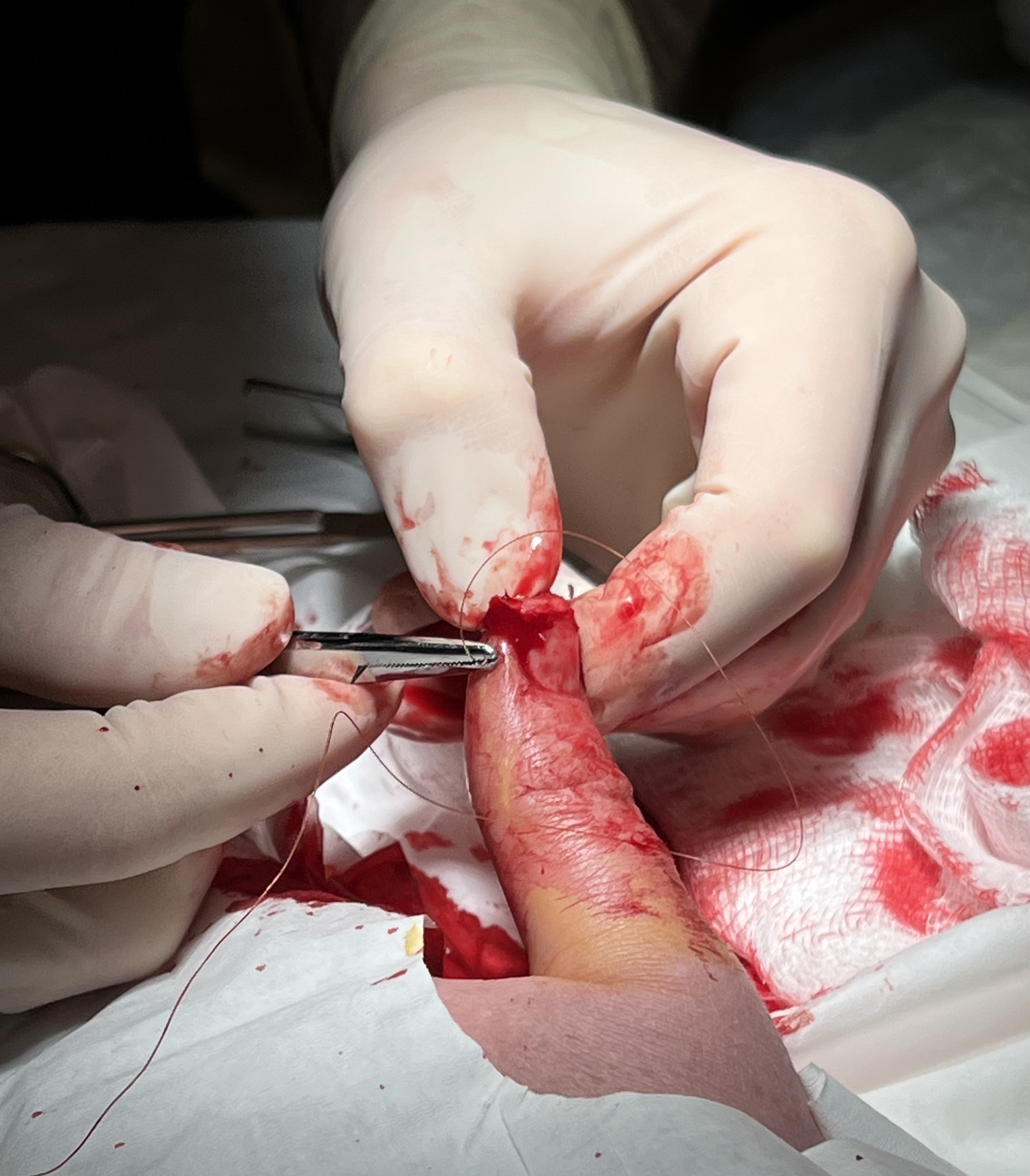
Surgical staples are specialized staples used in surgery in place of sutures to close skin wounds or connect or remove parts of the bowels or lungs. The use of staples over sutures reduces the local inflammatory response, width of the wound, and time it takes to close. A more recent development, from the 1990s, uses clips instead of staples for some applications; this does not require the staple to penetrate. History The technique was pioneered by "father of surgical stapling", Hungarian surgeon Hümér Hültl. Hultl's prototype stapler of 1908 weighed , and required two hours to assemble and load. The technology was refined in the 1950s in the Soviet Union, allowing for the first commercially produced re-usable stapling devices for creation of bowel and vascular anastomoses. Mark M. Ravitch brought a sample of stapling device after attending a surgical conference in USSR, and introduced it to entrepreneur Leon C. Hirsch, who founded the United States Surgical Corporation ... [...More Info...] [...Related Items...] OR: [Wikipedia] [Google] [Baidu] |
Surgical Suture
A surgical suture, also known as a stitch or stitches, is a medical device used to hold body tissues together and approximate wound edges after an injury or surgery. Application generally involves using a needle with an attached length of thread. There are numerous types of suture which differ by needle shape and size as well as thread material and characteristics. Selection of surgical suture should be determined by the characteristics and location of the wound or the specific body tissues being approximated. In selecting the needle, thread, and suturing technique to use for a specific patient, a medical care provider must consider the tensile strength of the specific suture thread needed to efficiently hold the tissues together depending on the mechanical and shear forces acting on the wound as well as the thickness of the tissue being approximated. One must also consider the elasticity of the thread and ability to adapt to different tissues, as well as the memory of the threa ... [...More Info...] [...Related Items...] OR: [Wikipedia] [Google] [Baidu] |
Brainstem
The brainstem (or brain stem) is the posterior stalk-like part of the brain that connects the cerebrum with the spinal cord. In the human brain the brainstem is composed of the midbrain, the pons, and the medulla oblongata. The midbrain is continuous with the thalamus of the diencephalon through the tentorial notch, and sometimes the diencephalon is included in the brainstem. The brainstem is very small, making up around only 2.6 percent of the brain's total weight. It has the critical roles of regulating cardiac, and respiratory function, helping to control heart rate and breathing rate. It also provides the main motor and sensory nerve supply to the face and neck via the cranial nerves. Ten pairs of cranial nerves come from the brainstem. Other roles include the regulation of the central nervous system and the body's sleep cycle. It is also of prime importance in the conveyance of motor and sensory pathways from the rest of the brain to the body, and from the body back to t ... [...More Info...] [...Related Items...] OR: [Wikipedia] [Google] [Baidu] |
Internal Capsule
The internal capsule is a white matter structure situated in the inferomedial part of each cerebral hemisphere of the brain. It carries information past the basal ganglia, separating the caudate nucleus and the thalamus from the putamen and the globus pallidus. The internal capsule contains both ascending and descending axons, going to and coming from the cerebral cortex. It also separates the caudate nucleus and the putamen in the dorsal striatum, a brain region involved in motor and reward pathways. The corticospinal tract constitutes a large part of the internal capsule, carrying motor information from the primary motor cortex to the lower motor neurons in the spinal cord. Above the basal ganglia the corticospinal tract is a part of the corona radiata. Below the basal ganglia the tract is called cerebral crus (a part of the cerebral peduncle) and below the pons it is referred to as the corticospinal tract. Structure The internal capsule consists of three parts and is V-shap ... [...More Info...] [...Related Items...] OR: [Wikipedia] [Google] [Baidu] |
Coagulopathy
Coagulopathy (also called a bleeding disorder) is a condition in which the blood's ability to coagulate (form clots) is impaired. This condition can cause a tendency toward prolonged or excessive bleeding (bleeding diathesis), which may occur spontaneously or following an injury or medical and dental procedures. Coagulopathies are sometimes erroneously referred to as "clotting disorders", but a clotting disorder is the opposite, defined as a predisposition to excessive clot formation (thrombus), also known as a hypercoagulable state or thrombophilia. Signs and symptoms Coagulopathy may cause uncontrolled internal or external bleeding. Left untreated, uncontrolled bleeding may cause damage to joints, muscles, or internal organs and may be life-threatening. People should seek immediate medical care for serious symptoms, including heavy external bleeding, blood in the urine or stool, double vision, severe head or neck pain, repeated vomiting, difficulty walking, convulsions, or se ... [...More Info...] [...Related Items...] OR: [Wikipedia] [Google] [Baidu] |
Mean Arterial Pressure
In medicine, the mean arterial pressure (MAP) is an average blood pressure in an individual during a single cardiac cycle. MAP is altered by cardiac output and systemic vascular resistance. Testing Mean arterial pressure can be measured directly or determined by using a formula. The least invasive method is the use of an blood pressure cuff which gives the values to calculate the mean pressure. A similar method is to use a oscillometric blood pressure device that works by a cuff only method where a microprocessor determines the systolic and diastolic blood pressure. Invasively, an arterial catheter with a transducer is placed and the mean pressure is determined by the subsequent waveform. Calculation While MAP can only be measured directly by invasive monitoring. The MAP can be estimated by using a formula in which the lower (diastolic) blood pressure is doubled and added to the higher (systolic) blood pressure and that composite sum then is divided by 3 to estimate MAP. Nor ... [...More Info...] [...Related Items...] OR: [Wikipedia] [Google] [Baidu] |
Cerebral Perfusion Pressure
Cerebral perfusion pressure, or CPP, is the net pressure gradient causing cerebral blood flow to the brain (brain perfusion). It must be maintained within narrow limits because too little pressure could cause brain tissue to become ischemic (having inadequate blood flow), and too much could raise intracranial pressure (ICP). Definitions The cranium encloses a fixed-volume space that holds three components: blood, cerebrospinal fluid (CSF), and very soft tissue (the brain). While both the blood and CSF have poor compression capacity, the brain is easily compressible. Every increase of ICP can cause a change in tissue perfusion and an increase in stroke events. From resistance CPP can be defined as the pressure gradient causing cerebral blood flow (CBF) such that : CBF = CPP/CVR where: :CVR is cerebrovascular resistance By intracranial pressure An alternative definition of CPP is: : CPP=MAP-ICP where: :MAP is mean arterial pressure :ICP is intracranial pressure :JVP is jugul ... [...More Info...] [...Related Items...] OR: [Wikipedia] [Google] [Baidu] |


