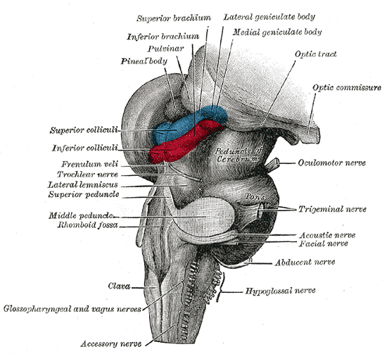
|
Brainstem
The brainstem (or brain stem) is the posterior stalk-like part of the brain that connects the cerebrum with the spinal cord. In the human brain the brainstem is composed of the midbrain, the pons, and the medulla oblongata. The midbrain is continuous with the thalamus of the diencephalon through the tentorial notch, and sometimes the diencephalon is included in the brainstem. The brainstem is very small, making up around only 2.6 percent of the brain's total weight. It has the critical roles of regulating cardiac, and respiratory function, helping to control heart rate and breathing rate. It also provides the main motor and sensory nerve supply to the face and neck via the cranial nerves. Ten pairs of cranial nerves come from the brainstem. Other roles include the regulation of the central nervous system and the body's sleep cycle. It is also of prime importance in the conveyance of motor and sensory pathways from the rest of the brain to the body, and from the body back to t ... [...More Info...] [...Related Items...] OR: [Wikipedia] [Google] [Baidu] |
Human Brain
The human brain is the central organ of the human nervous system, and with the spinal cord makes up the central nervous system. The brain consists of the cerebrum, the brainstem and the cerebellum. It controls most of the activities of the body, processing, integrating, and coordinating the information it receives from the sense organs, and making decisions as to the instructions sent to the rest of the body. The brain is contained in, and protected by, the skull bones of the head. The cerebrum, the largest part of the human brain, consists of two cerebral hemispheres. Each hemisphere has an inner core composed of white matter, and an outer surface – the cerebral cortex – composed of grey matter. The cortex has an outer layer, the neocortex, and an inner allocortex. The neocortex is made up of six neuronal layers, while the allocortex has three or four. Each hemisphere is conventionally divided into four lobes – the frontal, temporal, parietal, and occipital lo ... [...More Info...] [...Related Items...] OR: [Wikipedia] [Google] [Baidu] |
Midbrain
The midbrain or mesencephalon is the forward-most portion of the brainstem and is associated with vision, hearing, motor control, sleep and wakefulness, arousal (alertness), and temperature regulation. The name comes from the Greek ''mesos'', "middle", and ''enkephalos'', "brain". Structure The principal regions of the midbrain are the tectum, the cerebral aqueduct, tegmentum, and the cerebral peduncles. Rostrally the midbrain adjoins the diencephalon (thalamus, hypothalamus, etc.), while caudally it adjoins the hindbrain (pons, medulla and cerebellum). In the rostral direction, the midbrain noticeably splays laterally. Sectioning of the midbrain is usually performed axially, at one of two levels – that of the superior colliculi, or that of the inferior colliculi. One common technique for remembering the structures of the midbrain involves visualizing these cross-sections (especially at the level of the superior colliculi) as the upside-down face of a be ... [...More Info...] [...Related Items...] OR: [Wikipedia] [Google] [Baidu] |
Central Nervous System
The central nervous system (CNS) is the part of the nervous system consisting primarily of the brain and spinal cord. The CNS is so named because the brain integrates the received information and coordinates and influences the activity of all parts of the bodies of bilaterally symmetric and triploblastic animals—that is, all multicellular animals except sponges and diploblasts. It is a structure composed of nervous tissue positioned along the rostral (nose end) to caudal (tail end) axis of the body and may have an enlarged section at the rostral end which is a brain. Only arthropods, cephalopods and vertebrates have a true brain (precursor structures exist in onychophorans, gastropods and lancelets). The rest of this article exclusively discusses the vertebrate central nervous system, which is radically distinct from all other animals. Overview In vertebrates, the brain and spinal cord are both enclosed in the meninges. The meninges provide a barrier to chemicals dissolv ... [...More Info...] [...Related Items...] OR: [Wikipedia] [Google] [Baidu] |
Neck
The neck is the part of the body on many vertebrates that connects the head with the torso. The neck supports the weight of the head and protects the nerves that carry sensory and motor information from the brain down to the rest of the body. In addition, the neck is highly flexible and allows the head to turn and flex in all directions. The structures of the human neck are anatomically grouped into four compartments; vertebral, visceral and two vascular compartments. Within these compartments, the neck houses the cervical vertebrae and cervical part of the spinal cord, upper parts of the respiratory and digestive tracts, endocrine glands, nerves, arteries and veins. Muscles of the neck are described separately from the compartments. They bound the neck triangles. In anatomy, the neck is also called by its Latin names, or , although when used alone, in context, the word ''cervix'' more often refers to the uterine cervix, the neck of the uterus. Thus the adjective ''cervical'' ma ... [...More Info...] [...Related Items...] OR: [Wikipedia] [Google] [Baidu] |
Cranial Nerves
Cranial nerves are the nerves that emerge directly from the brain (including the brainstem), of which there are conventionally considered twelve pairs. Cranial nerves relay information between the brain and parts of the body, primarily to and from regions of the head and neck, including the special senses of vision, taste, smell, and hearing. The cranial nerves emerge from the central nervous system above the level of the first vertebra of the vertebral column. Each cranial nerve is paired and is present on both sides. There are conventionally twelve pairs of cranial nerves, which are described with Roman numerals I–XII. Some considered there to be thirteen pairs of cranial nerves, including cranial nerve zero. The numbering of the cranial nerves is based on the order in which they emerge from the brain and brainstem, from front to back. The terminal nerves (0), olfactory nerves (I) and optic nerves (II) emerge from the cerebrum, and the remaining ten pairs arise from the ... [...More Info...] [...Related Items...] OR: [Wikipedia] [Google] [Baidu] |
Sleep Cycle
The sleep cycle is an oscillation between the slow-wave and REM (paradoxical) phases of sleep. It is sometimes called the ultradian sleep cycle, sleep–dream cycle, or REM-NREM cycle, to distinguish it from the circadian alternation between sleep and wakefulness. In humans, this cycle takes 70 to 110 minutes (90 ± 20 minutes). Characteristics Electroencephalography shows the timing of sleep cycles by virtue of the marked distinction in brainwaves manifested during REM and non-REM sleep. Delta wave activity, correlating with slow-wave (deep) sleep, in particular shows regular oscillations throughout a good night's sleep. Secretions of various hormones, including renin, growth hormone, and prolactin, correlate positively with delta-wave activity, while secretion of thyroid-stimulating hormone correlates inversely. Heart rate variability, well known to increase during REM, predictably also correlates inversely with delta-wave oscillations over the ~90-minute cycle. In order t ... [...More Info...] [...Related Items...] OR: [Wikipedia] [Google] [Baidu] |
Corticospinal Tract
The corticospinal tract is a white matter motor pathway starting at the cerebral cortex that terminates on lower motor neurons and interneurons in the spinal cord, controlling movements of the limbs and trunk. There are more than one million neurons in the corticospinal tract, and they become myelinated usually in the first two years of life. The corticospinal tract is one of the pyramidal tracts, the other being the corticobulbar tract. Anatomy The corticospinal tract originates in several parts of the brain, including not just the motor areas, but also the primary somatosensory cortex and premotor areas. Most of the neurons originate in the primary motor cortex (precentral gyrus, Brodmann area 4) or the premotor frontal areas.Purves, D. et al. (2012). Neuroscience: Fifth edition. Sunderland, MA: Sinauer Associates, Inc.Kolb, B. & Whishaw, I. Q. (2014). An introduction to brain and behavior: Fourth edition. New York, NY: Worth Publishers. About 30% of corticospinal neurons origi ... [...More Info...] [...Related Items...] OR: [Wikipedia] [Google] [Baidu] |
Dorsal Column-medial Lemniscus Pathway
Dorsal (from Latin ''dorsum'' ‘back’) may refer to: * Dorsal (anatomy), an anatomical term of location referring to the back or upper side of an organism or parts of an organism * Dorsal, positioned on top of an aircraft's fuselage * Dorsal consonant, a consonant articulated with the back of the tongue * Dorsal fin A dorsal fin is a fin located on the back of most marine and freshwater vertebrates within various taxa of the animal kingdom. Many species of animals possessing dorsal fins are not particularly closely related to each other, though through conv ..., the fin located on the back of a fish or aircraft * Dorsal transcription factor, a maternally synthesized transcription factor {{disambig de:Dorsale fr:Dorsale it:Dorsale ... [...More Info...] [...Related Items...] OR: [Wikipedia] [Google] [Baidu] |
Brain
A brain is an organ that serves as the center of the nervous system in all vertebrate and most invertebrate animals. It is located in the head, usually close to the sensory organs for senses such as vision. It is the most complex organ in a vertebrate's body. In a human, the cerebral cortex contains approximately 14–16 billion neurons, and the estimated number of neurons in the cerebellum is 55–70 billion. Each neuron is connected by synapses to several thousand other neurons. These neurons typically communicate with one another by means of long fibers called axons, which carry trains of signal pulses called action potentials to distant parts of the brain or body targeting specific recipient cells. Physiologically, brains exert centralized control over a body's other organs. They act on the rest of the body both by generating patterns of muscle activity and by driving the secretion of chemicals called hormones. This centralized control allows rapid and coordinated respon ... [...More Info...] [...Related Items...] OR: [Wikipedia] [Google] [Baidu] |
Breathing Rate
The respiratory rate is the rate at which breathing occurs; it is set and controlled by the respiratory center of the brain. A person's respiratory rate is usually measured in breaths per minute. Measurement The respiratory rate in humans is measured by counting the number of breaths for one minute through counting how many times the chest rises. A fibre-optic breath rate sensor can be used for monitoring patients during a magnetic resonance imaging scan. Respiration rates may increase with fever, illness, or other medical conditions. Inaccuracies in respiratory measurement have been reported in the literature. One study compared respiratory rate counted using a 90-second count period, to a full minute, and found significant differences in the rates.. Another study found that rapid respiratory rates in babies, counted using a stethoscope, were 60–80% higher than those counted from beside the cot without the aid of the stethoscope. Similar results are seen with animals when t ... [...More Info...] [...Related Items...] OR: [Wikipedia] [Google] [Baidu] |
Fine Touch
In physiology, the somatosensory system is the network of Nervous system, neural structures in the brain and body that produce the perception of touch (haptic perception), as well as temperature (thermoception), body position (proprioception), and pain. It is a subset of the sensory nervous system, which also represents Visual system, visual, Auditory system, auditory, olfactory, and gustatory stimuli. Somatosensation begins when Mechanoreceptor, mechano- and Thermoreceptor, thermosensitive structures in the skin or internal organs sense physical stimuli such as pressure on the skin (see mechanotransduction, nociception). Activation of these structures, or receptors, leads to activation of Dorsal root ganglion, peripheral sensory neurons that convey signals to the spinal cord as patterns of action potentials. Sensory information is then processed Posterior horn of spinal cord, locally in the spinal cord to drive reflexes, and is also Dorsal column–medial lemniscus pathway, con ... [...More Info...] [...Related Items...] OR: [Wikipedia] [Google] [Baidu] |


