|
Venous Cutdown
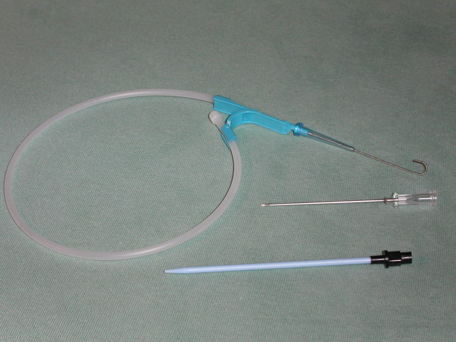
Venous cutdown is an emergency procedure in which the vein is exposed surgically and then a cannula is inserted into the vein under direct vision. It is used to get vascular access in trauma and hypovolemic shock patients when peripheral cannulation is difficult or impossible. The saphenous vein is most commonly used. This procedure has fallen out of favor with the development of safer techniques for central venous catheterization such as the Seldinger technique, the modified Seldinger technique, intraosseous infusion, as well as the use of ultrasound guidance for placement of central venous catheters without using the cutdown technique.Sabba JA, Hedges JR: Ultrasonographic guidance for internal jugular vein cannulation: An educational imperative; a desirable practice alternative. Ann Emerg Med 2006; 48:548-550 Procedure The skin is cleaned, draped, and anesthetized if time allows. The greater saphenous vein is identified on the surface above the medial malleolus, a full-thickness ... [...More Info...] [...Related Items...] OR: [Wikipedia] [Google] [Baidu] |
Cannula
A cannula (; Latin meaning 'little reed'; plural or ) is a tube that can be inserted into the body, often for the delivery or removal of fluid or for the gathering of samples. In simple terms, a cannula can surround the inner or outer surfaces of a trocar needle thus extending the effective needle length by at least half the length of the original needle. Its size mainly ranges from 14 to 24 gauge. Different-sized cannula have different colours as coded. Decannulation is the permanent removal of a cannula (extubation), especially of a tracheostomy cannula, once a physician determines it is no longer needed for breathing. Medicine Cannulas normally come with a trocar inside. The trocar is a needle, which punctures the body in order to get into the intended space. Many types of cannulas exist: Intravenous cannulas are the most common in hospital use. A variety of cannulas are used to establish cardiopulmonary bypass in cardiac surgery. A nasal cannula is a piece of plastic tu ... [...More Info...] [...Related Items...] OR: [Wikipedia] [Google] [Baidu] |
Venous Access
Venous access is any method used to access the bloodstream through the veins, either to administer intravenous therapy (e.g. medication, fluid), parenteral nutrition, to obtain blood for analysis, or to provide an access point for blood-based treatments such as dialysis or apheresis. Access is most commonly achieved via the Seldinger technique, and guidance tools such as ultrasound and fluoroscopy can also be used to assist with visualizing access placement. Methods Peripheral The most common form of venous access is a peripheral venous cannula which is generally inserted into veins of the hands, forearms, and occasionally feet. Healthcare providers may use a number of different techniques in order to improve the chances of successful access. Some techniques include using a tourniquet, tapping over the vein, warming the area to dilate the vein, or using an ultrasound to directly visualize the target vein. Near-infrared illumination devices can also be used to help identify ... [...More Info...] [...Related Items...] OR: [Wikipedia] [Google] [Baidu] |
Physical Trauma
An injury is any physiological damage to living tissue caused by immediate physical stress. An injury can occur intentionally or unintentionally and may be caused by blunt trauma, penetrating trauma, burning, toxic exposure, asphyxiation, or overexertion. Injuries can occur in any part of the body, and different symptoms are associated with different injuries. Treatment of a major injury is typically carried out by a health professional and varies greatly depending on the nature of the injury. Traffic collisions are the most common cause of accidental injury and injury-related death among humans. Injuries are distinct from chronic conditions, psychological trauma, infections, or medical procedures, though injury can be a contributing factor to any of these. Several major health organizations have established systems for the classification and description of human injuries. Occurrence Injuries may be intentional or unintentional. Intentional injuries may be acts o ... [...More Info...] [...Related Items...] OR: [Wikipedia] [Google] [Baidu] |
Hypovolemic Shock
Hypovolemic shock is a form of shock caused by severe hypovolemia (insufficient blood volume or extracellular fluid in the body). It could be the result of severe dehydration through a variety of mechanisms or blood loss. Hypovolemic shock is a medical emergency; if left untreated, the insufficient blood flow can cause damage to organs, leading to multiple organ failure. In treating hypovolemic shock, it is important to determine the cause of the underlying hypovolemia, which may be the result of bleeding or other fluid losses. To minimize ischemic damage to tissues, treatment involves quickly replacing lost blood or fluids, with consideration of both rate and the type of fluids used. Tachycardia, a fast heart rate, is typically the first abnormal vital sign. When resulting from blood loss, trauma is the most common root cause, but severe blood loss can also happen in various body systems without clear traumatic injury. The body in hypovolemic shock prioritizes getting oxygen ... [...More Info...] [...Related Items...] OR: [Wikipedia] [Google] [Baidu] |
Great Saphenous Vein
The great saphenous vein (GSV, alternately "long saphenous vein"; ) is a large, subcutaneous, superficial vein of the leg. It is the longest vein in the body, running along the length of the lower limb, returning blood from the foot, leg and thigh to the deep femoral vein at the femoral triangle. Structure The great saphenous vein originates from where the dorsal vein of the big toe (the hallux) merges with the dorsal venous arch of the foot. After passing in front of the medial malleolus (where it often can be visualized and palpated), it runs up the medial side of the leg. At the knee, it runs over the posterior border of the medial epicondyle of the femur bone. In the proximal anterior thigh inferolateral to the pubic tubercle, the great saphenous vein dives down deep through the cribriform fascia of the saphenous opening to join the femoral vein. It forms an arch, the saphenous arch, to join the common femoral vein in the region of the femoral triangle at the sapheno-femoral ... [...More Info...] [...Related Items...] OR: [Wikipedia] [Google] [Baidu] |
Seldinger Technique
The Seldinger technique, also known as Seldinger wire technique, is a medical procedure to obtain safe access to blood vessels and other hollow organs. It is named after Sven Ivar Seldinger (1921–1998), a Swedish radiologist who introduced the procedure in 1953. Uses The Seldinger technique is used for angiography, insertion of chest drains and central venous catheters, insertion of PEG tubes using the push technique, insertion of the leads for an artificial pacemaker or implantable cardioverter-defibrillator, and numerous other interventional medical procedures. Complications The initial puncture is with a sharp instrument, and this may lead to hemorrhage or perforation of the organ in question. Infection is a possible complication, and hence asepsis is practiced during most Seldinger procedures. Loss of the guidewire into the cavity or blood vessel is a significant and generally preventable complication. Description The desired vessel or cavity is punctured with a shar ... [...More Info...] [...Related Items...] OR: [Wikipedia] [Google] [Baidu] |
Intraosseous Infusion
Intraosseous infusion (IO) is the process of injecting medications, fluids, or blood products directly into the marrow of a bone; this provides a non-collapsible entry point into the systemic venous system. The intraosseous infusion technique is used to provide fluids and medication when intravenous access is not available or not feasible. Intraosseous infusions allow for the administered medications and fluids to go directly into the vascular system. The IO route of fluid and medication administration is an alternative to the preferred intravascular route when the latter cannot be established in a timely manner in emergent situations. Intraosseous infusions are used when people have compromised intravenous access and need immediate delivery of life-saving fluids and medications. Background The use of the IV route to administer fluids has been around since the 1830s, and, in 1922, Cecil K. Drinker et al. saw that bone, specifically the sternum, could also be used as a route o ... [...More Info...] [...Related Items...] OR: [Wikipedia] [Google] [Baidu] |
Anesthesia
Anesthesia is a state of controlled, temporary loss of sensation or awareness that is induced for medical or veterinary purposes. It may include some or all of analgesia (relief from or prevention of pain), paralysis (muscle relaxation), amnesia (loss of memory), and unconsciousness. An individual under the effects of anesthetic drugs is referred to as being anesthetized. Anesthesia enables the painless performance of procedures that would otherwise cause severe or intolerable pain in a non-anesthetized individual, or would otherwise be technically unfeasible. Three broad categories of anesthesia exist: * General anesthesia suppresses central nervous system activity and results in unconsciousness and total lack of sensation, using either injected or inhaled drugs. * Sedation suppresses the central nervous system to a lesser degree, inhibiting both anxiety and creation of long-term memories without resulting in unconsciousness. * Regional and local anesthesia, which blo ... [...More Info...] [...Related Items...] OR: [Wikipedia] [Google] [Baidu] |
Malleolus
A malleolus is the bony prominence on each side of the human ankle. Each leg is supported by two bones, the tibia on the inner side (medial) of the leg and the fibula on the outer side (lateral) of the leg. The medial malleolus is the prominence on the inner side of the ankle, formed by the lower end of the tibia. The lateral malleolus is the prominence on the outer side of the ankle, formed by the lower end of the fibula. The word ''malleolus'' (), plural ''malleoli'' (), comes from Latin and means "small hammer". (It is cognate with ''mallet''.) Medial malleolus The medial malleolus is found at the foot end of the tibia. The medial surface of the lower extremity of tibia is prolonged downward to form a strong pyramidal process, flattened from without inward - the medial malleolus. * The ''medial surface'' of this process is convex and subcutaneous. * The ''lateral'' or ''articular surface'' is smooth and slightly concave, and articulates with the talus. * The ''anterior bo ... [...More Info...] [...Related Items...] OR: [Wikipedia] [Google] [Baidu] |
Intravenous
Intravenous therapy (abbreviated as IV therapy) is a medical technique that administers fluids, medications and nutrients directly into a person's vein. The intravenous route of administration is commonly used for rehydration or to provide nutrients for those who cannot, or will not—due to reduced mental states or otherwise—consume food or water by mouth. It may also be used to administer medications or other medical therapy such as blood products or electrolytes to correct electrolyte imbalances. Attempts at providing intravenous therapy have been recorded as early as the 1400s, but the practice did not become widespread until the 1900s after the development of techniques for safe, effective use. The intravenous route is the fastest way to deliver medications and fluid replacement throughout the body as they are introduced directly into the circulatory system and thus quickly distributed. For this reason, the intravenous route of administration is also used for the consumpti ... [...More Info...] [...Related Items...] OR: [Wikipedia] [Google] [Baidu] |
Cellulitis
Cellulitis is usually a bacterial infection involving the inner layers of the skin. It specifically affects the dermis and subcutaneous fat. Signs and symptoms include an area of redness which increases in size over a few days. The borders of the area of redness are generally not sharp and the skin may be swollen. While the redness often turns white when pressure is applied, this is not always the case. The area of infection is usually painful. Lymphatic vessels may occasionally be involved, and the person may have a fever and feel tired. The legs and face are the most common sites involved, although cellulitis can occur on any part of the body. The leg is typically affected following a break in the skin. Other risk factors include obesity, leg swelling, and old age. For facial infections, a break in the skin beforehand is not usually the case. The bacteria most commonly involved are streptococci and '' Staphylococcus aureus''. In contrast to cellulitis, erysipelas is a bacte ... [...More Info...] [...Related Items...] OR: [Wikipedia] [Google] [Baidu] |
Hematoma
A hematoma, also spelled haematoma, or blood suffusion is a localized bleeding outside of blood vessels, due to either disease or trauma including injury or surgery and may involve blood continuing to seep from broken capillary, capillaries. A hematoma is benign and is initially in liquid form spread among the tissues including in sacs between tissues where it may coagulate and solidify before blood is reabsorbed into blood vessels. An ecchymosis is a hematoma of the skin larger than 10 mm. They may occur among and or within many areas such as skin and other organs, connective tissues, bone, joints and muscle. A collection of blood (or even a hemorrhage) may be aggravated by anticoagulant medication (blood thinner). Blood seepage and collection of blood may occur if heparin is given via an Intramuscular injection, intramuscular route; to avoid this, heparin must be given intravenously or subcutaneous injection, subcutaneously. Signs and symptoms Some hematomas are visible ... [...More Info...] [...Related Items...] OR: [Wikipedia] [Google] [Baidu] |


