|
Valvular Regurgitation
Regurgitation is blood flow in the opposite direction from normal, as the backward flowing of blood into the heart or between heart chambers. It is the circulatory equivalent of backflow in engineered systems. It is sometimes called reflux. Regurgitation in or near the heart is often caused by valvular insufficiency (insufficient function, with incomplete closure, of the heart valves); for example, aortic valve insufficiency causes regurgitation through that valve, called aortic regurgitation, and the terms ''aortic insufficiency'' and ''aortic regurgitation'' are so closely linked as usually to be treated as metonymically interchangeable. The various types of heart valve regurgitation via insufficiency are as follows: # Aortic regurgitation: the backflow of blood from the aorta into the left ventricle, owing to insufficiency of the aortic semilunar valve; it may be chronic or acute. # Mitral regurgitation: the backflow of blood from the left ventricle into the left atrium, ow ... [...More Info...] [...Related Items...] OR: [Wikipedia] [Google] [Baidu] |
Blood Flow
Hemodynamics or haemodynamics are the dynamics of blood flow. The circulatory system is controlled by homeostatic mechanisms of autoregulation, just as hydraulic circuits are controlled by control systems. The hemodynamic response continuously monitors and adjusts to conditions in the body and its environment. Hemodynamics explains the physical laws that govern the flow of blood in the blood vessels. Blood flow ensures the transportation of nutrients, hormones, metabolic waste products, oxygen, and carbon dioxide throughout the body to maintain cell-level metabolism, the regulation of the pH, osmotic pressure and temperature of the whole body, and the protection from microbial and mechanical harm. Blood is a non-Newtonian fluid, and is most efficiently studied using rheology rather than hydrodynamics. Because blood vessels are not rigid tubes, classic hydrodynamics and fluids mechanics based on the use of classical viscometers are not capable of explaining haemodynamics. The ... [...More Info...] [...Related Items...] OR: [Wikipedia] [Google] [Baidu] |
Mitral Valve
The mitral valve (), also known as the bicuspid valve or left atrioventricular valve, is one of the four heart valves. It has two cusps or flaps and lies between the left atrium and the left ventricle of the heart. The heart valves are all one-way valves allowing blood flow in just one direction. The mitral valve and the tricuspid valve are known as the atrioventricular valves because they lie between the atria and the ventricles. In normal conditions, blood flows through an open mitral valve during diastole with contraction of the left atrium, and the mitral valve closes during systole with contraction of the left ventricle. The valve opens and closes because of pressure differences, opening when there is greater pressure in the left atrium than ventricle and closing when there is greater pressure in the left ventricle than atrium. In abnormal conditions, blood may flow backward through the valve ( mitral regurgitation) or the mitral valve may be narrowed (mitral stenosis). Rh ... [...More Info...] [...Related Items...] OR: [Wikipedia] [Google] [Baidu] |
Stenosis
A stenosis (from Ancient Greek στενός, "narrow") is an abnormal narrowing in a blood vessel or other tubular organ or structure such as foramina and canals. It is also sometimes called a stricture (as in urethral stricture). ''Stricture'' as a term is usually used when narrowing is caused by contraction of smooth muscle (e.g. achalasia, prinzmetal angina); ''stenosis'' is usually used when narrowing is caused by lesion that reduces the space of lumen (e.g. atherosclerosis). The term coarctation is another synonym, but is commonly used only in the context of aortic coarctation. Restenosis is the recurrence of stenosis after a procedure. Types The resulting syndrome depends on the structure affected. Examples of vascular stenotic lesions include: * Intermittent claudication (peripheral artery stenosis) * Angina ( coronary artery stenosis) * Carotid artery stenosis which predispose to (strokes and transient ischaemic episodes) * Renal artery stenosis The types of sten ... [...More Info...] [...Related Items...] OR: [Wikipedia] [Google] [Baidu] |
Right Ventricular Volume Overload
Volume overload refers to the state of one of the chambers of the heart in which too large a volume of blood exists within it for it to function efficiently. Ventricular volume overload is approximately equivalent to an excessively high preload. It is a cause of cardiac failure. Pathophysiology In accordance with the Frank–Starling law of the heart, the myocardium contracts more powerfully as the end-diastolic volume increases. Stretching of the myofibrils in cardiac muscle causes them to contract more powerfully due to a greater number of cross-bridges being formed between the myofibrils within cardiac myocytes.Klabunde, Richard E. "Cardiovascular Physiology Concepts". Lippincott Williams & Wilkins, 2011, p. 74. This is true up to a point, however beyond this there is a loss of contractile ability due to loss of connection between myofibrils; see figure. Various pathologies, listed below, can lead to volume overload. Different mechanisms are involved depending on the cau ... [...More Info...] [...Related Items...] OR: [Wikipedia] [Google] [Baidu] |
Cardiac Muscle
Cardiac muscle (also called heart muscle, myocardium, cardiomyocytes and cardiac myocytes) is one of three types of vertebrate muscle tissues, with the other two being skeletal muscle and smooth muscle. It is an involuntary, striated muscle that constitutes the main tissue of the wall of the heart. The cardiac muscle (myocardium) forms a thick middle layer between the outer layer of the heart wall (the pericardium) and the inner layer (the endocardium), with blood supplied via the coronary circulation. It is composed of individual cardiac muscle cells joined by intercalated discs, and encased by collagen fibers and other substances that form the extracellular matrix. Cardiac muscle contracts in a similar manner to skeletal muscle, although with some important differences. Electrical stimulation in the form of a cardiac action potential triggers the release of calcium from the cell's internal calcium store, the sarcoplasmic reticulum. The rise in calcium causes the ... [...More Info...] [...Related Items...] OR: [Wikipedia] [Google] [Baidu] |
Framingham Heart Study
The Framingham Heart Study is a long-term, ongoing cardiovascular cohort study of residents of the city of Framingham, Massachusetts. The study began in 1948 with 5,209 adult subjects from Framingham, and is now on its third generation of participants. Prior to the study almost nothing was known about the epidemiology of hypertensive or arteriosclerotic cardiovascular disease. Much of the now-common knowledge concerning heart disease, such as the effects of diet, exercise, and common medications such as aspirin, is based on this longitudinal study. It is a project of the National Heart, Lung, and Blood Institute, in collaboration with (since 1971) Boston University. Various health professionals from the hospitals and universities of Greater Boston staff the project. History In 1948, the study was commissioned by Congress, with a choice made between Framingham, Massachusetts and Paintsville, Kentucky. Framingham was chosen when residents showed more general interest in heart rese ... [...More Info...] [...Related Items...] OR: [Wikipedia] [Google] [Baidu] |
Stroke Volume
In cardiovascular physiology, stroke volume (SV) is the volume of blood pumped from the left ventricle per beat. Stroke volume is calculated using measurements of ventricle volumes from an echocardiogram and subtracting the volume of the blood in the ventricle at the end of a beat (called end-systolic volume) from the volume of blood just prior to the beat (called end-diastolic volume). The term ''stroke volume'' can apply to each of the two ventricles of the heart, although it usually refers to the left ventricle. The stroke volumes for each ventricle are generally equal, both being approximately 70 mL in a healthy 70-kg man. Stroke volume is an important determinant of cardiac output, which is the product of stroke volume and heart rate, and is also used to calculate ejection fraction, which is stroke volume divided by end-diastolic volume. Because stroke volume decreases in certain conditions and disease states, stroke volume itself correlates with cardiac function. Calculati ... [...More Info...] [...Related Items...] OR: [Wikipedia] [Google] [Baidu] |
Mitral Insufficiency
Mitral regurgitation (MR), also known as mitral insufficiency or mitral incompetence, is a form of valvular heart disease in which the mitral valve is insufficient and does not close properly when the heart pumps out blood.Mitral valve regurgitation at Mount Sinai Hospital It is the abnormal leaking of blood backwards – regurgitation from the , through the mitral valve, into the |
Left Ventricle
A ventricle is one of two large chambers toward the bottom of the heart that collect and expel blood towards the peripheral beds within the body and lungs. The blood pumped by a ventricle is supplied by an atrium, an adjacent chamber in the upper heart that is smaller than a ventricle. Interventricular means between the ventricles (for example the interventricular septum), while intraventricular means within one ventricle (for example an intraventricular block). In a four-chambered heart, such as that in humans, there are two ventricles that operate in a double circulatory system: the right ventricle pumps blood into the pulmonary circulation to the lungs, and the left ventricle pumps blood into the systemic circulation through the aorta. Structure Ventricles have thicker walls than atria and generate higher blood pressures. The physiological load on the ventricles requiring pumping of blood throughout the body and lungs is much greater than the pressure generated by the atria ... [...More Info...] [...Related Items...] OR: [Wikipedia] [Google] [Baidu] |
Tricuspid Valve
The tricuspid valve, or right atrioventricular valve, is on the right dorsal side of the mammalian heart, at the superior portion of the right ventricle. The function of the valve is to allow blood to flow from the right atrium to the right ventricle during diastole, and to close to prevent backflow ( regurgitation) from the right ventricle into the right atrium during right ventricular contraction ( systole). Structure The tricuspid valve usually has three cusps or leaflets, named the anterior, posterior, and septal cusps. Each leaflet is connected via chordae tendineae to the anterior, posterior, and septal papillary muscles of the right ventricle, respectively. Tricuspid valves may also occur with two or four leaflets; the number may change over a lifetime. Function The tricuspid valve functions as a one-way valve that closes during ventricular systole to prevent regurgitation of blood from the right ventricle back into the right atrium. It opens during ventricular diastol ... [...More Info...] [...Related Items...] OR: [Wikipedia] [Google] [Baidu] |
Tricuspid Regurgitation
Tricuspid regurgitation (TR), also called tricuspid insufficiency, is a type of valvular heart disease in which the tricuspid valve of the heart, located between the right atrium and right ventricle, does not close completely when the right ventricle contracts (systole). TR allows the blood to flow backwards from the right ventricle to the right atrium, which increases the volume and pressure of the blood both in the right atrium and the right ventricle, which may increase central venous volume and pressure if the backward flow is sufficiently severe. The causes of TR are divided into ''hereditary'' and ''acquired''; and also ''primary'' and ''secondary''. Primary TR refers to a defect solely in the tricuspid valve, such as infective endocarditis; secondary TR refers to a defect in the valve as a consequence of some other pathology, such as left ventricular failure or pulmonary hypertension. The mechanism of TR is either a dilatation of the base (annulus) of the valve due to righ ... [...More Info...] [...Related Items...] OR: [Wikipedia] [Google] [Baidu] |
Pulmonary Artery
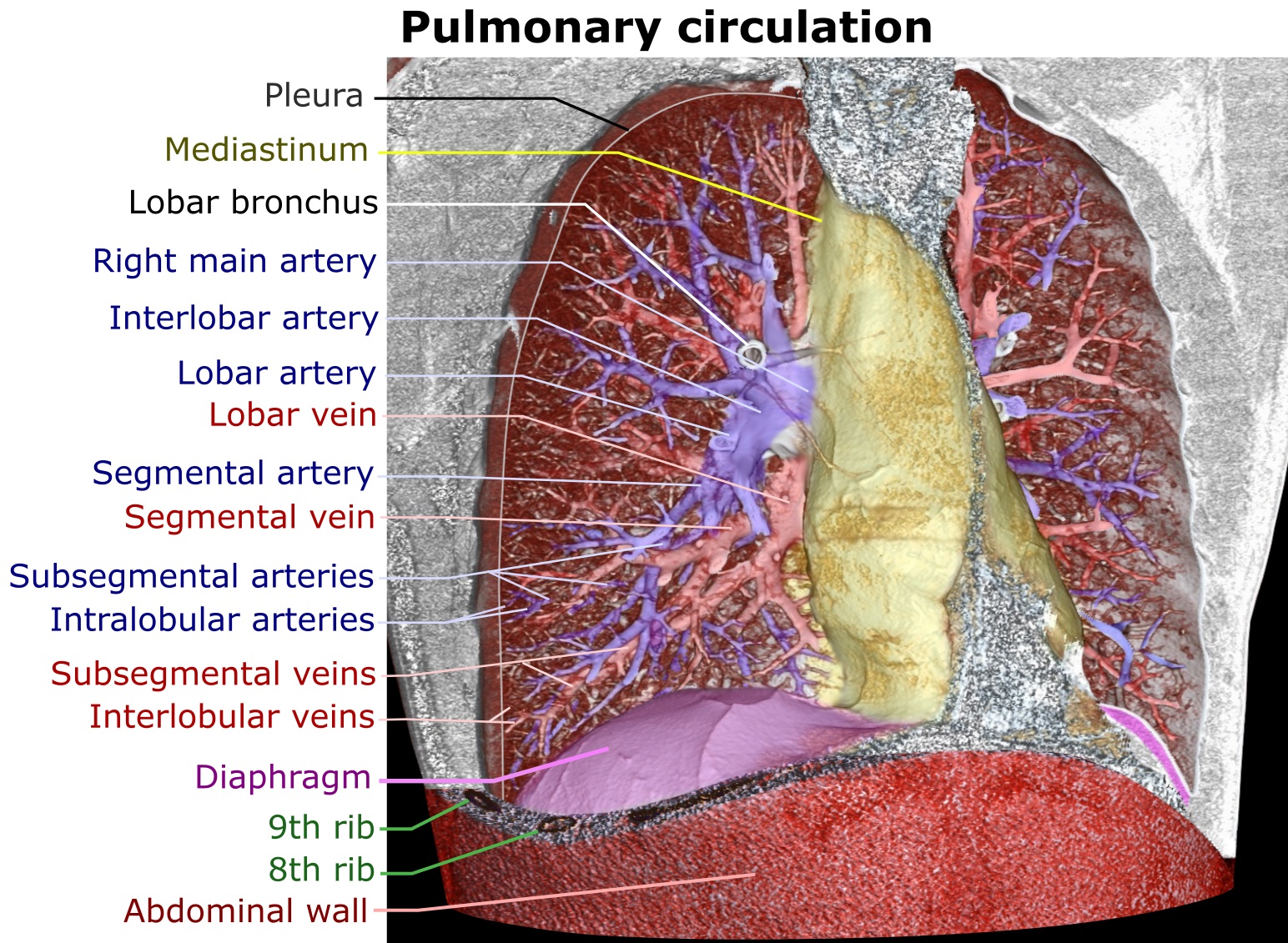
A pulmonary artery is an artery in the pulmonary circulation that carries deoxygenated blood from the right side of the heart to the lungs. The largest pulmonary artery is the ''main pulmonary artery'' or ''pulmonary trunk'' from the heart, and the smallest ones are the arterioles, which lead to the capillaries that surround the pulmonary alveoli. Structure The pulmonary arteries are blood vessels that carry systemic venous blood from the right ventricle of the heart to the microcirculation of the lungs. Unlike in other organs where arteries supply oxygenated blood, the blood carried by the pulmonary arteries is deoxygenated, as it is venous blood returning to the heart. The main pulmonary arteries emerge from the right side of the heart, and then split into smaller arteries that progressively divide and become arterioles, eventually narrowing into the capillary microcirculation of the lungs where gas exchange occurs. Pulmonary trunk In order of blood flow, the pulmonary art ... [...More Info...] [...Related Items...] OR: [Wikipedia] [Google] [Baidu] |


