|
Ultramicroscopic
An ultramicroscope is a microscope with a system that lights the object in a way that allows viewing of tiny particles via light scattering, and not light reflection or absorption. When the diameter of a particle is below or near the wavelength of visible light (around 500 nanometers), the particle cannot be seen in a light microscope with the usual methods of illumination. The ''ultra-'' in ''ultramicroscope'' refers to the ability to see objects whose diameter is shorter than the wavelength of visible light, on the model of the ''ultra-'' in ''ultraviolet''. Synopsis In the system, the particles to be observed are dispersed in a liquid or gas colloid (or less often in a coarser suspension). The colloid is placed in a light-absorbing, dark enclosure, and illuminated with a convergent beam of intense light entering from one side. Light hitting the colloid particles will be scattered. In discussions about light scattering, the converging beam is called a "Tyndall cone". The scene is ... [...More Info...] [...Related Items...] OR: [Wikipedia] [Google] [Baidu] |
Microscope
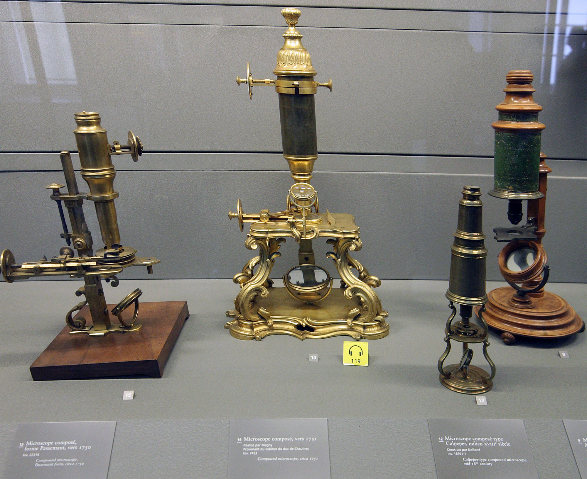
A microscope () is a laboratory instrument used to examine objects that are too small to be seen by the naked eye. Microscopy is the science of investigating small objects and structures using a microscope. Microscopic means being invisible to the eye unless aided by a microscope. There are many types of microscopes, and they may be grouped in different ways. One way is to describe the method an instrument uses to interact with a sample and produce images, either by sending a beam of light or electrons through a sample in its optical path, by detecting photon emissions from a sample, or by scanning across and a short distance from the surface of a sample using a probe. The most common microscope (and the first to be invented) is the optical microscope, which uses lenses to refract visible light that passed through a thinly sectioned sample to produce an observable image. Other major types of microscopes are the fluorescence microscope, electron microscope (both the transmi ... [...More Info...] [...Related Items...] OR: [Wikipedia] [Google] [Baidu] |
Colloids
A colloid is a mixture in which one substance consisting of microscopically dispersed insoluble particles is suspended throughout another substance. Some definitions specify that the particles must be dispersed in a liquid, while others extend the definition to include substances like aerosols and gels. The term colloidal suspension refers unambiguously to the overall mixture (although a narrower sense of the word ''suspension'' is distinguished from colloids by larger particle size). A colloid has a dispersed phase (the suspended particles) and a continuous phase (the medium of suspension). The dispersed phase particles have a diameter of approximately 1 nanometre to 1 micrometre. Some colloids are translucent because of the Tyndall effect, which is the scattering of light by particles in the colloid. Other colloids may be opaque or have a slight color. Colloidal suspensions are the subject of interface and colloid science. This field of study was introduced in 1845 by Italian ... [...More Info...] [...Related Items...] OR: [Wikipedia] [Google] [Baidu] |
Optical Microscopy Techniques
Optics is the branch of physics that studies the behaviour and properties of light, including its interactions with matter and the construction of instruments that use or detect it. Optics usually describes the behaviour of visible, ultraviolet, and infrared light. Because light is an electromagnetic wave, other forms of electromagnetic radiation such as X-rays, microwaves, and radio waves exhibit similar properties. Most optical phenomena can be accounted for by using the classical electromagnetic description of light. Complete electromagnetic descriptions of light are, however, often difficult to apply in practice. Practical optics is usually done using simplified models. The most common of these, geometric optics, treats light as a collection of rays that travel in straight lines and bend when they pass through or reflect from surfaces. Physical optics is a more comprehensive model of light, which includes wave effects such as diffraction and interference that cannot be acc ... [...More Info...] [...Related Items...] OR: [Wikipedia] [Google] [Baidu] |
Microscopes
A microscope () is a laboratory instrument used to examine objects that are too small to be seen by the naked eye. Microscopy is the science of investigating small objects and structures using a microscope. Microscopic means being invisible to the eye unless aided by a microscope. There are many types of microscopes, and they may be grouped in different ways. One way is to describe the method an instrument uses to interact with a sample and produce images, either by sending a beam of light or electrons through a sample in its optical path, by detecting photon emissions from a sample, or by scanning across and a short distance from the surface of a sample using a probe. The most common microscope (and the first to be invented) is the optical microscope, which uses lenses to refract visible light that passed through a thinly sectioned sample to produce an observable image. Other major types of microscopes are the fluorescence microscope, electron microscope (both the transmi ... [...More Info...] [...Related Items...] OR: [Wikipedia] [Google] [Baidu] |
Light Sheet Fluorescence Microscopy
Light or visible light is electromagnetic radiation that can be perceived by the human eye. Visible light is usually defined as having wavelengths in the range of 400–700 nanometres (nm), corresponding to frequencies of 750–420 terahertz, between the infrared (with longer wavelengths) and the ultraviolet (with shorter wavelengths). In physics, the term "light" may refer more broadly to electromagnetic radiation of any wavelength, whether visible or not. In this sense, gamma rays, X-rays, microwaves and radio waves are also light. The primary properties of light are intensity, propagation direction, frequency or wavelength spectrum and polarization. Its speed in a vacuum, 299 792 458 metres a second (m/s), is one of the fundamental constants of nature. Like all types of electromagnetic radiation, visible light propagates by massless elementary particles called photons that represents the quanta of electromagnetic field, and can be analyzed as both waves and particl ... [...More Info...] [...Related Items...] OR: [Wikipedia] [Google] [Baidu] |
Dark-field Microscopy
Dark-field microscopy (also called dark-ground microscopy) describes microscopy methods, in both light and electron microscopy, which exclude the unscattered beam from the image. As a result, the field around the specimen (i.e., where there is no specimen to scatter the beam) is generally dark. In optical microscopes a darkfield condenser lens must be used, which directs a cone of light away from the objective lens. To maximize the scattered light-gathering power of the objective lens, oil immersion is used and the numerical aperture (NA) of the objective lens must be less than 1.0. Objective lenses with a higher NA can be used but only if they have an adjustable diaphragm, which reduces the NA. Often these objective lenses have a NA that is variable from 0.7 to 1.25. Light microscopy applications In optical microscopy, dark-field describes an illumination technique used to enhance the contrast in unstained samples. It works by illuminating the sample with light that wil ... [...More Info...] [...Related Items...] OR: [Wikipedia] [Google] [Baidu] |
Electron Microscope
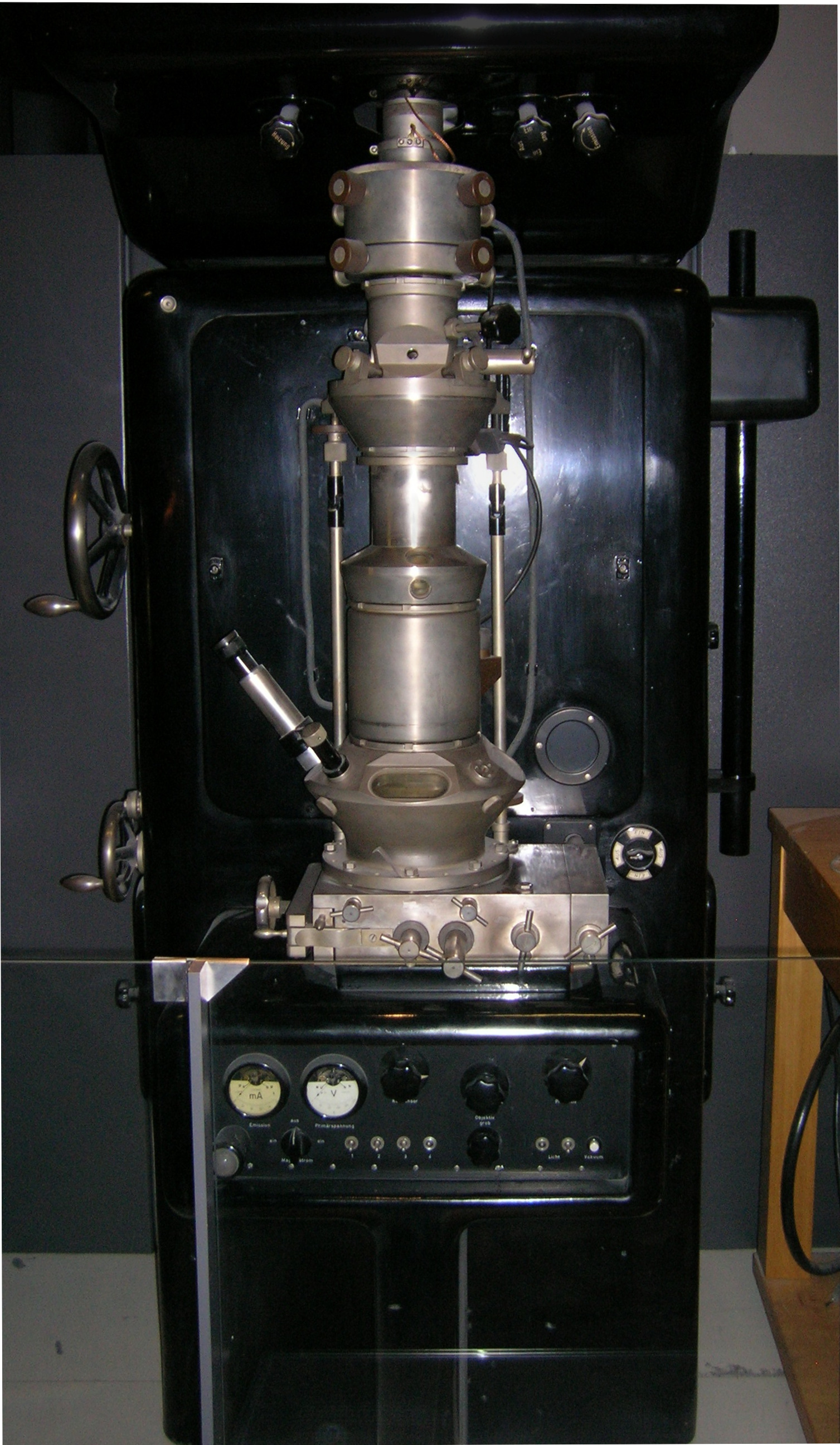
An electron microscope is a microscope that uses a beam of accelerated electrons as a source of illumination. As the wavelength of an electron can be up to 100,000 times shorter than that of visible light photons, electron microscopes have a higher resolving power than light microscopes and can reveal the structure of smaller objects. A scanning transmission electron microscope has achieved better than 50 pm resolution in annular dark-field imaging mode and magnifications of up to about 10,000,000× whereas most light microscopes are limited by diffraction to about 200 nm resolution and useful magnifications below 2000×. Electron microscopes use shaped magnetic fields to form electron optical lens systems that are analogous to the glass lenses of an optical light microscope. Electron microscopes are used to investigate the ultrastructure of a wide range of biological and inorganic specimens including microorganisms, cells, large molecules, biopsy samples, ... [...More Info...] [...Related Items...] OR: [Wikipedia] [Google] [Baidu] |
Cranberry Glass
Cranberry glass or Gold Ruby glass is a red glass made by adding gold salts or colloidal gold to molten glass. Tin, in the form of stannous chloride, is sometimes added in tiny amounts as a reducing agent. The glass is used primarily in expensive decorations. Production Cranberry glass is made in craft production rather than in large quantities, due to the high cost of the gold. The gold chloride is made by dissolving gold in a solution of nitric acid and hydrochloric acid ( aqua regia). The glass is typically hand blown or molded. The finished, hardened glass is a type of colloid, a solid phase (gold) dispersed inside another solid phase (glass). History The origins of cranberry glass making are unknown, but many historians believe a form of this glass was first made in the late Roman Empire. This is evidenced by the British Museum's collection Lycurgus Cup, a 4th-century Roman glass cage cup made of a dichroic glass, which shows a different colour depending on whet ... [...More Info...] [...Related Items...] OR: [Wikipedia] [Google] [Baidu] |
Nanoparticle
A nanoparticle or ultrafine particle is usually defined as a particle of matter that is between 1 and 100 nanometres (nm) in diameter. The term is sometimes used for larger particles, up to 500 nm, or fibers and tubes that are less than 100 nm in only two directions. At the lowest range, metal particles smaller than 1 nm are usually called atom clusters instead. Nanoparticles are usually distinguished from microparticles (1-1000 µm), "fine particles" (sized between 100 and 2500 nm), and "coarse particles" (ranging from 2500 to 10,000 nm), because their smaller size drives very different physical or chemical properties, like colloidal properties and ultrafast optical effects or electric properties. Being more subject to the brownian motion, they usually do not sediment, like colloidal particles that conversely are usually understood to range from 1 to 1000 nm. Being much smaller than the wavelengths of visible light (400-700 nm), nano ... [...More Info...] [...Related Items...] OR: [Wikipedia] [Google] [Baidu] |
Carl Zeiss AG
Carl Zeiss AG (), branded as ZEISS, is a German manufacturer of optical systems and optoelectronics, founded in Jena, Germany in 1846 by optician Carl Zeiss. Together with Ernst Abbe (joined 1866) and Otto Schott (joined 1884) he laid the foundation for today's multi-national company. The current company emerged from a reunification of Carl Zeiss companies in East and West Germany with a consolidation phase in the 1990s. ZEISS is active in four business segments with approximately equal revenue (Industrial Quality and Research, Medical Technology, Consumer Markets and Semiconductor Manufacturing Technology) in almost 50 countries, has 30 production sites and around 25 development sites worldwide. Carl Zeiss AG is the holding of all subsidiaries within Zeiss Group, of which Carl Zeiss Meditec AG is the only one that is traded at the stock market. Carl Zeiss AG is owned by the foundation Carl-Zeiss-Stiftung. The Zeiss Group has its headquarters in southern Germany, in the smal ... [...More Info...] [...Related Items...] OR: [Wikipedia] [Google] [Baidu] |
Henry Siedentopf
Henry Friedrich Wilhelm Siedentopf (22 September 1872 in Bremen – 8 May 1940 in Jena) was a German physicist and pioneer of microscopy. Biography Siedentopf worked in Carl Zeiss company from 1899 to 1938. In 1907 he was nominated as the head of the microscopy department. In 1902 the ultramicroscope was developed by Richard Adolf Zsigmondy (1865–1929) and Siedentopf, working for Carl Zeiss AG. The ultramicroscope was suitable for the determination of small particles and became the most important instrument of colloid research in colloid chemistry. In 1925, Zsigmondy received the Nobel Prize for Chemistry also for this work. From 1919 till 1940 he was a. o. Professor for microscopy at the University of Jena. He also worked on the development of micro photography and slow motion and fast motion in the cinephotomicrography. In 1908 he invented together with August Köhler the fluorescence microscope. In 1930 he was elected a member of the German National Academy of Sciences Le ... [...More Info...] [...Related Items...] OR: [Wikipedia] [Google] [Baidu] |
Richard Adolf Zsigmondy
Richard Adolf Zsigmondy ( hu, Zsigmondy Richárd Adolf; 1 April 1865 – 23 September 1929) was an Austrian-born chemist. He was known for his research in colloids, for which he was awarded the Nobel Prize in chemistry in 1925, as well as for co-inventing the slit-ultramicroscope, and different membrane filters. The crater Zsigmondy on the Moon is named in his honour. Biography Early years Zsigmondy was born in Vienna, Austrian Empire, to a Hungarian gentry family. His mother Irma Szakmáry, a poet born in Martonvásár, and his father, Adolf Zsigmondy Sr., a scientist from Pressburg (Pozsony, today's Bratislava) who invented several surgical instruments for use in dentistry. Zsigmondy family members were Lutherans. They originated from Johannes ( hu, János) Sigmondi (1686–1746, Bártfa, Kingdom of Hungary) and included teachers, priests and Hungarian freedom-fighters. Richard was raised by his mother after his father's early death in 1880, and received a comprehensive e ... [...More Info...] [...Related Items...] OR: [Wikipedia] [Google] [Baidu] |


.jpg)


.jpg)
