|
Transverse Ligament
A transverse ligament is a ligament on a transverse plane, orthogonal to the anteroposterior or oral-aboral axiscan of the body. In human anatomy, examples are: * Flexor retinaculum of the hand or transverse carpal ligament (ligamentum carpi transversum) * Inferior transverse ligament of scapula (ligamentum transversum scapulae inferius) * Inferior transverse ligament of the tibiofibular syndesmosis * Superior transverse ligament of the scapula (ligamentum transversum scapulae superius) * Superior extensor retinaculum of foot or transverse crural ligament (ligamentum transversum cruris) * Transverse acetabular ligament (ligamentum transversum acetabuli) * Transverse humeral ligament (ligamentum transversum humeri) * Transverse ligament of the atlas In anatomy, the transverse ligament of the atlas is a ligament which arches across the ring of the atlas (the topmost cervical vertebra, which directly supports the skull), and keeps the odontoid process in contact with the atlas. ... [...More Info...] [...Related Items...] OR: [Wikipedia] [Google] [Baidu] |
Ligament
A ligament is the fibrous connective tissue that connects bones to other bones. It is also known as ''articular ligament'', ''articular larua'', ''fibrous ligament'', or ''true ligament''. Other ligaments in the body include the: * Peritoneal ligament: a fold of peritoneum or other membranes. * Fetal remnant ligament: the remnants of a fetal tubular structure. * Periodontal ligament: a group of fibers that attach the cementum of teeth to the surrounding alveolar bone. Ligaments are similar to tendons and fasciae as they are all made of connective tissue. The differences among them are in the connections that they make: ligaments connect one bone to another bone, tendons connect muscle to bone, and fasciae connect muscles to other muscles. These are all found in the skeletal system of the human body. Ligaments cannot usually be regenerated naturally; however, there are periodontal ligament stem cells located near the periodontal ligament which are involved in the adult regener ... [...More Info...] [...Related Items...] OR: [Wikipedia] [Google] [Baidu] |
Transverse Plane
The transverse plane (also known as the horizontal plane, axial plane and transaxial plane) is an anatomical plane that divides the body into Anatomical terms of location#Superior and inferior, superior and inferior sections. It is perpendicular to the coronal plane, coronal and sagittal plane, sagittal planes. List of clinically relevant anatomical planes * Transverse ''thoracic plane'' * ''Xiphosternal plane'' (or xiphosternal junction) * ''Transpyloric plane'' * ''Subcostal plane'' * ''Umbilical plane'' (or transumbilical plane) * ''Supracristal plane'' * ''Intertubercular plane'' (or transtubercular plane) * ''Interspinous plane'' Clinically relevant anatomical planes with associated structures * The transverse ''thoracic plane'' ** Plane through T4 & T5 vertebral junction and Sternal angle, sternal angle of Louis. ** Marks the: *** Attachment of costal cartilage of rib 2 at the sternal angle; *** Aortic arch (beginning and end); *** Upper margin of Superior vena cava, SVC ... [...More Info...] [...Related Items...] OR: [Wikipedia] [Google] [Baidu] |
Human Anatomy
The human body is the structure of a human being. It is composed of many different types of cells that together create tissues and subsequently organ systems. They ensure homeostasis and the viability of the human body. It comprises a head, hair, neck, trunk (which includes the thorax and abdomen), arms and hands, legs and feet. The study of the human body involves anatomy, physiology, histology and embryology. The body varies anatomically in known ways. Physiology focuses on the systems and organs of the human body and their functions. Many systems and mechanisms interact in order to maintain homeostasis, with safe levels of substances such as sugar and oxygen in the blood. The body is studied by health professionals, physiologists, anatomists, and by artists to assist them in their work. Composition The human body is composed of elements including hydrogen, oxygen, carbon, calcium and phosphorus. These elements reside in trillions of cells and non-cellula ... [...More Info...] [...Related Items...] OR: [Wikipedia] [Google] [Baidu] |
Flexor Retinaculum Of The Hand
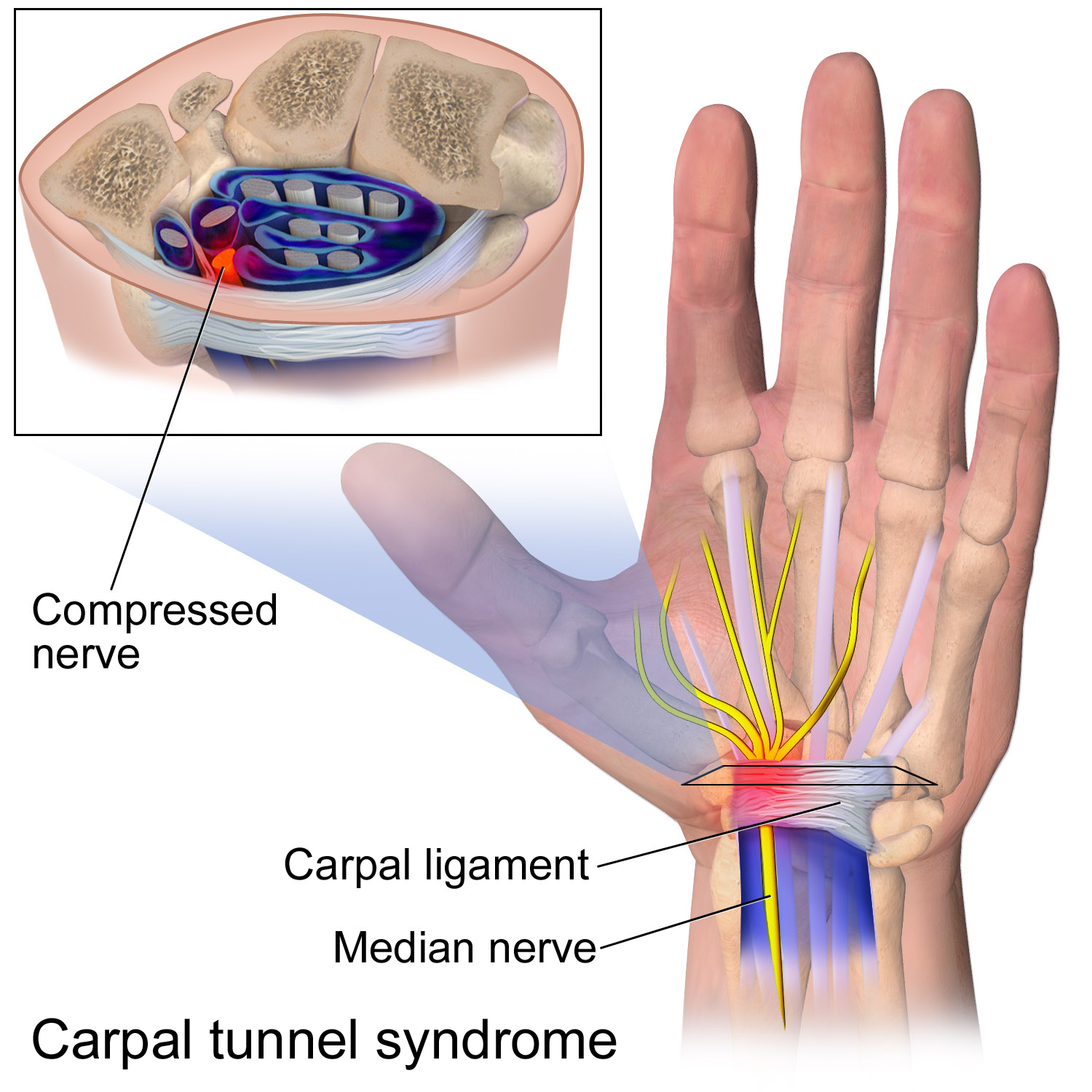
The flexor retinaculum (transverse carpal ligament, or anterior annular ligament) is a fibrous band on the palmar side of the hand near the wrist. It arches over the carpal bones of the hands, covering them and forming the carpal tunnel. Structure The flexor retinaculum is a strong, fibrous band that covers the carpal bones on the palmar side of the hand near the wrist. It attaches to the bones near the radius and ulna. On the ulnar side, the flexor retinaculum attaches to the pisiform bone and the hook of the hamate bone. On the radial side, it attaches to the tubercle of the scaphoid bone, and to the medial part of the palmar surface and the ridge of the trapezium bone. The flexor retinaculum is continuous with the palmar carpal ligament, and deeper with the palmar aponeurosis. The ulnar artery and ulnar nerve, and the cutaneous branches of the median and ulnar nerves, pass on top of the flexor retinaculum. On the radial side of the retinaculum is the tendon of the flexor c ... [...More Info...] [...Related Items...] OR: [Wikipedia] [Google] [Baidu] |
Inferior Transverse Ligament Of Scapula
The inferior transverse ligament (spinoglenoid ligament) is a weak membranous band, situated behind the neck of the scapula and stretching from the lateral border of the spine to the margin of the glenoid cavity. It forms an arch under which the transverse scapular vessels and suprascapular nerve enter the infraspinatous fossa The infraspinous fossa (infraspinatus fossa or infraspinatous fossa) of the scapula The scapula (plural scapulae or scapulas), also known as the shoulder blade, is the bone that connects the humerus (upper arm bone) with the clavicle (coll .... References Ligaments of the upper limb {{ligament-stub ... [...More Info...] [...Related Items...] OR: [Wikipedia] [Google] [Baidu] |
Inferior Transverse Ligament Of The Tibiofibular Syndesmosis
The inferior transverse ligament of the tibiofibular syndesmosis is a connective tissue structure in the lower leg that lies in front of the posterior ligament. It is a strong, thick band, of yellowish fibers which passes transversely across the back of the ankle joint, from the lateral malleolus to the posterior border of the articular surface of the tibia, almost as far as its malleolar process. This ligament projects below the margin of the bones, and forms part of the articulating surface for the talus. It is not included in Terminologia Anatomica ''Terminologia Anatomica'' is the international standard for human anatomical terminology. It is developed by the Federative International Programme on Anatomical Terminology, a program of the International Federation of Associations of Anatomi ..., but it still appears in some anatomy textbooks. References Ligaments Lower limb anatomy {{Portal bar, Anatomy ... [...More Info...] [...Related Items...] OR: [Wikipedia] [Google] [Baidu] |
Superior Transverse Ligament Of The Scapula
The superior transverse ligament (transverse or suprascapular ligament) converts the suprascapular notch into a foramen or opening. It is a thin and flat fascicle, narrower at the middle than at the extremities, attached by one end to the base of the coracoid process and by the other to the medial end of the scapular notch. The suprascapular nerve always runs through the foramen; while the suprascapular vessels cross over the ligament in most of the cases. The suprascapular ligament can become completely or partially ossified Ossification (also called osteogenesis or bone mineralization) in bone remodeling is the process of laying down new bone material by Cell (biology), cells named osteoblasts. It is synonymous with bone tissue formation. There are two processes .... The ligament also been found to split forming doubled space within the suprascapular notch. References External links * Ligaments of the upper limb {{ligament-stub ... [...More Info...] [...Related Items...] OR: [Wikipedia] [Google] [Baidu] |
Superior Extensor Retinaculum Of Foot
The superior extensor retinaculum of the foot (transverse crural ligament) is the upper part of the extensor retinaculum of foot which extends from the ankle to the heelbone. The superior extensor retinaculum binds down the tendons of extensor digitorum longus, extensor hallucis longus, peroneus tertius, and tibialis anterior as they descend on the front of the tibia and fibula; under it are found also the anterior tibial vessels and deep peroneal nerve. It is found on the lateral side of the lower leg, attached laterally to the lower end of the fibula, and medially to the tibia; above it is continuous with the fascia of the leg. Additional images File:Gray437.png, Muscles of the front of the leg. See also * Peroneal retinacula The fibular retinacula (also known as peroneal retinacula) are fibrous retaining bands that bind down the tendons of the fibularis longus and fibularis brevis muscles as they run across the side of the ankle. (''Retinaculum'' is Latin for "retaine ... [...More Info...] [...Related Items...] OR: [Wikipedia] [Google] [Baidu] |
Transverse Acetabular Ligament
The transverse acetabular ligament (transverse ligament or Tunstall’s ligament) is a portion of the acetabular labrum, though differing from it in having no cartilage cells among its fibers. It consists of strong, flattened fibers, which cross the acetabular notch, and convert it into a foramen through which the nutrient vessels enter the joint. It is an intra-articular structure of the hip. Function The transverse acetabular ligament prevents inferior displacement of head of femur The femur (; ), or thigh bone, is the proximal bone of the hindlimb in tetrapod vertebrates. The head of the femur articulates with the acetabulum in the pelvic bone forming the hip joint, while the distal part of the femur articulates with .... Additional Images File:Slide2DAD.JPG, Hip joint. Lateral view. Transverse acetabular ligament File:Slide2DADA.JPG, Hip joint. Lateral view. Transverse acetabular ligament References External links * Ligaments of the lower limb {{l ... [...More Info...] [...Related Items...] OR: [Wikipedia] [Google] [Baidu] |
Transverse Humeral Ligament
The transverse humeral ligament (Brodie's ligament) forms a broad band bridging the lesser and greater tubercle of the humerus. Its attachments are limited superior to the epiphysial line. By enclosing the canal of the bicipital groove The bicipital groove (intertubercular groove, sulcus intertubercularis) is a deep groove on the humerus that separates the greater tubercle from the lesser tubercle. It allows for the long tendon of the biceps brachii muscle to pass. Structure ... (intertubercular groove), it functions to hold the long head of the biceps tendon within the bicipital groove. References Ligaments of the upper limb {{ligament-stub ... [...More Info...] [...Related Items...] OR: [Wikipedia] [Google] [Baidu] |
Transverse Ligament Of The Atlas
In anatomy, the transverse ligament of the atlas is a ligament which arches across the ring of the atlas (the topmost cervical vertebra, which directly supports the skull), and keeps the odontoid process in contact with the atlas. Anatomy It is concave in front, convex behind, broader and thicker in the middle than at the ends, and firmly attached on either side to a small tubercle on the medial surface of the lateral mass of the atlas.Gray's anatomy, 1918 As it crosses the odontoid process, a small fasciculus (''crus superius'') is prolonged upward, and another (''crus inferius'') downward, from the superficial or posterior fibers of the ligament. The former is attached to the basilar part of the occipital bone, in close relation with the membrana tectoria; the latter is fixed to the posterior surface of the body of the axis; hence, the whole ligament is named the cruciate ligament of the atlas. The transverse ligament divides the ring of the atlas into two unequal parts: of the ... [...More Info...] [...Related Items...] OR: [Wikipedia] [Google] [Baidu] |


